GERD (reflux esophagitis) is one of the common diseases of the digestive system.
If the disease is combined with an inflammatory process affecting the lower esophagus, gastroesophageal reflux with esophagitis develops. A disease such as GERD with esophagitis, the treatment of which must be timely, is caused by frequently repeated reflux of stomach contents, as well as enzymes involved in the digestive process, into the esophagus.
If such a reflux occurs after eating, then this is a normal phenomenon and is not a pathology. But when such reflux of stomach contents occurs, regardless of food intake, then these are already prerequisites for the disease.
The mucous membrane of the esophageal tube is susceptible to the acidic environment of gastric secretions, so inflammation occurs with corresponding symptoms.
Gastroesophageal reflux with esophagitis: what kind of disease is it?
Gastroesophageal reflux disease is a disease of the esophagus, which is characterized by the presence of an inflammatory process in the mucous membrane of the distal esophageal tube. It is also called reflux esophagitis, Barrett's esophagus, gastroesophageal reflux.
Normally, the esophagus should not contain gastric contents, as well as its secretions, which have an acidic environment and negatively affect the epithelium of the esophageal tube. When these substances frequently enter the esophagus, irritation, swelling and inflammation of the mucous membrane of the organ initially occurs.
With further progression of the disease, erosive and ulcerative defects appear , which subsequently lead to the formation of scars and stenosis of the esophageal tube.
If this disease is left untreated for a long time, Barrett's esophagus may develop. This is a very serious complication of esophagitis, in which multilayered squamous cells of the esophageal epithelium are replaced by single-layer cylindrical ones.
Such an esophagus requires serious treatment and constant monitoring, since it is considered a precancerous condition.
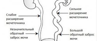
Frequent reflux of gastric secretions into the distal esophagus occurs as a result of insufficient function of the cardia - the muscular ring that separates the stomach from the esophageal tube. Through a loosely closed sphincter, secretion flows back into the esophagus.
GERD is not an independent disease, but a consequence of other disorders in the body.
The causes of such a disease as gastroesophageal reflux disease with esophagitis are considered:
- hiatal hernia;
- stomach and duodenal ulcers;
- congenital pathology of the esophagus;
- increased body weight;
- cholecystitis;
- surgical interventions.
Provoking factors for the development of this disease are:
- stress;
- work associated with constant bending of the body forward;
- pregnancy;
- spicy, fatty foods;
- smoking;
- pregnancy.
Gastroesophageal disease has two types of course: with and without esophagitis. Quite often, gastroesophageal reflux with esophagitis is diagnosed, a description of which is given below.
Causes
A number of factors contribute to the development of reflux of gastric contents into the esophagus. Among them:
- incompetence of the lower esophageal sphincter;
- transient episodes of relaxation of the lower esophageal sphincter;
- insufficiency of esophageal clearance;
- painful changes in the stomach that increase the severity of physiological reflux.
The protective, “anti-reflux” function of the lower esophageal sphincter is ensured by maintaining the tone of its muscles, the sufficient length of the sphincter zone and the location of part of the sphincter zone in the abdominal cavity. In a fairly large proportion of patients, a decrease in pressure in the lower esophageal sphincter is detected; in other cases, episodes of transient relaxation of his muscles are observed.
It has been established that hormonal factors play a role in maintaining the tone of the lower esophageal sphincter. A number of medications and certain foods help reduce pressure in the lower esophageal sphincter and develop or maintain reflux.
The location of part of the sphincter zone in the abdominal cavity, below the diaphragm, serves as a wise adaptive mechanism that prevents the reflux of gastric contents into the esophagus at the height of inspiration, at a time when this is facilitated by increasing intra-abdominal pressure.
At the height of inspiration, under normal conditions, “squeezing” of the lower segment of the esophagus occurs between the legs of the diaphragm. In cases of hiatal hernia formation, the final segment of the esophagus is displaced above the diaphragm. “Pressing” of the upper part of the stomach by the legs of the diaphragm interferes with the evacuation of acidic contents from the esophagus.
Thanks to the contraction of the esophagus, the natural cleansing of the esophagus from acidic contents is maintained, and normally the indicator of intraesophageal acidity does not exceed 4. The natural mechanisms through which cleansing is carried out are as follows:
- motor activity of the esophagus;
- salivation; The bicarbonates contained in saliva neutralize the acidic contents.
Disturbances on the part of these links contribute to a decrease in the “cleaning” of the esophagus from acidic or alkaline contents that have entered it.
GERD with reflux esophagitis
GERD with esophagitis: what it is, we have already figured it out. It is important to know that the disease has an acute and chronic course, accompanied by damage to the mucous membrane of the esophageal tube. There are different degrees of damage to the esophageal mucosa.
Grade 1 - characterized by the presence of single ulcers or erosive defects. They are small and do not exceed half a centimeter in size. Only the lower part of the esophagus is affected.
Degree 2 - has more extensive lesions, in which not only the upper layer of the epithelium is involved in the process, but also the tissues underlying it. Ulcerations are single or multiple, capable of merging. Erosions or ulcers are larger than half a centimeter. In this case, the lesion is located within one fold. Symptoms appear after eating.
Degree 3 - erosive or ulcerative defects extend beyond one fold, spread around the circumference of the inner wall of the esophagus, but do not affect more than 75% of the mucous membrane in a circle. Symptoms do not depend on whether the patient has eaten or not.
Grade 4 - ulcers and erosions can spread throughout the entire circumference of the esophagus. This is a very severe degree of the disease, which causes complications in the form of stenosis, bleeding, suppuration, and the development of Barrett's esophagus.
Depending on the degree of pathological changes in the epithelium of the esophagus, the disease has the following classification by type.
Catarrhal appearance - hyperemia of the epithelium without ulcers and erosions. Develops when exposed to rough food, spicy, hot food, strong drinks. May occur after mechanical trauma (fish and fruit bones).
Edema – the presence of edema of the esophagus, accompanied by a narrowing of the lumen of the organ.
Erosive - erosions and ulcers appear on the inflamed areas of the epithelium, the esophageal glands enlarge, and cysts form.
A characteristic symptom of this period is a cough with mucous secretion. Pseudomembranous - fibrous formations appear on the mucosa.
After their separation, ulcers and erosions form on the esophageal mucosa. Characteristic symptoms: cough and vomiting mixed with fibrin films. Exfoliative – separation of fibrin films from the walls of the esophagus. This causes the patient to have a severe cough, pain, and bleeding.
Necrotic - necrosis of parts of the tissues of the esophagus, a precancerous condition.
Phlegmatous - purulent inflammation caused by infection of nearby organs.
Prevention measures
GERD is quite difficult to treat and can cause serious complications. Therefore, it is better to prevent the development of this pathology in advance.
- Consume less carbonated drinks, fatty foods, chocolate and alcohol.
- Don't pull the belt on your skirt or pants too tight.
- After a meal, try not to bend over or lie down for 1-2 hours.
There are also ways to help prevent another flare-up of GERD:
- Sleep on a fairly high headboard.
- Stop smoking. Smoking on an empty stomach is especially harmful.
- If you are overweight, try to get rid of it.
- Try not to overeat. It is better to eat often, but in small portions.
- Don't talk while eating. Chew your food well.
- Don't lift anything heavy.
- Try to take less medications that can relax the esophageal sphincter.
These simple measures will help you consolidate the results of treatment and minimize the likelihood of relapses.
GERD is a chronic disease, so it cannot be completely cured. Erosion and ulcers of the esophagus heal with treatment within 1-2 months. If the regime or diet is violated, the probability of relapse within a year reaches 98%.
To prevent exacerbations, maintenance therapy is prescribed, the duration of which is determined individually. Within 3 years from the date of the last relapse, patients should be observed by a gastroenterologist.
Symptoms of GERD with esophagitis
The clinical picture of this disease has esophageal and non-esophageal symptoms . The first category includes:
- dysphagia;
- pain;
- heartburn;
- belching.
The most characteristic manifestation of exophagitis is heartburn , which is accompanied by a painful syndrome localized behind the sternum. Such unpleasant sensations appear during physical work associated with constant bending of the torso forward, as well as in a lying position, with a reflex contraction of the esophagus due to nervous spasm.
Soreness and burning appear as a result of the negative effect of the acidic environment on the esophageal mucosa when gastric secretions are thrown back into the distal region of the esophageal tube.
But often patients do not pay attention to this symptom and consult a doctor. Then the disease enters the second phase of development.
With further progression of the disease, patients may experience belching, which indicates dysfunction of the sphincter located between the stomach and esophagus. Most often it occurs during sleep.
This symptom is dangerous because food masses can enter the respiratory tract and lead to suffocation. Also, food entering the respiratory tract provokes the development of aspiration pneumonia.
Dysphagia appears later in the development of the disease and is characterized by impaired swallowing.
Non-esophageal symptoms are the appearance of:
- caries;
- reflux laryngitis and pharyngitis;
- sinusitis.
With GERD, chest pain is of the “cardiac” type and can be confused with an attack of angina, but it will not be relieved by nitroglycerin, and the appearance of the pain syndrome is not associated with physical activity or stress.
If the symptoms include shortness of breath, cough, suffocation, then the disease develops according to the bronchial type.
Treatment of GERD with esophagitis
What is the treatment regimen for GERD with esophagitis? Therapy for this disease consists of:
- drug treatment;
- surgical intervention;
- not drug treatment.
How to treat GERD reflux esophagitis? Treatment with medications is aimed at reducing the negative impact of the acidic environment on the mucous membrane of the esophagus, accelerating regenerative processes and preventing relapses of the disease.
Antacids - reduce the acidity of gastric juice (phosphalugel, almagel). Read more about this in the article Antacids for reflux esophagitis.
Alginates - form a protective film on the surface of the food mass, which neutralizes hydrochloric acid, which is part of the gastric juice. When food is thrown back into the esophagus, the epithelium is not irritated by gastric contents ( Gaviscon ).
Prokinetics - improve the contractile function of the esophagus, promote the rapid movement of food through the esophageal tube, increase the force of contraction of the sphincter muscles, which prevents the reflux of stomach contents (cerucal, mothylium).
Proton pump inhibitors - reduce the production of gastric juice, which will reduce the negative effect on the mucous membrane of the esophagus (omez, omeprazole, pantoprazole).
For the speedy restoration of the affected epithelium, solcoseryl and alanton .
After treatment with medications, an endoscopic examination should be performed to confirm the positive effect of the therapy.
Surgical treatment
If symptoms persist after treatment, and there are other indications for surgery, then surgery is performed.
Surgical treatment is carried out in the presence of:
- stenosis;
- Barrett's esophagus;
- frequent bleeding;
- ineffectiveness of conservative therapy;
- frequent aspiration pneumonia.
Surgery is performed using the classical method (an incision is made on the abdomen or chest), as well as laparoscopy (a minimally invasive method that minimally affects healthy tissue).
Non-drug treatment
Such therapy includes:
- dietary nutrition;
- weight loss;
- rejection of bad habits.
In addition, you need to limit your intake of medications that increase the risk of developing reflux ( theophylline, tranquilizers ). It is also necessary to avoid stress on the abdominal area, and not to wear tight clothing, corsets or belts.
Dietary nutrition excludes hot, spicy, fatty foods. Include vegetables, fruits, cereals, and vegetable stews in your diet.
Gastroesophageal reflux without esophagitis
Gastroesophageal reflux without esophagitis: what is it and how to treat? It should be noted that a disease such as gastroesophageal reflux disease without esophagitis develops due to the reflux of stomach contents into the esophagus, but there are no erosive and ulcerative lesions of the mucosa.
The clinical picture of a disease such as reflux without esophagitis is marked by the following symptoms:
- heartburn;
- belching;
- sore throat;
- inflammation of the throat, nasopharynx, oral cavity;
- tooth decay.
The reasons for the development of GERD without esophagitis are:
- poor nutrition;
- frequent vomiting (toxicosis, poisoning, taking medications);
- obesity;
- bad habits;
- coffee addiction.
The main methods of treating this disease are taking medications (antacids and alginates) and following a diet.
Diagnostic methods
In order to confirm the fact of the patient’s illness and prescribe rational therapy, it is necessary to undergo additional examinations. The most informative are the following research methods:
- Esophagoscopy.
- 24-hour intraesophageal pH-metry.
- Provocative tests.
- Esophagomonometry.
Esophagoscopy
The examination allows you to identify and evaluate the degree of development of reflux esophagitis, possible complications, and exclude the presence of an esophageal tumor.
With reflux esophagitis, the following changes in the internal wall of the organ are noted:
- Redness and swelling of the mucous membrane.
- The presence of submucosal hemorrhages and superficial erosions.
- Increased vulnerability of the mucous membrane and easy bleeding when touched with a probe.
The extent of damage to the esophagus, which was mentioned above, is determined by the prevalence of the pathological process.
The diagnosis must be confirmed by performing a biopsy. A situation often occurs when a patient has severe symptoms of the disease, but there are no signs of pathology on the endoscopic picture.
24-hour intraesophageal pH-metry
The study is carried out using a special electrode that measures the pH of the environment. The device is lowered using a probe and fixed 5 cm above the LES. The normal pH level in the esophagus is 7.0-8. 0. At the moment of reflux of the acidic contents of the stomach, this indicator decreases and becomes less than 4.0.
The technique allows you to assess the frequency, duration and daily dynamics of reflux within 24 hours. For this purpose, the following indicators are determined:
- The total time the pH decreases below 4.0 in the horizontal and vertical position of the patient.
- Number of refluxes per day.
- The number of surges of gastric juice lasting more than 5 minutes.
- Duration of the longest reflux
- The value of the “symptom index” is the ratio of the number of GERD symptoms (for example, heartburn), which coincides with a decrease in pH less than 4.0 to the total number of manifestations of the disease that arose during the examination.
Provocative tests
One of the options for pH measurement is the use of provocative tests. They help to identify disturbances in the functioning of the esophagus and LES, since it is not always possible to see the spontaneous appearance of signs of pathology using the above methods.
The most commonly used test is the acid test. For this purpose, a pH electrode is installed on the patient, as in the previous method. Using a catheter, dilute hydrochloric acid is injected into the stomach, after which the change in pH of the esophagus is observed. During the examination, the doctor asks the patient to breathe deeply and cough.
The examination is performed in a supine position, on the right and left side, with the head bowed. Patients with GERD experience a decrease in pH below 4.0 and the appearance of symptoms of the disease.
Esophagomonometry
This method is used to measure LES pressure and the contractility of the esophageal muscles. If the sphincter tone decreases below 10 mm Hg, reflux esophagitis can be assumed. This study does not reflect the degree of damage to the mucous membrane or whether the patient has complications of GERD. In this regard, esophagomonometry is recommended in combination with previous examinations.