Structure of the cornea
The diameter of the cornea averages 11.5 mm vertically and up to 12 mm horizontally; its thickness is heterogeneous: in the center it is approximately 500 microns, and at the periphery it can reach 1 mm.
The cornea consists of 5 layers: the anterior epithelial layer, Bowman's membrane, stroma, Descemet's membrane and the inner endothelial layer.
- The anterior epithelial layer is a flat stratified non-keratinizing epithelium endowed with a protective function. It is resistant to mechanical stress and quickly recovers when damaged. Due to the ability of the epithelium to quickly regenerate, scars do not form on it.
- Bowman's membrane is an acellular layer of the surface of the stroma. Its damaged surface undergoes scarring.
- Stroma is corneal tissue, occupying about 90% of its thickness. It is composed of correctly oriented collagen fibers, in which the intercellular space is filled with keratan sulfate and chondroitin sulfate.
- Descemet's membrane is the basement membrane of the corneal endothelium, which is a network of thin collagen fibers. Serves as a reliable barrier to infection.
- The corneal endothelium is a monolayer of cells that have a hexagonal shape. It plays one of the main roles in nourishing and maintaining the functions of the cornea, preventing its swelling under the influence of IOP. Does not have the ability to regenerate. With age, the number of its cells gradually decreases.
The endings of the first branch of the trigeminal nerve take part in the innervation of the cornea. The process of nutrition of the cornea is carried out due to the network of blood vessels, as well as nerves, tear film and moisture of the anterior chamber.
Eyeball
The eyeball occupies the main place in the orbit or orbit, which is the bony seat of the eye and also serves to protect it. Between the orbit and the eyeball there is fatty tissue, which performs shock-absorbing functions and contains blood vessels, nerves and muscles. The eyeball weighs about 7 grams.
Eyeball
is a sphere with a diameter of about 25 mm, consisting of three shells.
The outer, fibrous membrane consists of an opaque sclera
about 1 mm thick, which passes into the cornea in front.
On the outside, the sclera is covered with a thin transparent mucous membrane - the conjunctiva.
.
The middle layer is called choroid
. From its name it is clear that it contains a lot of vessels that nourish the eyeball. It forms, in particular, the ciliary body and the iris. The inner layer of the eye is the retina.
Eye muscles
The eye also has an appendage
, in particular, the eyelids and lacrimal organs.
Eye movements are controlled by six muscles
- four rectus muscles and two oblique muscles. In terms of their structure and functions, the eyes can be compared to the optical system of, for example, a camera. The image on the retina (analogue of photographic film) is formed as a result of the refraction of light rays in the system of lenses located in the eye (cornea and lens) (analogue of a lens). Let's look at how this happens in more detail.
Protective function of the cornea
The cornea is the outer protective shell of the eye, and therefore is the first to be exposed to the harmful effects of the environment: the ingress of mechanical particles onto its surface, the influence of chemicals suspended in the air, air movement, temperature, etc.
The properties of the protective function of the cornea are determined by its high sensitivity. The slightest irritation of its surface, for example by a particle of dust, causes an instant unconditioned reflex in a person, expressed in the closing of the eyelids, increased lacrimation and photophobia. Likewise, the cornea protects the eye from possible damage. When the eyelids close, the eyeballs simultaneously roll up and there is a profuse secretion of tears, which wash away small mechanical particles or chemicals from the surface of the eye.
How to treat
To avoid dangerous consequences leading to partial or complete loss of vision, it is necessary to carry out treatment on time. For many diseases of the cornea, surgical intervention is indicated. In rare cases, eye drops that provide more symptomatic treatment may be used. The following are considered effective:
Some symptoms of the pathology can be eliminated with the help of Taufon drops.
- "Balarpan";
- "Taufon";
- "Emoxipin";
- "Actovegin";
- "Solcoseryl".
In case of disease, corneal crosslinking is performed, which is indicated in case of progression of keratoconus. Keratotomy is also often performed, which is required to level the horny surface. In the process of keratoplasty, tissues of the corneal layer are transplanted in order to eliminate deformation processes. Before performing any surgical intervention, it is necessary to undergo a comprehensive examination in order to prevent dangerous complications.
Symptoms of corneal damage in various diseases
Changes in the shape of the cornea and its refractive power
- Myopia makes the shape of the cornea steeper in relation to the norm, this determines its greater refractive power.
- Farsightedness, on the contrary, flattens the cornea and its optical power decreases.
- Astigmatism accompanies the irregular shape of the cornea, manifesting itself in various planes.
- There are congenital changes in the corneal form - megalocornea and microcornea.
Damage to the surface of the corneal epithelium:
- Point erosions are small defects in the epithelium, revealed by fluorescein staining. This nonspecific sign of corneal diseases can be observed with spring catarrh, dry eye syndrome, inadequate selection of contact lenses, keratitis, lagophthalmos, and sometimes it is caused by the toxic effect of local ophthalmic drugs.
- Swelling of the corneal epithelium is evidence of damage to the endothelial layer or a rapid and significant increase in IOP.
- Punctate epithelial keratitis is a manifestation of viral infections of the eyeball. It is characterized by granular, swollen epithelial cells.
- Threads are thin, comma-shaped, mucous strands connected on one side to the surface of the cornea. They are detected with keratoconjunctivitis, dry eye syndrome, and recurrent erosion of the cornea.
Damage to the corneal stroma:
- Infiltrates are areas of active inflammation in the cornea. They can have a non-infectious (when wearing contact lenses) and infectious nature - bacterial, fungal, viral keratitis.
- Swelling of the stroma, manifested by an increase in the thickness of the cornea and a decrease in its transparency. Observed in keratitis, Fuchs' dystrophy, keratoconus, endothelial damage due to ophthalmological operations.
- Ingrowth of blood vessels (vascularization) becomes a manifestation of the outcome of an inflammatory disease of the cornea.
- Damage to Descemet's membrane.
- The folds are the result of surgical trauma.
- Tears can appear due to injury to the cornea, and also occur with keratoconus.
Consequences, complications
Even a minor injury to the cornea can lead to complications; patients with weakened immune systems and metabolic disorders are at risk. The consequences of injury can be quite serious; the patient faces complete irreversible loss of visual function. Such complications are often observed against the background of penetrating wounds, as well as due to chemical burns of the cornea. Seeking medical help will help you avoid negative consequences.
Against the background of deep burns, secondary glaucoma often occurs; this category includes a group of diseases that arise as a result of the outflow of intraocular fluid. After injury, rough scars, corneal swelling, clouding of the vitreous, pupil displacement, and increased intraocular pressure may also appear on the cornea.
There is a risk of traumatic cataracts, accompanied by clouding of the lens and deterioration of visual function. Resorption of cataracts can cause aphakia, in which the lens is missing from the eye.
Severe consequences are quite rare; timely medical care plays an important role. If the processing rules are violated, the following consequences may occur:
- Sepsis - this condition occurs due to the penetration of infections into the circulatory system and toxicity of the body;
- Complete loss of the eyeball;
- Persistent decrease in visual function;
- Brain abscess - accompanied by an accumulation of purulent fluid in the skull;
- Scarring of tissue;
- Sympathetic inflammation - accompanied by damage to the second eyeball;
- Fibroplastic iridocyclitis;
- Endophthalmitis;
- Panophthalmitis;
- Deformation of facial tissues;
- Pathologies of the lacrimal apparatus.
Diagnosis of corneal pathologies
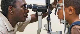
- Biomicroscopy is an examination of the cornea in the light of a slit lamp, which makes it possible to identify almost the entire range of diseases.
- Pachymetry is the measurement of the size of the cornea using an ultrasound device.
- Mirror microscopy is a photographic scanning of the corneal endothelial layer, counting the number of cells and analyzing its shape. Normally, the cell density is 3000 per 1 mm2.
- Keratometry is the study of the curvature of the anterior corneal surface.
- Topography is a computer study concerning the entire corneal surface, with an accurate analysis of its shape and refractive power capabilities.
- Microbiological studies - scraping from the surface (under drip anesthesia). If scraping results are inconclusive, a corneal biopsy may be performed.
Developmental anomalies subject to correction
A large cornea or megalocornea is found in babies at birth. The norm is considered to be a shell diameter of 10 mm. If it exceeds by at least 1 mm, then this is a corneal symptom. The refraction of the eye is impaired; the larger the radius of curvature of the lens, the lower the refractive power.
The defect is dangerous for a number of complications - glaucoma, cataracts, problems with the retina. Megalocornea is often a symptom of complex genetic diseases (Markesani syndrome, Marfan syndrome, etc.). The disease has no cure. To improve vision, corrective glasses and lenses are prescribed.
Microcornea or small cornea. The opposite case. The shell, on the contrary, is less than 10 mm in diameter. The pupil appears unnaturally small. A consequence of microcornea, farsightedness. Glaucoma, cataracts, etc. develop against the background of the disease. If the cornea remains transparent, the doctor will prescribe symptomatic treatment and telescopic devices for vision correction.
Return to contents
| Both cases (macro-, microcornea) are of genetic origin and are inherited. |
Treatment of corneal diseases
When changes in the shape, as well as the refractive power of the cornea accompanying myopia, farsightedness, and astigmatism, vision correction should be carried out using glasses, contact lenses or refractive surgery.
Persistent opacities and corneal cataracts are eliminated by keratoplasty surgery, corneal endothelium transplantation.
In case of corneal infection, antibacterial, antiviral, or antifungal drugs may be used, depending on the nature of the infectious agent.
In addition, topical glucocorticoids are recommended to suppress the inflammatory response while limiting the scarring process. For superficial damage to the cornea, drugs that accelerate regeneration are also needed. When the tear film is depleted, moisturizers and tear replacement agents are used. Molchanova Anna Alexandrovna
Allergic keratitis
Eye health is a necessary condition for human well-being. Keratitis - inflammation of the cornea of the eye - puts this well-being at risk. In some cases, this disease is caused by an allergic reaction.
When we talk about keratitis, we mean an inflammatory process that affects the cornea of the eye. This disease is very dangerous and requires immediate medical intervention. There are fungal, bacteriological, herpetic, syphilitic, tuberculosis varieties. Next to them, an allergic form seems like a mere trifle. But it also causes the patient a lot of suffering.
Examination methods
Examination methods include examination of R. under lateral illumination using two magnifying glasses, biomicroscopy (see Biomicroscopy of the eye), keratoscopy (see Ophthalmometry), keratography (see), mirror contact and non-contact microscopy of the endothelium using a mirror endothelial microscope, determination of R permeability using local electrophoresis with fluorescein (keratopenetrofluorimetry), as well as determining the sensitivity of R. with various algesimeters (metal hairs of different diameters).
A mirror-like shiny and moist surface is one of the properties of normal R. Thanks to it, it is possible to obtain a mirror image of various objects placed in front of the eye. The use of various devices (keratoscope, photokeratoscope, ophthalmometer, etc.) is based on this principle, with the help of which the shape, curvature and refraction of the cornea are determined. Photokeratoscope is a device created on the basis of the Placido keratoscope, which allows you to photograph the keratoscope rings on film and subsequently carry out a mathematical analysis of the resulting image.
See also Examination of the patient, ophthalmological.
First aid
First aid when an erosive defect of the cornea appears is to contact a specialist. If any foreign substances get into the mucous membranes of the eyes, you need to wash them under running water as soon as possible. An ophthalmologist will assess the degree of tissue damage, conduct an examination in a short time and make an accurate diagnosis. Only after this can a treatment regimen be prescribed.
Important! To prevent complications, it is prohibited to use medications without prior consultation with a professional. Medicines can muffle the characteristic symptoms of the disease and complicate the upcoming diagnosis. Corneal scars can be removed using laser treatment
It is effective for superficial defects and avoids keratoplasty and the use of a graft. An ophthalmologist performs laser ablation of the upper layers of the cornea, which allows you to remove scar tissue
Corneal scars can be removed using laser treatment. It is effective for superficial defects and avoids keratoplasty and the use of a graft. An ophthalmologist performs laser ablation of the upper layers of the cornea, which allows the removal of scar tissue.
