The macula of the eye, or the central macula of the retina, as it is also called, is responsible for central vision. Thanks to this element of the visual apparatus, we easily distinguish small details and can see the world around us in bright light. This feature is due to the high concentration of cones and rods in the center of the retinal tissue.
Degeneration or dysfunction of this spot can significantly impair the functioning of the entire visual system. Therefore, it is important to remember that the loss of the central sector from the image may indicate a violation of the structure of the macula area.
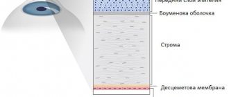
Structure
Functions
Symptoms
Treatment
Causes of retinal macular degeneration
Unfortunately, the causes of macular degeneration of the retina have not been fully studied by science to date. However, doctors still identify some unfavorable factors that may contribute to the development of the disease:
- Aging of the human body;
- Genetic predisposition;
- Poor environmental conditions;
- Problems with excess weight;
- Bad habits;
- Deficiency of vitamins and microelements;
- The presence of a number of chronic diseases.
Although macular degeneration of the retina can (albeit very rarely) occur at a young age. People of mature and retirement age are at particular risk. According to recent studies conducted on 2,000 volunteers, it was found that less than 3% of people between 25 and 35 years of age have a certain risk of developing the disease, while in older people over 70 years of age this percentage increased 10 times.
Causes of macular degeneration
In fact, the disease is multifactorial, since so far no direct causes have been identified. Factors include the following:
- Age-related macular degeneration occurs against the background of age-related changes. The disease develops starting at the age of 50.
- Female. For unknown reasons, women are most susceptible to developing macular degeneration of the retina.
- The hereditary factor plays an important role, since pathology is transmitted at the genetic level.
- Abuse of ultraviolet radiation. This means frequent visits to the solarium and prolonged exposure to the open sun without sunglasses. But everyone knows that ultraviolet radiation has a negative effect on the retina of the eyes.
- Tobacco smoking, alcohol abuse, drug use.
- Diseases of the heart and blood vessels. For example, strokes, atherosclerosis, blood pressure changes, myocardial infarction, etc.
- Obesity and poor nutrition, vitamin deficiencies.
- Diseases of the visual apparatus and consequences after eye surgery.
Types of retinal macular degeneration
In ophthalmology, it is customary to distinguish 2 forms of this disease:
- Dry macular degeneration. Among all patients suffering from degenerative changes in the retina, 10% have the dry form of macular degeneration. It is characterized by structural changes in the retina, as well as its thinning at the molecular level, characteristic of the natural aging of the human body. Due to degenerative processes, the blood supply to the retina is disrupted, and in the absence of supportive treatment, visual acuity decreases.
- Wet macular degeneration. This form of the disease is much less common, only in 10% of all cases. It is characterized by a rapid course, as a result of which pathological vessels appear in the eyeball. New vessels are characterized by increased fragility, so very often they rupture in place of which scars form. With this form of the disease, patients experience a rapid decrease in visual acuity up to its complete loss.
Treatment with leeches for glaucoma: setting points
Treatment with leeches for glaucoma is also used to relieve an attack in small children, even infants with congenital glaucoma, 1-2 pieces for 2-3 minutes. In general, patients with an acute attack receive relief from the first application of leeches for glaucoma - after 20-30 minutes, unbearable pain in the eye and temple, vomiting, nausea, and fog in the eyes disappear.
After a course of leeches for glaucoma in patients with various abnormalities, inflammation is relieved, visual acuity increases, the eye muscle relaxes, the field of vision expands, intraocular pressure decreases, and overall well-being improves. In patients with diabetes, the walls of blood vessels become more permeable, so the most common consequence of this disease is retinal hemorrhage, which in turn leads to further deterioration of vision and the emergence of a number of diagnoses. It is hirudin in leech saliva that can significantly improve the condition of the circulatory system.
First of all, the healing saliva of the leech acts on the optic nerve, the optic tract and its roots, reaching the anterior tubercles of the quadrigemina, where the primary visual centers are located, as well as in the cortical visual centers, where the fibers of the optic tract pass as part of the posterior part of the occipital part.
There are a number of standard points to which leeches are applied in ophthalmic patients. This is the upper part of the back, the “withers”, behind the ears (the mastoid processes are the raised part of the skull behind the ear - we place the bones on the “corner” immediately behind the ear), around the eyes on the bones, the point between the eyebrows is the “third eye” - it is easily determined by increased sensitivity to pressure.
Tags: eye, treatment, macular degeneration, folk, retina, remedy
About the author: admin4ik
« Previous entry
Symptoms of the disease
Macular degeneration in both forms is completely painless, but the symptoms of the disease differ significantly.
The dry form is characterized by:
- The appearance of blurred vision;
- Difficulties arise when reading, knitting, working at the computer and other activities that require increased visual load;
- Objects and images begin to be perceived in a distorted form;
- Confusion appears in recognizing people's faces.
All these signs appear due to destruction of light-sensitive cells in the macula. Deterioration of vision directly depends on the number of functioning cells, so if their death is rapid, then the symptoms of the disease will appear one after another just as quickly: the patient will feel that he needs more light to perform his usual work, although this will not solve the problem. The wet form of macular degeneration is characterized by:
- Rapid loss of central vision;
- Distortion of the visibility of straight lines. Distorted perception occurs due to a violation of the integrity of blood vessels and accumulation of fluid, resulting in increased pressure in the center of the retina;
- Enlargement of the blind spot. A particularly dangerous symptom, as it can lead to complete loss of vision.
What kind of disease is this?
At the center of the retina is the macula, the light-sensitive element. Macular degeneration is a disease of the retina that occurs due to vascular pathology and disruption of their nutrition. Due to these reasons, central vision is damaged.
Macular degeneration is considered an age-related disease, which most often causes blindness in people after 50 years of age.
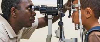
Diagnosis of retinal macular degeneration
Macular degeneration is fraught with serious complications, so it is very important to visit an ophthalmologist for an initial examination when the first symptoms of the disease appear: the doctor will examine the retina and measure visual acuity. If, based on the results of the initial examination, there is a suspicion of the presence of this disease, the doctor will prescribe additional examinations:
- Fluorescein angiography. The examination involves the introduction of a special contrast agent into a vein, a fluorescent dye to examine the vessels of the retina. If there are specific indications, both contrast agents can be administered on the same day. This method of diagnosing eye diseases is considered reliable and safe, and has been used in ophthalmology for more than half a century.
- Indocyanine – green angiography. This type of examination using indocyanine green plays a big role in the diagnosis of macular degeneration: the doctor can assess the functioning of blood flow in the choroid and identify the presence of degenerative changes in the macula.
These diagnostic methods can be performed simultaneously by sequentially administering drugs. Indocyanine green angiography and fluorescein angiography are considered absolutely safe research methods and have been successfully used in ophthalmology for more than half a century.
However, before prescribing these procedures, the doctor must check with the patient’s medical history for the presence of diseases:
- Liver;
- Of cardio-vascular system;
- Diabetes mellitus;
- Hypertension;
- The presence of allergic reactions to sodium fluorescein and iodine.
Examination during pregnancy is also not recommended, since there is currently no data on the safety of such diagnostic methods for the unborn child.
Methods for diagnosing the macula
A lot of different diagnostic techniques and tests are used to examine the macular area.:
- Ophthalmoscopy in various variations using special magnifying lenses.
- Optical coherence tomography allows you to obtain an image of all layers of the macular area with an accurate assessment of all parameters.
- Fluorescein angiography is performed using contrast agents that stain the retinal vasculature, which allows the detection of various diseases of the macular area.
- Computer perimetry reveals loss in the central field of vision - scotoma.
Now you know what the macula is and what problems arise with it. We hope this information was useful and interesting.
We recommend that you familiarize yourself with: the structure of the sclera of the eye.
Treatment of macular degeneration of the retina
Unfortunately, today there is no effective way to treat retinal macular degeneration. The disease is irreversible and it is not possible to restore vision - medicine does not know of cases of recovery of patients with this diagnosis. All methods of therapy are aimed exclusively at improving the nutrition of the retina, supporting the normal functioning of the blood vessels of the eye and slowing down their rapid growth.
The success of stopping the pathologically irreversible process that occurs under the influence of this disease directly depends on the time of seeking medical help and correctly prescribed treatment.
Therapy for the disease directly depends on its type - dry or wet and is treated differently, so ophthalmologists prescribe individual types and treatment regimens, according to the diagnosis and stage of macular degeneration:
- Conservative therapy. The wet form of macular degeneration is treated with vascular drugs, antioxidants and immunomodulators. In most cases, treatment is ineffective, since this form is already advanced, which patients treat when it is too late to do anything;
- Intravitreal administration of drugs. Treatment of the dry form of macular degeneration involves a course of intravitreal injections of Lucentis or Eilia, which slow down degenerative processes in the retina and normalize its thickness, activate blood supply, and relieve swelling. This method of treatment stops the pathological process, which allows the patient’s vision to be preserved without deterioration. The duration of such treatment is 2 calendar years. During this time, about 8 injections are performed, the cost of intravitreal treatment is about 60,000 rubles;
- Surgical intervention. The laser coagulation method is very common in ophthalmology in the treatment of many eye diseases. But in this case, surgical intervention is limited. During the operation, the doctor must block the pathological vessels of the eye that put pressure on the retina. Laser coagulation allows achieving the desired effect in 70% of cases. However, the positive dynamics are temporary: there is a high risk of the appearance of new pathological vessels, which will also have to be “clogged” during the next operation.
Often, patients with macular degeneration even resort to traditional medicine in the fight against their illness. But is it so effective and safe? Let's try to find answers to these difficult questions.
Question answer
Acceptable, but the Amsler test is only an overview additional check of vision status
Before making a diagnosis, it is important to undergo a complete examination by an ophthalmologist. Independent conclusions and self-analysis can lead to false conclusions
As for the test, its essence is that the patient puts on glasses, if they are prescribed to him, and looks at the center of a black grid depicted on a white background. Then he closes one eye, and meanwhile, with the other, he fixates on the central point and slowly brings his face closer to the monitor to a distance of 20-30 cm. Then he repeats the same with the other eye. If the lines on the grid, when approaching the drawing, look distorted, tortuous instead of straight, then this may indicate retinal dystrophy or changes in the fundus.
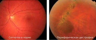
This is the image you see with normal vision:
And with retinal dystrophy, the pattern takes on the following forms:
The Amsler grating was developed by Swiss professor of ophthalmology Mark Amsler back in the 20th century and has already become a proven optical classic.
There are a number of risk factors for the development of retinal dystrophy:
- diabetes
- atherosclerosis
- obesity
- hypertonic disease
- lack of microelements and vitamins in the diet combined with regular stress
- excessive exposure to sunlight
- genetic predisposition
“Dry” macular degeneration (which is also called non-exudative) - it is characterized by the accumulation of cell waste products and residual chemicals (drusen) between the blood vessels and the retina. These chemical deposits collect under the retina as yellow bumps. The “dry” version of dystrophy prevails over its wet variety. Over time, the dry form can transform into a wet form (20% of the total number of dysfunctions)
“Wet” macular degeneration occurs on average in 10% of cases of the total number of pathologies. But against this background there is a significant risk of developing retinal detachment and macular edema. In this form, new blood vessels grow under the retina of the eye. The vessels are thin, liquid and blood penetrate through them. And this substance is called exudate. It is this that puts pressure on the retina and contributes to its detachment. Visual acuity decreases very sharply.
Treatment of the disease using traditional methods
Traditional methods of treatment are very popular among many patients. I would like to warn readers: traditional medicine is effective only for the dry form of the disease. Wet due to rapid progression should be treated exclusively through intravitreal injections and microsurgery. The dry form of the disease can be stopped using folk methods and prevent further deterioration.
Before starting treatment, you should reconsider your diet: give preference to fresh fruits and vegetables, herbs, and nuts. You should avoid eating high-calorie and fatty foods. Without meeting this condition, it is unlikely that you will be able to achieve improvements in your health.
We bring to your attention effective recipes for the treatment of macular degeneration:
- Wheat water. Grind the wheat sprouts in a blender, then take 50 g, add 200 ml of water and let it brew for several hours until completely swollen. Eat every morning for 4 calendar weeks, then take a break;
- Tincture of calendula. Pour 200 ml of boiling water over 1 tablespoon of flowers, cover with a saucer and allow to cool. After this, the resulting infusion must be filtered through a sieve and placed in a cool place. Take 50 g 3 times a day. It is also recommended to use the strained decoction as eye drops - every morning, 2 drops in each eye. Duration of treatment - 6 calendar months;
- Red rowan, sea buckthorn and blueberry. Take 50 g of red rowan, 50 g of blueberries, 50 g of sea buckthorn and grind on a sieve, add 20 g of honey and put in the refrigerator. Take 40 g before breakfast, lunch and dinner. Duration of treatment - 4-6 calendar weeks;
- Tincture of mumiyo and aloe. Dissolve 50 g of purified mumiyo in 100 g of aloe juice. Use 1 tablespoon morning and evening for 2 weeks, then take a break from treatment for 2 calendar weeks, and then you can repeat the course;
- Cumin decoction. Pour 1 tablespoon of caraway seeds, 1 tablespoon of cornflower flowers into 200 ml of boiling water, place in a water bath and boil for 5 minutes. Strain the resulting broth and let it cool. The finished infusion is instilled into the eyes in the morning and evening, 2 drops each, the duration of treatment should be at least 2 weeks. The decoction is stored exclusively in the refrigerator for no more than 48 hours; before use, it is recommended to warm it to room temperature.
Treatment of the wet form
There are three types of treatment for wet AMD:
- Photodynamic therapy.
Using a cold laser, the doctor clogs the blood vessels that are leaking. However, proper treatment requires frequent repetitions of the procedure and the use of extremely expensive drugs, so such treatment is not available in Ukraine.
Abroad, PDT is used in combination with the administration of VEGF factor inhibitors.
- Photocoagulation.
The doctor uses a laser to block the fragile blood vessels that are leaking. The high-energy laser beam targets the newly formed blood vessels directly and destroys them, stopping further vision loss. Laser treatment does not reduce the risk of new blood vessels, so treatment is often repeated.
Such treatment is aimed at the consequences of AMD, and not at the cause of its occurrence. Additionally, this therapy can only be used on vessels that are not in the macula. That is, it is impossible to achieve stabilization of vision or even improvement of visual functions only through photocoagulation.
- Endovitreal administration of VEGF inhibitors is used to treat wet AMD.
High levels of VEGF, a protein that causes new blood vessels to grow, cause blood vessels to break down and leak fluid. Due to the accumulation of fluid under the retina, it bends and rises from its usual position. Therefore, AMD patients often complain that lines appear distorted and objects appear irregular in shape.
In order to overcome the disease, it is necessary to stop the production of abnormal protein and “dissolve” the fragile vessels through the walls of which fluid enters under the retina.
It is for this purpose that during the operation the surgeon injects a drug into the eye, the action of which is aimed at a protein involved in the formation of new blood vessels. This procedure is carried out in the operating room.
Before the injection, your eye will be numbed with special drops, so you will not feel pain during the operation. After the painkiller wears off, you may feel some discomfort.
The eye may hurt, you may feel that something is floating in front of your eye, you may see non-existent stripes, circles, lines. Often patients complain of a feeling of a veil before their eyes.
This is due to the fact that the drug mixes very slowly with the internal structures of the eye; As soon as the drug is evenly distributed, all these sensations will disappear. Your vision may be somewhat blurry for a few days after the procedure.
Complications
The main complications after such an operation are increased intraocular pressure and eye infection. It is to avoid these complications that after the procedure you will be given anti-inflammatory and other special drops that will reduce the risk of deterioration to a minimum.
Lucentis and Eylia have been proven to help patients improve and maintain vision, stopping the destructive effects of the disease and changing its course. "Makugen" allows you to stabilize vision and stop its further deterioration.
Treatment of dry AMD with antioxidants and zinc
Classical medicine recommends treating the dry form of AMD with antioxidants, vitamins, zinc, immunomodulators and peptides such as Retinalamin. They are prescribed in impressive dosages in the form of tablets, drops and injections.
These drugs are aimed at stimulating photoreceptors, improving their nutrition and actually temporarily improving vision.
However, macular degeneration is an irreversible, daily process of loss of pigment epithelium. Together with the pigment epithelium, photoreceptors and vision cells also die.
Neither vitamins, nor antioxidants, nor peptides stop the process of destruction of the pigment epithelium, much less restore it. Stimulation only causes the weakened organ to wear out more, and even with temporary improvement, you continue to lose vision.
Medical statistics are such that not a single person has been able to preserve their vision using these means.
Despite all its safety, such drug treatment has one very big danger. Unfounded hope for improvement is the main reason for the loss of precious time and the delay of patients seeking truly effective treatment.
Prevention of macular degeneration of the retina
Since it is impossible to overcome macular degeneration, and therefore restore vision completely, it is important to prevent the onset of the disease by following a number of recommendations:
- Give up bad habits. Smoking and alcohol are the causes of many serious diseases, including macular degeneration;
- Protect your eyes from exposure to sunlight. To do this, choose high-quality sunglasses from good manufacturers, and wear a brimmed hat while on the beach;
- Eat right. A balanced diet is very important to maintain the health of the whole body. With a deficiency of vitamins and microelements, metabolic processes are disrupted, internal organs suffer, and as a result, sooner or later the human body fails. Include greens, tomatoes, bell peppers, seaweed, bananas, apples and other healthy foods in your diet;
- Don't ignore exercise. Simple walks in the morning, light jogging in the park on weekends, swimming in the pool several times a week or doing fitness classes will help significantly reduce the risk of illness;
- Get periodic medical examinations. Visit an ophthalmologist at least once a year, and do not hesitate to talk to your doctor about your complaints. A competent specialist will determine the presence of the disease at the very first stages, which will prevent the development of irreversible processes.
Each of us must monitor our health and respond to the slightest changes in its condition, because the quality of our life and longevity depend to a large extent on timely access to a doctor. In conclusion to the article, we quote the words of Academician N.I. Pirogov: “The future belongs to preventive medicine.”
We invite readers to also read reviews from patients with macular degeneration of the retina.
Reviews from patients with macular degeneration of the retina
Shabanova Victoria, Alekseevka. My vision began to deteriorate sharply after retirement. I changed my glasses, but this did not solve the problem of vision distortion and severe photosensitivity. Then I decided to see an ophthalmologist, where I was diagnosed with macular degeneration of the retina. When I found out that it would not be possible to completely cure the disease, I was shocked and confused, but after some time I pulled myself together and began to make attempts to somehow preserve my vision. The doctor prescribed intravitreal injections of Lucentis, which continued at wide intervals for 2 years. It was not possible to restore my vision, and I did not hope for a miracle. But thanks to a competent doctor, I managed to maintain my vision at least at this level.
Sapryakina Elena, Yegoryevsk. Macular degeneration of the retina is a hereditary disease in our family: my grandmother lost her sight from the disease, a significant decrease in visual acuity was observed in my mother, and my sister also began having problems after 45 years. To preserve my vision, I took preventive measures very seriously. So, once every six months I undergo an examination by an ophthalmologist, pay attention to proper nutrition, and additionally drink dietary supplements high in vitamins A, C, E and omega-3. As the doctor assures, so far he has not observed any abnormalities in the functioning of the retina, so it is not advisable to prescribe medications. I really hope that the preventive measures being taken will be sufficient to prevent this serious disease.
Sharaponova Irina, Samara. At the time of seeking medical help, the stage of macular degeneration of the retina had reached the point at which it was no longer possible to do without laser coagulation of the retina. Since I didn’t have much of a choice, I had to agree and hope for the best. The operation was very quick and painless, vision was preserved and further decline was prevented. At the moment I am under observation in a paid clinic by a very competent ophthalmologist, whom I visit monthly. It is good that in our time it is possible, if not to cure, but at least to stop the progression of the disease.
Shapovalova Galina, St. Petersburg. Vision problems began to appear to me after I was 45 years old, then I limited myself to choosing the right glasses. Over time, my vision deteriorated and I had to change my glasses more and more often. As a result, at my next visit to the doctor, I was diagnosed with macular degeneration of the retina, although it is very strange that the disease was not recognized earlier. To stop the progression of the disease, very painful intravitreal injections had to be performed, but in my case it was impossible to do without treatment. Now I periodically check my vision, wear contact lenses and try not to think about my illness, because it will not be possible to completely overcome it, and unnecessary disorders will only worsen the situation.
Kotlyarova Daria, Yaroslavl. I have had vision problems since childhood, periodically I visited the doctor for the correct selection of lenses, but they did not offer me any measures to correct my vision. Perhaps due to an incorrect approach to treatment, I developed macular degeneration of the retina by the age of 40; my vision became much worse, especially at dusk. Conservative treatment methods turned out to be powerless, so I had to resort to intravitreal injection of the drug “Lucentis”. I managed to maintain my vision at the same level, and now I try to regularly visit the doctor and follow all the prescribed recommendations.
Zakharov Konstantin, Surgut. I have been diagnosed with macular degeneration of the retina for 5 years. During this time, I tried a huge number of medications, tried to be treated even with traditional methods and all to no avail: my vision is gradually deteriorating, now it has reached the point that I can’t do without two glasses at the same time! The doctor suggests an operation to at least stop the onset of complete loss of vision, but I am very afraid of surgery and have not yet agreed. I really hope that one day we will be able to overcome this disease, because medicine does not stand still.
Batrakova Ekaterina, Valuyki. When a “blind spot” appeared in my right eye, I was very scared and immediately consulted a doctor. At my first appointment, macular degeneration was suspected; the diagnosis was confirmed after fluorescein angiography. I couldn’t understand where such a serious illness came from, because none of my relatives had this disease. Treatment was required immediately, so there was no time to panic. That same month I had an operation that helped save the vision in my right eye. I am very grateful to my doctor for his competent approach to treatment and the successful outcome of the operation. I would like to say to everyone who is faced with the same misfortune: do not be afraid of the disease, be afraid of the consequences and illiterate specialists.
Sikacheva Svetlana, St. Petersburg. After reading many reviews, I recognized myself in these stories: the disease did not appear immediately, but for some reason I did not pay timely attention to it. As a result, I changed not a single attending physician, not a single clinic or treatment regimen, and went through the laser coagulation procedure twice. I just couldn’t get my sight back; it’s good that I didn’t go blind at all. Now I don’t spend money on expensive drops, but try to maintain my vision in the intervals between drug courses using traditional methods; calendula and aloe are especially effective. The doctor approves of this approach to treatment, I completely trust him: after all, from my own example I can say that herbs can really help in this problem.
How to determine the disease diagnosis
The latest ophthalmological equipment makes it possible to detect retinal disease at the initial stage of development. If you have alarming symptoms, contact an ophthalmologist who will select the necessary treatment for wet or dry macular degeneration of the retina. To make an accurate diagnosis, the following diagnostic procedures are performed:
- Ophthalmoscopic examination. The method is aimed at examining the fundus of the eye using ophthalmological equipment.
- Fluorescein angiography. A new diagnostic test helps evaluate the condition of the choroid plexuses using cameras and a contractile substance.
- Perimetry. Electrophysiological features are assessed.
The Amsler test allows a person to identify a problem while still at home.
Some methods for diagnosing macular degeneration of the retina can be carried out at home. These include performing a test with an Amsler grid. To diagnose visual function, you need to take a sheet of paper with a size of 10x10 cm, on which a bold black dot is drawn in the center. Attach the drawing to the wall at arm's length. They try to focus as much as possible on the black dot, gradually approaching the leaf. If the lines become distorted and dark spots appear, this may indicate that age-related macular degeneration of the retina is occurring.