The placenta is an important organ, the condition of which plays a big role in the course of pregnancy and its outcome. The key point is the location of attachment. After all, the higher the location, the more favorable the pregnancy will be. The ideal option is a placenta along the posterior wall. In this case, the fetus develops and grows normally. This arrangement is good because there is sufficient blood supply and the placenta is protected from injury. In this case, pregnancy will proceed with the least risk of possible complications.
There is another opinion, according to which it is not important where the placenta is attached, but at what distance from the internal pharynx the so-called “baby place” is located. Each case should be considered individually.
Is this normal?
The placenta forms during pregnancy to provide nutrition to the fetus. It is a temporary organ that can be called a connecting link between mother and child. Thanks to the placenta, the fetus receives all the necessary nutrients, as well as oxygen. The baby's lungs are not yet functioning, and nature has come up with a simple method of life support.
The attachment of the placenta matters - there are several options. The most optimal of them is along the back wall, at the bottom of the uterus. Read more about the placenta during pregnancy→
The more the pregnancy progresses, the more the walls stretch, and the process occurs unevenly. The front is more stretchable, while the back is less elastic. Thanks to this fact, the fetus is well supported and protected.
It is still unknown why the placenta attaches along the back wall and closer to the fundus of the uterus. But there are several assumptions:
- This area is equipped with a large number of vessels, and the temperature there is higher than anywhere else.
- Nearby is the exit from the fallopian tubes. The egg cannot move on its own, so it remains where the contractions of the fallopian tubes brought it.
- Inside it are the mechanisms that are responsible for choosing a place for fastening.
Advantages of this location
It is easy for obstetricians to control the pregnancy process if the placenta is located on the posterior wall of the uterus - the fetus is accessible for palpation, ultrasound and a stethoscope. Even if there are some physical impacts on this area, amniotic fluid will soften them.
There are several points according to which it is proven that the placenta along the back wall of the uterus is the best option:
- The immobility of the placenta is ensured. The back wall can remain dense for a long time and is little subject to change. It increases slightly in size, which reduces the level of stress on the placenta.
- The risk of injury is reduced. If the placenta is localized along the posterior wall, then we can talk about less susceptibility to external factors and pushes from the child.
- The risk of placenta previa is reduced. Very often, in the early stages of pregnancy, posterior placenta previa is detected by ultrasound. She gradually rises up and assumes a normal position. When attachment occurs to the anterior wall, this process does not exist.
- The risk of premature detachment is reduced.
- The likelihood of placenta accreta and tight attachment is reduced. This point applies only to those cases where a woman had to undergo surgery with the formation of a scar on the anterior wall. If during pregnancy it is discovered that the placenta is attached there, there is a risk of true accreta.
In all respects, the location of the placenta along the posterior wall is better than along the anterior wall. Indeed, in the second case, it may not have time to react to changes, and hematomas may form. These 2-3 cm compactions interfere with listening to the fetal heartbeat, and the woman later begins to feel movements.
Features of the condition
It happens that the placenta is located low on the back wall. The doctor understands that its edge lags behind the internal pharynx by less than 6 cm. The reasons for this condition are frequent pregnancies, the presence of abortions, and inflammatory diseases of the endometrium of an infectious nature. A dangerous diagnosis is posterior placenta previa. In this case, the distance between its edge and the internal os is less than 6 cm. Because of this, there is a risk of premature placental abruption. As a result of this condition, heavy bleeding occurs.
Women whose placenta is low-lying should undergo an ultrasound scan at certain dates. Sometimes this is necessary more often than during normal pregnancy. If the diagnosis is confirmed at 36 weeks, hospitalization and surgical delivery are required. However, most cases end favorably.
There are factors that prevent the placenta from attaching to the optimal location:
- Defects in the area of the egg shell.
- The presence of fibroids, inflammatory, purulent phenomena in a woman, the presence of physical deformations of the uterus.
- An unproven factor is the effect of gravity during sleep.
More often, abnormal attachment is observed in women who have given birth.
It is important to remember that the posterior location of the placenta is not something for which all expert recommendations should be avoided. An ultrasound scan once per trimester allows you to determine whether a woman has a problem. If presentation is diagnosed, the doctor carefully plans monitoring of the pregnant woman.
As the size of the uterus increases, presentation may go away on its own, but how the placenta behaves cannot be predicted or controlled. It is important that there is no tone in the area where the fetus is attached, as this increases the likelihood of abruption.
In case of pathology, it is necessary to identify the problem in time and constantly see a doctor. Even with full presentation, women have the opportunity to hear the cry of their baby. But to do this, you need to follow the advice of your doctor and check your condition more often using ultrasound. It is impossible to “feel” the placenta on your own - it can only cause harm.
It is possible to save the life of the baby and mother by caesarean section if the presentation continues to persist. In this case, none of the dangers that exist during natural childbirth will be avoided. In most women, the placenta is attached along the posterior wall, and its development occurs normally. But even if a pathological picture is identified, in 90% of cases the diagnosis is removed by the end of pregnancy due to the fact that the uterus is actively growing.
Author: Irina Levchenko, doctor, especially for Mama66.ru
Complications
It is worth noting that gynecologists pay more attention not to the location of the placenta, but to its height along the lower edge above the os of the uterus. When indicators are no more than six centimeters, they speak of normal condition. In certain situations, the site is lower on the first ultrasound, but subsequently it rises.
Also, in 5% of clinical cases, there is an overlap of the uterine pharynx with the placenta. And this condition is considered pathological, since the tissues will block the path of the child during labor. Experts identify several reasons under the influence of which the embryo attaches to the wrong place:
- The egg shell had certain defects;
- The woman has a history of fibroids, inflammation or structural abnormalities of the uterus;
- The effect of gravity on the fetus during night sleep.
If the placenta along the posterior wall of the uterus is located below the permissible level, then we speak of posterior placenta previa. This condition develops in women who have previously undergone gynecological curettage, had abortions, or suffered endometrial diseases of an infectious or inflammatory nature.
The danger in this case is that there is a high risk of bleeding, the pregnant woman may develop anemia, and premature placental abruption may occur. With this diagnosis, the patient should be under constant medical supervision in a hospital setting. Ultrasound is performed in such patients more often than in healthy ones.
If the placenta has completely blocked the uterine os, then delivery is performed by cesarean section. If the exit from the uterus is partially closed, then childbirth is allowed to occur naturally. However, specialists will be required to monitor the condition of the child and the contractility of the uterus.
The location of the placenta is determined by ultrasound. Source: okeydoc.ru
It is possible to definitively say whether placenta previa exists or is absent only after reaching the period of 33-34 weeks. If a specialist has only suggested such a condition, then there is no need to panic in advance. However, a woman will need to visit a gynecologist more often and be more attentive to her health.
In extremely rare cases, placenta accreta may occur, which is considered a rather complex and severe pathology. This condition occurs in patients who had scars or defects in the uterine wall, for example, after surgical removal of a cyst or fibroid, or a cesarean section.
The danger is that after the baby is born, the placenta does not come out on its own. In this case, specialists perform manual separation of the child's seat. With this procedure, there is a risk of bleeding, and some women even undergo amputation of the uterus later.
Girls need to know that it is not possible to independently determine where the placenta is. That is why you should not try to feel it, because the risk of injury to the child cannot be ruled out. When the site is located on the posterior wall, doctors do not consider this a pathology, since such localization is more favorable for the fetus.
What does it mean?
Placental tissue is formed quite early during pregnancy. Already in the second trimester of pregnancy, it begins to function fully. The placenta contains various blood vessels through which the fetus receives the nutrients necessary for its growth and development, as well as dissolved oxygen.
How the placenta is attached to the uterine wall determines how the baby’s intrauterine development will proceed, as well as the course of pregnancy as a whole.
The location of the placenta and its initial localization are determined almost from the first days after conception. It all depends on where the fertilized egg will be located. In most cases, it is implanted (tightly attached) into the inner wall of the uterus in the area of its fundus along the back wall. This feature of implantation is due to nature. It has been established that this area has the best blood flow.
The presence of blood vessels in this anatomical zone of the uterus also contributes to the physiological growth of the chorion. In such a situation, it grows and develops quite quickly and fully. Let us note that in most clinical cases, the placenta on the posterior wall of the uterus is located quite high - almost in the area of the uterine fundus, that is, in its upper section. The distance to the internal uterine os is quite large.
In certain situations, the fertilized egg changes its attachment site and is implanted in the lower parts of the uterus. This situation can be dangerous and usually leads to the development of a low-lying placenta or previa.
Normally, there is a certain distance between the placental tissue and the internal os. It is different at each stage of pregnancy. So, normally in the 2nd trimester of pregnancy it is 5 cm, and by the third it changes to 7 cm.
If the placental tissue is almost completely adjacent to the internal uterine os and even directly touches it, then this pathology is called presentation.
Doctors distinguish several clinical variants of placenta previa: it can be central, lateral or marginal. It all depends on the zone in which the placental tissue shifts to the area of the internal uterine os.
Thus, central presentation is characterized by a displacement of the central part of the placenta to the area of the internal os. With lateral presentation, the placenta comes into contact with the pharynx from the area of the lateral walls, and with marginal presentation, only with separate edges.
Also, placenta previa can be complete or partial. With complete presentation, almost all the placental tissue is located in the area of the internal uterine os. If the placenta is in contact only in separate areas (parts), then such a presentation is called partial or incomplete.
The severity of adverse symptoms and possible complications in this case significantly depends on how the placental tissue is located relative to the internal uterine os. This also determines the nature of the pregnancy. Experts note that attachment of the placenta to the posterior wall of the uterus occurs in most clinical cases.
Features of pregnancy
With the normal location of the placental tissue along the posterior wall of the uterus at a considerable distance from the uterine os, the course of pregnancy usually proceeds quite physiologically. Good blood flow in the area of the uterine fundus and its posterior wall ensures optimal fetal growth. In such a situation, the risk of developing any complications and adverse consequences is quite low.
If for some reason the placental tissue moves lower along the back wall and reaches the internal os, then the course of pregnancy is already complicated by the development of presentation. In such a situation, the risk of developing unwanted complications increases significantly.
It is important to note that placenta previa along the posterior wall of the uterus is more favorable. The prognosis for pregnancy in this case is quite good.
Thus, the risk of developing mechanical damage to the placental tissue, which is located on the posterior wall, is much lower. This is due to certain anatomical features of the structure of the female body. The placenta is protected in front by the anterior abdominal wall and pelvic bones, and in the back by the skeleton of the spine. Such reliable protection minimizes possible trauma to delicate placental tissue.
Removal of fibroids on the back wall of the uterus
If the decision is made to remove the myomatous formation, then the next question is how to do it.
It is currently possible to remove fibroids in several ways:
EMA – under X-ray control. More specifically, an angiograph reveals the vessel feeding the pathological neoplasm. Using a catheter, an embolus is inserted into this artery, which blocks the lumen of this vessel and turns it off from the bloodstream, causing tumor necrosis;
- FUS ablation of a node – heating of the tumor with ultrasound and its necrosis as a result of such thermal effects;
- Conservative myomectomy - removal of only the node can be performed either laparotomically or laparoscopically.
Is placenta migration possible?
Even more interesting:
Apple cider vinegar for warts
Ovaries during menstruation
A change in the initial localization of placental tissue is called migration by specialists. It usually lasts for several weeks and is not accompanied by the development of adverse symptoms. However, during pregnancy occurring with placenta previa along the posterior wall of the uterus, the possibility of migration of placental tissue, unfortunately, is extremely low.
Many obstetricians and gynecologists believe that in this position, the placental tissue practically does not change its original location. Only in extremely rare cases is migration possible.
In this case, it is very important for a pregnant woman to monitor her well-being. The appearance of bleeding during the 2-3 trimester of pregnancy, especially if it develops spontaneously, should be a good reason for urgently contacting a specialist. In this case, there is a high risk of bleeding or even placental abruption.
How to prevent the development of tone in the posterior wall of the uterus during pregnancy
Experienced obstetricians and gynecologists say that pregnant women who sleep enough, eat a nutritious and varied diet, try not to get into stressful situations and carefully dose physical activity are less at risk of developing local hypertonicity.
Finally, here are some useful recommendations for expectant mothers:
- attend all scheduled examinations at the antenatal clinic;
- avoid high physical activity, especially those that strain the abdominal muscles;
- maintain an even emotional state;
- eliminate the risk of hypothermia;
- plan your pregnancy responsibly: undergo a full medical examination and treat all pathologies of the reproductive system before the date of expected conception.
How to determine?
The location of the placenta can be determined in various ways. Thus, the localization of the placental tissue is determined by conducting a routine vaginal examination. When conducting such an examination, the doctor must evaluate where the placenta is located.
If the placental tissue is too low and its presentation develops, then too frequent vaginal examinations should not be performed. In this case, the delicate tissue of the placenta can be easily damaged. It is also very important that vaginal examinations are performed by a qualified professional. Accuracy in carrying out such gynecological examinations is very important.
A more accurate way to determine the location of the placenta is to conduct an ultrasound examination. With the help of modern equipment, it is quite easy and accurate to determine where the placental tissue is located. An experienced and qualified specialist can also quite easily determine the distance from the placenta to the uterine os.
If during pregnancy a diagnosis of “placenta previa” is established, then in this situation the expectant mother is prescribed several more repeated ultrasound examinations. This allows doctors to monitor the dynamics of pregnancy, as well as timely determine the onset of possible complications. Such dynamic ultrasound examinations also make it possible to evaluate the migration of placental tissue, if it does occur.
If there is placenta previa, then preference is given to transabdominal ultrasound techniques. During these procedures, the ultrasound sensor is located on the surface of the anterior abdominal wall.
Performing a transvaginal ultrasound, when the sensor is inserted into the vagina, can lead to bleeding. As a rule, this examination is not carried out during presentation.
Treatment
Treatment of this disease can be carried out both conservatively and surgically. It all depends on the degree of neglect. Conservative therapy is indicated for minor prolapse, when there are practically no complaints, and the only thing that worries the patient is a feeling of discomfort in the abdomen. Treatment begins with physical exercises (using the Kegel method), the purpose of which is to strengthen the muscles of the perineum and pelvis. At the same time, special attention is paid to nutrition: the consumption of heavy foods, which provoke constipation, is excluded. For menopausal women, doctors prescribe hormone replacement therapy.
In cases where conservative treatment does not help, silicone uterine rings (pessaries) are used, which are installed in the vagina for several months. The selection of these devices is carried out for each patient individually. After installation, observation by a gynecologist is necessary for preventive purposes. Uterine rings made of rubber can be installed (they differ from silicone ones in that they must be removed at night).
If the above treatment methods do not justify themselves or the pathological process is initially started, surgical intervention is prescribed. The purpose of the operation is to fix the uterus to adjacent anatomical structures (hysteropexy). The method of attaching the uterus to the sacral longitudinal anterior ligament has proven itself, and the uterosacral ligaments are fixed to the sacrum with special suture material.
When the patient’s only complaint is urinary incontinence, and no visible pathological changes are observed, it is rational to use a different tactic of surgical treatment. The operation is called free synthetic loop plastic surgery (TVT/TVT-O). Surgical intervention is aimed at alleviating the course of the disease, but does not eliminate the main problem - prolapse of the genitals. The procedure itself takes about 30 minutes and is performed under local anesthesia.
What should you consider?
Pregnancies in which the placenta is located on the back wall of the uterus usually proceed well. However, this does not at all exclude scheduled visits to the doctor and undergoing screening examinations recommended by him.
During pregnancy, numerous changes can occur in a woman's body. At any time, the course of even a physiological pregnancy can be complicated by the development of complications. In order for them to be identified on time, the expectant mother should regularly visit her obstetrician-gynecologist.
If, during examinations, the doctor determines placenta previa, then, first of all, you should be very careful about this. The behavior of the expectant mother and her attitude towards her own health are a very important component for carrying such a pregnancy to term.
The appearance of bloody discharge from the genital tract, the occurrence of sudden and severe pain in the abdomen should be important reasons for contacting a specialist for advice.
When placenta previa occurs on the posterior wall of the uterus, doctors make a number of recommendations. They are mainly aimed at reducing the development of dangerous complications, such as bleeding or placental abruption. An expectant mother who has placenta previa should not lift heavy objects or engage in vigorous physical activity. Also, to improve her overall well-being, she should eat well and get enough sleep, and, if possible, limit as much as possible any stress and nervous shock.
You will learn more about the importance of the location of the placenta in the following video.
Expectant mothers, while carrying a baby, often ask questions about the normal and peculiarities of pregnancy. It also does not go unnoticed that during routine ultrasound examinations, doctors must look at the condition and location of the placenta. What is this miracle organ that was not previously in the female body? What functions does it perform and what is the area where the placenta is attached?
How does the tone of the posterior wall of the uterus manifest during pregnancy?
Staying the posterior wall of the reproductive organ in good shape is most often an asymptomatic phenomenon. In such cases, pathology is detected only after ultrasound. However, according to observations of a certain part of pregnant women, this disorder is accompanied by clearly defined symptoms:
- painful sensations in the lower abdomen, which radiate to the lower back and intensify significantly as soon as the woman physically strains. The pains are very similar to those that accompany menstruation;
- feeling of tension in the uterus;
- a feeling of “fossilization” of the uterus in late pregnancy;
- spotting mixed with blood or heavy bleeding from the genital tract.
The last sign of tone along the posterior wall is the most dangerous. If blood appears, you should immediately call an ambulance and remain calm until it arrives. The developments of modern medicine make it possible to maintain pregnancy in most cases.
What is the placenta and why is it needed?
Despite the availability of information on any issues related to pregnancy, many expectant mothers still believe that the placenta is the baby’s “house”. What misleads women is that it is often called a “children’s place.” In fact, this is not so; the child’s temporary “home” is the uterus and the bladder with amniotic fluid. The placenta is a unique organ that forms in a woman’s body only during pregnancy, which has the shape of a flat disk or a thick pancake. It is densely supplied with blood vessels through which the fetus is nourished.
In the “baby place” the blood of mother and child circulates simultaneously, but it does not mix due to the placental barrier
The placenta begins to form from the moment the embryo attaches to the uterine cavity. At first it looks like villi, which penetrate the mucous membrane and continue to develop until 15–16 weeks of pregnancy. After the 20th week, the most active exchange begins with the help of its blood vessels. Through the placenta, oxygen flows from the mother's blood to the fetus, and carbon dioxide flows back, replacing the baby's breathing through the lungs. Also, thanks to this organ, the child is supplied with the necessary nutrients, ensuring his growth and development.
Although the blood of the pregnant woman and the fetus are exchanged in the “baby place”, they do not mix, thanks to a special membrane called the placental barrier. Thanks to this, the placenta also acts as a filter, protecting the baby from harmful substances and viruses that can come from the mother’s blood, and also prevents the penetration of antibodies during Rh conflict.
In addition to the above functions, this organ is assigned the important hormonal task of maintaining pregnancy. About 15 types of different hormones are synthesized in the placenta, among them human chorionic gonadotropin (hCG), prolactin and others.
How can the placenta be located?
"Children's place" has several location options. Normally, it can be attached to the back wall of the uterus, along the front and in the fundus. There is also the concept of placenta previa, when it is located at the bottom of the uterus, blocking the exit to the cervical canal. This arrangement is considered pathological.
It is impossible to independently determine where exactly this organ is located. The attachment site is determined exclusively by ultrasound. Most often, the placenta is located on the back wall, less often - on the front.
It should be noted that the placenta is not always located along one wall. It can be slightly shifted to the left or right, and also located simultaneously on part of the back or front wall and in the bottom; less often, a completely lateral attachment is observed. These variations are normal and should not cause concern.
Photo gallery: placenta location options
During the examination, the main attention of doctors is drawn to the proximity of the location of the “baby spot” to the internal os of the cervix. If the presentation is marginal, that is, the placenta only touches the pharynx on the edge, then this situation is not too critical and the expectant mother should not panic ahead of time. You need to undergo examinations more carefully, and not overload yourself with physical labor, because as the uterus grows, the situation may change and the placenta will rise to a distance sufficient for a safe natural birth.
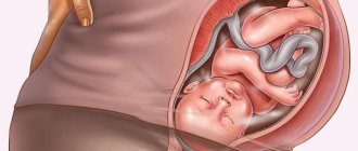
The placenta is located on the posterior wall - is it normal or not?
Attachment of the placenta to the posterior wall is one of the normal options. In addition, many doctors consider this arrangement to be the most physiological and safe. Why is embryo implantation into the posterior surface of the uterus more common? There is no proven information on this yet, but there are a number of factors that can contribute to the attachment of the placenta in this particular place. Thus, the posterior wall of the uterus has the greatest thickness of the endometrium and high temperature due to the abundant blood supply, which has a beneficial effect on the development of pregnancy in the short term. Also, do not forget that the egg itself cannot move and the fallopian tubes, contracting, push it into the uterine cavity, precisely closer to the back wall and bottom.
Another assumption is that in the fertilized egg there are certain mechanisms responsible for choosing the most successful and favorable place for attachment.
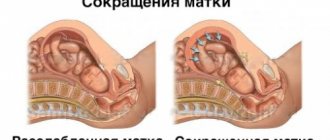
Why does the tone of the posterior wall of the uterus develop during pregnancy?
The uterus is formed by muscle tissue, so minimal tension is present in the organ, even when the woman is completely healthy. And if a slight tone is detected at the point of contact of the embryo with the wall of the uterus, this indicates that implantation is successful.
When the entire uterus is tense, we talk about general tone, and if the tension covers a separate area of it, we talk about local tone. The latter is classified into hypertonicity along the posterior and anterior wall.
Obstetricians-gynecologists regard this pathology as a fairly serious disorder. A large number of factors can cause the development of tone in the posterior wall of the uterus in an expectant mother:
- diseases that directly affect the tissues of the uterus - fibroids, endometriosis. Problems with bearing a child due to high tone can also arise due to the unusual anatomical structure of the reproductive organ (saddle-shaped, bicornuate uterus);
- progesterone deficiency. Hormone deficiency occurs if a pregnant woman’s genitals are underdeveloped or her body has too high a level of male sex hormones (androgens);
- viral infections. The aggressive activity of pathogenic microorganisms weakens general immunity, on which the normal state of the uterus directly depends. Due to the presence of infection in the external genital area, itching and burning occurs, the composition of vaginal discharge changes;
- polyhydramnios and oligohydramnios. Deviation of the volume of amniotic fluid from normal indicators has a bad effect on the condition of the muscle layer specifically on the posterior wall of the uterus;
- regular exposure to stress. A depressed state and nervousness not only puts the uterus in a state of constant tension, but also disrupts the general well-being of the pregnant woman;
- stretching of the muscles of the reproductive organ. Almost always present if the fruit is too large, if there are several fruits, as well as with severe polyhydramnios;
- foci of inflammation in the area of the uterus and ovaries. The condition is accompanied by alternating tension and relaxation of different parts of the uterus;
- too much physical activity. Obsessive exercise or heavy lifting puts the posterior wall of the uterus under constant tension;
- Rh conflict between mother and fetus. If the mother’s body begins to reject the child, the first thing that will happen is the tone of the uterus will increase;
- past abortions, miscarriages and premature births;
- slowing of intestinal motility due to pregnancy. Gases accumulate in its cavity and the swollen organ puts pressure on the uterus, which provokes an increase in its tone.
Advantages of posterior placement, impact on childbirth
Placing a child's seat on the rear wall has a number of advantages compared to its attachment in front:
- This attachment reduces the risk of injury to the placenta in the event of an external impact on the expectant mother’s abdomen. Of course, this does not mean that pregnancy with an anterior position of the “baby seat” is more dangerous and it can be damaged if a woman, for example, hits her stomach. No, and in this case the placenta and the fetus itself are well protected. But still, the further location of this organ from external stimuli can play a decisive role in severe abdominal injuries.
- When located posteriorly, placenta previa is less likely to occur. In the case of a low attachment, there is a chance that during the growth of the uterus and the increase in the “baby spot” itself, it will rise to a height sufficient for a safe natural birth. This process usually occurs before 32 weeks of pregnancy. Since the anterior wall stretches much faster, such active migration of the placenta, alas, is not observed.
- The posterior wall of the uterus is less susceptible to stretching, which reduces the risk of abruption. This is especially important with low placentation or posterior placenta previa.
- If a woman has had surgical interventions in the uterus and scars remain on it, then they are located in the front part of it. In this case, the attachment of the embryo in this place can be dangerous due to placenta accreta - when the chorionic villi (placenta in the initial stage) grow not only to the mucous membrane of the uterus, but also to its walls and even to the abdominal cavity. This may lead to complications in the form that after childbirth the placenta does not separate on its own, and there is a risk of bleeding. This situation is life-threatening for the woman and may require removal of the uterus.
- An important confirmation of the advantage of placing the placenta in the back is the convenience for the obstetrician-gynecologist during examination. So, with this attachment, the vessels of the “baby seat” will not interfere with listening to the child’s heart, as well as more accessible palpation of parts of the body, determining its presentation by external palpation of the abdomen.
- If a cesarean section is necessary, the risk of damage to the placenta during the uterine incision is reduced.
When the placenta is located along the posterior wall, the doctor is less likely to damage this organ when making an incision - Some pregnant women note that with posterior attachment, fetal movements can be felt earlier and much more clearly. This advantage cannot be proven, since, in addition to the location of the placenta, this is influenced by the individual sensitivity of the mother and what kind of child she is carrying.
During the second pregnancy, the first movements of the baby were felt at the 17th week. In this case, the location of the placenta was along the posterior wall and in the bottom. At the same time, a friend with the same term and anterior placenta began to feel movements at the 16th week, although this was her first pregnancy. Unfortunately, no one can confirm whether a woman really feels movements at the moment. Here you have to rely only on your own feelings and try not to confuse the first light kicks with intestinal peristalsis.
Causes and degrees of uterine prolapse
Since uterine prolapse develops due to weakening of the pelvic floor muscles, the following main reasons :
- change in pressure in the abdominal cavity (an increase forces the organ to move down);
- weakening of not only the muscular system, but also the pelvic floor ligaments leads to the inability to maintain the uterus in the correct position.
Here are some factors that in a certain way influence the tendency to develop pathology:
- difficult childbirth, multiple pregnancies (especially with trauma to the female genital organs during childbirth and deep lacerations of the perineum);
- surgical interventions on the organs of a woman’s reproductive system;
- sudden lifting of weights and excessive physical activity;
- advanced age of the woman;
- hormonal imbalance in the body, causing estrogen deficiency.
IMPORTANT!
In addition, heredity also plays an important role - if a woman’s grandmother or mother suffered from the disease in her family, then the likelihood of developing uterine prolapse increases several times. This becomes a recommendation to the “heiress” to carefully monitor the condition of the reproductive organ and be sure to have regular examinations with a gynecologist.
Doctors determine 4 degrees of development of the disease - the stage of the pathological process is decisive when choosing treatment:
- 1st degree - prolapse of the uterus (external os) no further than the middle of the vagina (1);
- 2nd degree - the uterus is lowered to the entrance to the vagina, but does not protrude from the genital slit (2);
- 3rd degree or incomplete uterine prolapse - the external os protrudes beyond the genital slit, but the uterus itself is located in the vagina (3);
- Stage 4 - the organ falls out completely.
Video: location of the placenta, placenta previa
The most important thing for a successful and easy pregnancy is the health and peace of mind of the expectant mother. The location of the placenta not on the back wall should not cause concern for a woman, this is just her feature. Currently, all pregnant women undergo mandatory examinations, during which the condition and place of attachment of the placenta are established; this information is very important for the obstetrician-gynecologist who delivers the baby. And it is important to remember that even the presence of complete placenta previa does not exclude a successful pregnancy; you just need to be more attentive to your health during this period. Enjoy your position, capture wonderful moments, feeling the life inside you, and in due time you will definitely hold your baby in your arms. Be healthy!