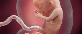
PGD (preimplantation genetic diagnosis) is the study of an embryo for chromosomal abnormalities and genetic diseases before transferring it into the uterine cavity. Used in IVF and ICSI, when fertilization occurs outside the woman's body.
- What diseases can be detected with PGD?
- Indications and timing of genetic diagnostics of the embryo
- Do I need to prepare for PGD?
- Embryo diagnostic technique
- Method safety
- Benefits of PGD
- Doctors performing PGD
- Cost of PGD
Purpose of the procedure:
- Preventing the birth of a child with genetic disorders, which is especially important for families at high risk.
- Increasing the effectiveness of assisted reproductive technologies.
- Reducing the risk of miscarriage or missed pregnancy.
IVF and PGD: concepts
First, let's remember what IVF is? IVF is an in vitro fertilization procedure. It differs from natural conception in that the embryos are fertilized outside the woman's body and then placed inside the uterus. The success of IVF largely depends on the skills and experience of doctors. Last but not least in importance is the health of the expectant mother and genetic father.
PGD with Eco what is it? Let's spell it out: preimplantation genetic diagnosis. With its help, the embryo is diagnosed before being transferred into the woman’s body. PGD is carried out in conjunction with an IVF program. Carrying out diagnostics, the doctor excludes in advance the presence of genetic pathologies in the unborn child. Since a practically healthy embryo is implanted into the uterine cavity, the risk of spontaneous miscarriage is reduced to a minimum.
Under what conditions is it necessary to conduct PGD?
Such diagnostics are not always carried out. This significantly increases the cost of IVF. But under certain conditions, it is mandatory. Let's list them:
- The expectant mother is over 35 years old. The proportion of genetically normal embryos decreases with age, while the proportion of abnormal embryos increases greatly.
- The genetic father is over 45 years old.
- There have already been deaths of children from incompatible Rh factor.
- Future parents have genetic diseases, which means there is a serious risk of getting a genetically unhealthy baby.
- The childless couple had already had several unsuccessful attempts at artificial insemination.
- There have been cases of spontaneous abortion at 4-8 weeks.
- The man was found to have serious spermatogenesis disorders or a high content of abnormal sperm.
The PGD procedure helps to identify a sufficient number of genetic diseases. By examining the chromosome set of an embryo, a doctor can detect more than 150 diseases. These include: Gaucher disease, myodystrophy, achondroplasia, hemophilia, Gettington's disease, fragile X chromosome, pemphigus, retinoblastoma, neurofibromatosis, Fanconi anemia, Down syndrome, torsion dystonia, polycystic kidney disease, myopathy, cystic fibrosis, Alpert, retinitis pigmentosa, Edwards , phenylketonuria.
Diagnosis requires a significant number of follicles. Therefore, with PGD, a woman undergoes increased stimulation of the ovaries.
What diseases are diagnosed?
PGD is used to diagnose a huge number of different diseases. These are chromosomal or monogenic pathologies. Diagnosis is carried out both empirically and purposefully. Empirical diagnosis is used in cases where parents or their relatives do not have any hereditary diseases. Then embryologists look for those diseases that are most common. In other cases, through genetic counseling, doctors determine that parents may be carriers of the following genes:
- cystic fibrosis;
- hemophilia;
- phenylketonuria;
- hearing loss;
- sickle cell anemia;
- Duchenne muscular dystrophy;
- Gaucher disease, etc.
The number of gene diseases for which an embryo is examined is in the hundreds.
All couples are examined for chromosomal abnormalities. This could be extra or missing chromosomes, a change in their position, the exchange of fragments of genetic material, the absence of parts of chromosomes, etc.
The most common pathologies are Patau, Down, Edwards, cat cry, Klinefelter, Shereshevsky-Turner, Prader-Willi syndromes.
When is genetic diagnosis performed?
Different parts of female germ cells or embryos can be examined. Three approaches are used:
- Study of polar bodies. These are cellular formations that arise during oocyte maturation. They play no role in fertilization. Therefore, polar bodies can be removed and examined without harming the egg. The method allows you to obtain information about the chromosome set of the female gamete. But it does not guarantee the usefulness of the genetic material of the embryo.
- Study of the blastomere. An embryo cell is taken on the 3rd day of its development. At this time, it usually has from 6 to 8 cells (blastomeres). The study allows us to identify most mutations. But it has one weak point - mosaicism cannot be determined. This is a mutation in which different cells carry different genetic material.
- Study of trophectoderm. These are cells of embryonic origin from which the outer shell of the embryo is formed. This diagnostic method is carried out on day 5, when the embryo is at the blastocyst stage. In this case, PGD during IVF can also detect mosaicism, because several cells are examined at once. But the disadvantage of such diagnostics is the need to freeze the embryos, because the analysis takes from 1 to 3 days. Thus, the transfer has to be postponed until the next cycle. However, although this results in additional and financial costs for the couple, the effect of cryopreservation of embryos on the likelihood of pregnancy is only positive. There is a greater chance of getting pregnant in a cryocycle than in a “fresh” cycle, because doctors manage to achieve the best synchronization of the state of the endometrium and the stage of embryo development.
Thus, PGD during IVF can be carried out before fertilization, on the 3rd day of embryo development, as well as on 5 days. In practice, the last option is most often used. Because on day 5 the research will be the most reliable and informative.
Goals of PGD
If the described diagnosis is necessary according to indications, then the doctors’ further actions will be aimed at achieving the following goals:
- Significant reduction in the risk of having a genetically unhealthy child.
- Determining the sex of the child in order to exclude the conception of a child with a disease associated with gender.
- Establishing the Rh factor, which will help eliminate hemolytic abnormalities in the embryo.
- Identifying information why implantation did not occur in previous IVF attempts.
- Immediately weed out embryos that are affected by incurable diseases.
- Selection of the necessary genetic information, which in the future will help the child act as a donor to a sick sister or brother.
In what cases is PGD performed for genetic pathologies?
- Indications for preimplantation diagnostics from the genetic point of view:
- Carriage of autosomal recessive diseases;
- Pathology linked to the X chromosome;
- Couples with significant chromosomal translocations that prevent implantation, cause miscarriages, or imply mental or physical problems in the offspring;
- Carriage of autosomal dominant diseases.
Preimplantation diagnosis is used for sex-related pathologies, single gene defects and chromosomal disorders.
Gender-related disorders
X-linked diseases are transmitted to the child through the mother, who is a carrier. An abnormal X chromosome causes the disease in sons who do not inherit a normal X chromosome from their father, but instead receive a Y chromosome. In the future, the birth of healthy boys is expected, but the probability of carriage in daughters is 50%, provided the mother is healthy.
Important
X-linked diseases are never passed from father to son.
Autosomal recessive pathologies include hemophilia, most neuromuscular dystrophies, and hundreds of other diseases.
Autosomal dominant disorders will manifest themselves as Rett syndrome, pseudohyperparathyroidism, rickets resistant to vitamin D, Bloch-Sulzberger syndrome (hereditary disorder of skin pigmentation with pathology of the visual apparatus, teeth and central nervous system), etc.
The Y chromosome carries a small number of genes, but some pathologies of sexual differentiation are associated with it.
In severe forms of spermatogenesis disorders (a critically small number or complete absence of male germ cells), microdeletions in the azoospermia factor gene, localized on the long arm of the Y chromosome, are diagnosed in 18% of cases.
Single gene defects
PGD is used to identify individual gene defects that cause a number of diseases:
- Cystic fibrosis;
- Tay-Sachs disease;
- Sickle cell anemia;
- Huntington's disease.
For these pathologies, genetic diagnostics detects a defect using molecular methods using polymerase chain reaction (PCR) amplification (multiple replication) of DNA from one cell.
Chromosomal disorders
As a result of translocation, inversion and deletion, chromosomal damage occurs, which can be detected using fluorescent hybridization. For some couples, the chances of achieving a viable pregnancy without using PGD are negligible, as previous attempts have resulted in embryos with a chromosomal mutation and resulted in spontaneous miscarriages.
How accurate is the PGD procedure?
Preimplantation diagnosis has proven its accuracy. The probability of a possible error does not exceed 5%. In addition, after pregnancy, a woman is prescribed and undergoes combined screenings. If suddenly they give an unacceptable percentage of chromosomal abnormalities, she undergoes invasive prenatal diagnosis.
Two sides of PGD: positive and negative
If we talk about the positive side, then this:
- The ability to immediately determine and “order” the gender of the child.
- Getting a healthy baby thanks to the addition of only good material.
- It is very difficult to damage the embryo; the risk is less than 1%.
- Possibility of preventing conflict between operational factors.
- Abortion due to identified genetic abnormalities is excluded.
- The procedure is completely harmless to the embryo. At this stage of development, cells are completely interchangeable. The blastomeres taken for the procedure are easily replaced with new ones.
- The success of the IVF program increases several times.
- Congenital defects are almost completely excluded.
- An excellent opportunity to help your sister or brother by selecting the necessary compatible material.
The advantages of PGD over IVF are impressive, but the disadvantages of IVF are also significant. The negative side is manifested in the following factors:
- A small, but still risk of damage to the embryo. You can’t get rid of it and it can come.
- Possibility of obtaining incorrect results. Sometimes healthy embryos are mistaken for unhealthy ones. They are weeded out. It turns out that material with incorrect information is implanted into the uterus. To exclude such a negative factor, screening is carried out, which allows one to identify such information at a fairly early stage of pregnancy.
- There is a 3-5% risk of getting an unhealthy baby.
- The PGD procedure is very expensive.
Benefits of SGP/CCS/PGDA
The benefits of PGD for the analysis of chromosomal mutations (SGP) are still being debated: does the percentage of successful embryo implantation increase with assisted fertilization methods? Next, the main pros and cons of diagnostics are presented.
Improving embryo selection
Only embryos with a normal number of chromosomes guarantee the birth of a healthy child. Thus, in the case of high-quality embryos, with the help of SGP analysis, doctors can detect and select embryos with a normal number of chromosomes, and exclude all those that are not recommended for transfer and do not guarantee the birth of a healthy child, even if they are high-quality embryos.
Increased success of infertility treatment
There are genetic mutations that are incompatible with life and make it difficult for the embryo to develop at an early stage and even implantation into the uterus. SGP allows you to exclude these embryos, optimizing the number of transfers.
Avoid transfer of embryos that cause abortion or the birth of children with various syndromes
Among chromosomal mutations, there are several that are less destructive for embryo implantation, but which impede the healthy development of the embryo and can cause abortion, or various diseases will manifest in the child, such as Down, Patau or Edwards syndrome. All embryos that could lead to these diseases or situations will be excluded thanks to SGP.
Reduce waiting time for a successful pregnancy
With SGP, we avoid transferring embryos that do not guarantee the birth of a healthy child, as they will be excluded. After the study, we will select only those embryos that guarantee a successful pregnancy, and will not waste time transferring embryos that will not develop and do not guarantee the birth of a healthy child.
Low cost
Many may believe that the cost of preimplantation genetic diagnosis of embryos is high, but careful testing of each embryo helps avoid freezing and retaining embryos that are apparently healthy but genetically not. Thanks to this analysis, you can avoid the costs of embryo transfers that do not guarantee a healthy and successful pregnancy.
Improve psychological well-being
Preimplantation genetic diagnosis of embryos can reduce the level of doubts of patients. On the one hand, diagnosis guarantees a healthy embryo and the use of advanced technology. On the other hand, emotional stress reduces the likelihood of abortion, especially in the case of patients with miscarriage or repeated unsuccessful attempts.
PGD price
Preimplantation genetic diagnosis costs a lot. The price range starts from 50,000 and ends at 130,000. The price includes expensive equipment and modern, expensive materials. The cost also depends on the number of embryos that will be examined. Having decided on the PGD procedure, you can order an examination for one pathology or carry out a full diagnosis. If the procedure is performed by top-class specialists using professional equipment, the likelihood of the desired positive outcome increases.
You can look at the high cost of PGD from the other side. If there is a high risk of getting a genetically unhealthy baby, then it may be more likely to be born. An unhealthy child will need special care, expensive treatment and general maintenance for life. Plus, the mental anguish of parents suffering for their child will always bother them. All this together significantly exceeds the stated cost of PGD. It turns out that it is better to carry out PGD.
When did the use of PGD in medicine begin?
The use of PGD became possible only after the polymerase chain reaction was developed, which made it possible to analyze the structure of DNA. In addition, this additional assisted reproductive technology is not possible without IVF.
Therefore, PGD was first used in 1989. An analysis of the blastomere (cell) of an embryo at the cleavage stage was carried out. It consisted of 6-8 identical cells. The procedure was completed successfully. A couple with an increased risk of an X-linked disease had a healthy child in 1990.
In 1992, PGD of a monogenic disease (cystic fibrosis) was performed for the first time. The low-quality embryo was discarded. An embryo without genetic diseases or mutations was transferred to the woman, and she gave birth to a healthy child.
In 2012, the FISH method began to be widely used to diagnose chromosomal abnormalities. From that moment on, the method of comparative hybridization began to be forced out of medical practice.
Who needs PGD?
Every year in Russia thousands of children are born after IVF. But preimplantation genetic diagnosis is not always performed during this procedure. PGD is carried out according to indications. The main ones:
- carriage of a chromosomal aberration or genetic disease in one or both future parents;
- woman's age over 35 years;
- male age 40 years or more;
- severe disorders of spermatogenesis in men;
- recurrent miscarriage (2 or more cases of perinatal losses in history);
- 3 or more unsuccessful IVF attempts;
- the need to prevent possible Rh conflict;
- burdened family history (miscarriages, stillbirths, chromosomal mutations, etc. among the spouses’ immediate relatives).
A woman’s age is one of the significant risk factors for genetic abnormalities in a child. Therefore, even in the absence of a history of perinatal losses and unsuccessful IVF, after 35 years it is better to perform PGD.
According to statistics, the risk of having a child with chromosomal aberrations for women of different ages is:
- at 30 years old – 0.26%;
- at 40 years old – 1.6%;
- at 45 years old – 5.26%.
Thus, at 45 years of age, one out of 19 pregnancies ends in miscarriage or the birth of a sick child due to chromosomal rearrangements. These consequences can be prevented with PGD.
PGD methods
Doctors practice four types of PGD. These are FISH, CGH, PCR, NGS. Let's look at them separately.
FISH method
It stands for fluorescent in situ hybridization. Or in another way it can be called the molecular cytogenetic method. It is often used in the CIS countries. FISH is used for interphase and metaphase nuclei. The method allows you to identify the DNA sequence on the chromosome under study. In terms of price, it is the most inexpensive diagnostic method. The study takes about 4-5 hours. FISH has the disadvantage that it cannot test all chromosomes.
The fish method also helps:
- Carry out PGD of embryos that are carriers of chromosomal rearrangements.
- Conduct a screening study for frequently occurring chromosome aneuploidies.
- Immediately determine the gender of the planned child.
CGH method (comparative genomic hybridization)
It can be used to check all chromosomes. The research takes quite a long period of time. For the price it is expensive. Its peculiarity: the resulting blastocysts are frozen, and further transfer is carried out using a cryoprotocol. In addition, this method tests embryos that have a strong potential for further implantation. Also, with CGH, the risk of misdiagnosis is significantly reduced, thanks to the biopsy of a large number of stem cells.
PCR method
It allows you to correctly diagnose diseases that arise due to dominant and recessive mutations (for example, diseases such as cystic fibrosis, hemophilia). PCR detects chromosomal abnormalities, determines HLA antigens and the Rhesus affiliation of embryos. The PCR technique also tests close relatives. That is, in addition to diagnosing parents, they do research on relatives for the presence of monogenic diseases. Such actions are necessary to identify gene disorders in embryos.
NGS method
Let's decipher: Now-Generation Sequencing. Translated as high-throughput sequencing. This is the most innovative diagnostics. With NGS, the DNA sequence is checked, thereby accurately identifying genetic pathology. The new technology is gradually replacing the first FISH technique and is widely used in foreign IVF centers. Practice has shown that success occurs in 70% of cases.
Negative aspects of genetic diagnosis of SGP/CCS/PGDA
Invasive procedure
In SGP, an embryo biopsy must be performed for genetic analysis. However, recent advances in this area minimize the possible side effects of biopsy. A biopsy on the 5th day of embryo development, rather than on the 3rd, is less traumatic for the embryo, and allows you to use this method to select a viable embryo.
Loop without carry
In some cases, with age, the number of genetically abnormal embryos increases significantly, and the proportion of normal ones decreases. In these cases, there is a possibility that, after SGP analysis, all embryos are abnormal and cannot be transferred. And in addition to the inconvenience caused due to the interruption of the procedure, the emotional pain of loss is added.
Embryo mosaicism
It is widely known that human fetuses exhibit some degree of mosaicism, but it is difficult to diagnose. Nowadays, thanks to the development of various genetic tests, we can determine whether the cells of an embryo are genetically different (mosaic). It remains to be determined whether this fact affects the embryo in any way. To evaluate this fact, the Instituto Bernabeu carried out various research projects.
SGP as screening
SGP examines the external tissues of the embryo and does not affect the tissues (inner cell mass) that affect the proper development of the child, since there are scientific works that have proven a high degree of correlation between them. Therefore, we believe that the material taken during the biopsy provides information about the entire embryo.
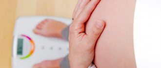
Preparing for PGD
If there is a need to combine IVF with PGD, a geneticist will consult with the couple. Next, specialists prescribe a full examination to the future parents to identify all changes, karyotyping the spouses and detecting mutations. Next, the exact indications for PGD are established. At the last stage of preparation, the doctor explains the entire essence of the PGD program and describes the stages of the procedure.
Method safety
During gene diagnostics there is no interference in the functioning of the body of the father and mother. The maximum that is required from the spouses is to donate blood from a vein. Therefore, in this regard, the method is absolutely safe.
The issue of the safety of PGD for the embryo has also been deeply studied by scientists. Based on the studies, experts came to the conclusion that the technique does not affect the risk of developing congenital malformations or other anomalies that may appear after birth. The procedure is carried out at that stage of development when the cells of the embryo have not acquired specific functions, so they can easily replace each other in the process of subsequent division.
Stages of PGD
Let's consider the stages of PGD:
- The first stage begins with the standard IVF procedure. The process ends with the stage of egg puncture.
- Carrying out embryological procedures for eggs and sperm, performing micromanipulations. On the third day of cultivation, a biopsy procedure is performed. To do this, one cell is taken from the embryo using a special microsurgical instrument. Biopsy uses mechanical, chemical and laser methods. Next, the blastomere is fixed. In addition, a trophectoderm biopsy and a biopsy of the polar body of the oocyte can be performed. In the first case, 3-5 cells are taken from the trophectoderm (usually they are on the outer layer of the blastocyst). A biopsy of the trophectoderm is performed on days 5 or 6 and is a reliable option for identifying the karyotype of abnormal development of embryos. Oocyte cell biopsy is the best diagnostic test for female infertility. The genetic information in the polar body and in the nucleus of the egg is the same.
- Carrying out hybridization with DNA probes. Probes mean a special mark. Each single chromosome has its own label. It is illuminated with color under the influence of fluorescent. Therefore, their identification is easy.
- Obtaining visualized information on a computer monitor screen. Conducting diagnostics of blastmasters that have been detected. Obtaining final results by the 5th day of cultivation. Here doctors count the number of chromosomes. If their number corresponds to the accepted value, then the material is recommended for further transfer. If instead of three chromosomes there are two, or instead of the required two, one is visualized on the screen, then such material should not be transferred, it is abnormal.
- Genetic transfer of healthy embryos on day 5 of cultivation. This time is considered the most optimal. The material for use is provided in sufficient quantity and the biopsy procedure for the embryo is not so traumatic. If you transfer the material to the third day, there is a high probability of stopping the development process. In addition, the genetic cells of a three-day-old embryo are very different. The result may be false studies.
- Carrying out cryopreservation after the transfer procedure.
- Diagnosis of a long-awaited pregnancy using a test (usually two weeks should pass after replantation).
Possible consequences of IVF with PGD
In addition to a screening study, a pregnant woman after IVF undergoes an analysis of amniotic fluid and an ultrasound of the fetal nervous system.
Depending on the PGD method, diagnosis will take from several hours to several days. The entire IVF cycle lasts 4-6 weeks. This period also includes PGD.
The PGD procedure is performed on an outpatient basis. Sedation will only be required for egg retrieval.
IVF with PGD carries the same risks as IVF without PGD. This:
- Multiple pregnancy.
- Damage to the embryo at the stage of cell removal.
- Reactions to drugs that may be used during the IVF procedure. These are mainly headaches, flushing and mood changes for the worse.
- Ovarian hyperexcitation syndrome is observed.
- Bleeding after egg retrieval.
- Sensitive spasms after the removal procedure.
- PGD research does not give a hundred percent result.
- Getting a conception outside the uterus.
How is the research conducted?
Monogenic PGD / PGD-M
First of all, in order to conduct a PGD analysis, it is necessary to have a genetic analysis of the future parents. For this reason, the first stage of analysis is a genetic analysis of the parents. That is, to detect genetic risks (mutations) that caused the disease. After receiving the result, we must develop the correct method for diagnosing future embryos in the in vitro fertilization (IVF) laboratory (informativeness).
Once the previous stages of PGD are completed (genetic analysis and information content), we can begin conducting the PGD cycle. To do this, the couple will have to undergo in vitro fertilization (IVF). We will have to wait until the embryos produced in this process have split before we can collect a few cells from the embryo (embryo biopsy). We will analyze the collected material in a molecular biology laboratory to find out whether the biopsy embryos are healthy. The purpose of the analysis is to select an embryo for transplantation without genetic pathologies carried by the parents, and to have a healthy pregnancy.
SGP/CCS/PGD-A:
In this case, a biopsy is taken of the trophectoderm (the area of embryonic tissue from which the placenta develops) of the blastocyst, and the number of embryo chromosomes is counted to select embryos with a normal number of chromosomes.
Contraindications
PGD, despite its necessity, effectiveness and painlessness, has several contraindications:
- The resulting embryo on the 2nd day of its development has less than 6 blastomeres.
- The formation of embryo fragments is more than 30% (normal value is 25%).
- The embryo contains multinucleated blastomeres.
Preimplantation genetic screening: relative indications
According to studies, most early pregnancy terminations occur due to aneuploidy - a violation of the number of chromosomes. Since healthy embryos are transferred into the uterus, the likelihood of miscarriage in the first and second trimester is reduced.
There is currently no specific list of indications for PGD.
Preimplantation genetic screening is considered desirable in the following cases:
- Planned pregnancy in an older woman.
- A burdened obstetric and gynecological history with repeated miscarriages.
- Unsuccessful multiple IVF attempts, including ICSI.
- Severe male factor infertility.
It is a proven fact that the risk of aneuploidy increases with a woman's age . Chromosomes are less likely to divide properly, resulting in an extra or missing chromosome in the embryo. The pathological number of chromosomes is more than 20% in mothers aged 35-39 years, and at the age of 40 years and older it reaches 40%.
The frequency of aneuploidy in children born to mothers aged 35-39 years is 0.6-1.4%, over 39 years old - 1.6-10%.
Important
Not every pregnancy will result in the birth of a viable child; sometimes self-abortion occurs earlier than the woman would suspect her condition.
An example of a common aneuploidy is trisomy 21, which leads to Down syndrome. The frequency of trisomy 21 in a viable fetus was assessed depending on the age of the mother. According to the results, the optimal age for childbearing is from 20-24 years (the lowest risk is 1/1400). Over the age of 40 - 1/100, over 45 years - 1/25.
Recurrent miscarriages and PGD diagnosis
If a woman has 2 or more consecutive pregnancy losses before 20 weeks, PGD before IVF is justified. The cause is often unknown, but chromosomal abnormalities in aborted fetuses are diagnosed in 50-80%.
note
Studies have shown that couples with multiple miscarriages are susceptible to a higher percentage of aneuploid embryos.
Unsuccessful IVF several times in a row
Three or more unsuccessful IVF attempts involving high-quality embryos may indicate chromosomal abnormalities. In addition, it is worth remembering the immunological and uterine factors that contribute to implantation failure.
Male factor infertility
Hypogonadism in men leads to the formation of embryos with chromosomal abnormalities. In normal sperm, about 3-8% of pathologically altered sperm are present. Their number increases significantly in men with severe infertility (decreased number, poor morphology and impaired motor activity) up to 27-74%.
With the help of intracytoplasmic injection of sperm into the egg, poor sperm quality is no longer an obstacle to conceiving a biological child. Various genetic defects have been found to be associated with male factor infertility. More often these are aneuploidies, Klinefelter syndrome, microdeletions of the Y chromosome, damage to androgen receptors and other mutations of autosomal genes (cystic fibrosis).
Important
During the ICSI procedure, natural selection is turned off, so the risk of transmitting genetic mutations to offspring increases.
Gender selection
PGD can determine the sex of an embryo, but wanting to have a boy or a girl without indication (sex-linked genetic diseases) is not ethical.
Human leukocyte antigen (HLA) matching
New indications for PGD include HLA matching. This method can be used to exclude genetic pathology. In situations where the family already has a child with a recessive disease and treatment with stem cell or bone marrow transplantation is needed (thalassemia, leukemia), it is possible to select a “suitable” embryo, which after birth will become a donor for the proband (brother or sister). The first case was described in a family with a child suffering from Fanconi anemia, and PGD was performed to select a healthy embryo with the same HLA type as the affected brother. The use of PGD in such situations is controversial for moral and ethical reasons.
Genetic diagnostics as a perfect method
Of course, a woman’s biggest fear is giving birth to a genetically unhealthy baby. The PGD method allows us to weed out genetically diseased embryos, thereby immediately giving the child health. Parents do not have to fear for the health of their long-awaited children. If someone believes that genetic research makes a child according to a template, then this is not so. You cannot create a design project for a future baby. Character and individuality do not depend at all on the genes received. Even artificially cloned animals are not similar to each other. Genes are responsible for external data.
IVF combined with PGD greatly influenced the verdict of “infertile”. More precisely, this is no longer a sentence, but a special disease that can be easily treated with the help of IVF. The method allows you to overcome a number of health difficulties, namely:
- Obstruction of the fallopian tubes.
- Absence of fallopian tubes.
- Ovarian exhaustion.
- Lack of ovulation for a long time.
- Ovarian dysfunction.
- Adhesions in the pelvis.
- Absence of gonads.
Even if a woman does not have a uterus, the problem can still be solved. But at the same time, her ovaries should function normally. To do this, eggs are taken from a woman, fertilized with her husband's sperm, and the embryo is placed in the body of another woman (for example, a relative or surrogate mother).
PGD is an absolutely safe procedure. The diagnostic procedure is carried out at the shortest possible time, when each collected cell can produce a full-fledged organism. There will be no harm to the health of the embryo; accordingly, the unborn child will also be healthy.
Genetics continues to evolve. It allows you to get healthy offspring and future parents in advance, who are more likely to be free of genetic diseases. An adult really evaluates the future; he wants in advance to give the baby a full life and in the future to receive offspring (grandchildren) from him. Genetics certainly helps with this.
So, the method of preimplantation genetic diagnosis is an addition to the entire IVF program. Do not pay attention to the cost of the procedure. If money has already been found for IVF itself, then the remaining funds needed are meager. The child's health is not worth such savings.
Similar articles
No similar articles
How are genetic diseases inherited?
In the diagrams below, D or d represents the defective gene, and N or n represents the normal gene. Mutations do not always lead to disease.
Dominant diseases:
One of the parents has one defective gene that dominates its normal pair. Since offspring inherit half of their genetic material from each parent, there is a 50% risk of inheriting the defective gene, and therefore the disease.
Recessive diseases:
Both parents are carriers of one defective gene, but at the same time have a normal gene pair. To inherit the disease, two defective copies of the gene are required. Each offspring has a 50% chance of being a carrier, and a 25% chance of inheriting the disease.
X-linked diseases:
Normal women have XX chromosomes, and normal men have XY chromosomes. Women who have a normal gene on one X chromosome are protected from a defective gene on their other X chromosome. However, a man lacks such protection due to the presence of only one X chromosome. Each male offspring of a mother who carries the defect has a 50% chance of inheriting the defective gene and disease. Each female offspring has a 50% chance of being a carrier, just like her mother. (in the picture below, X represents a normal gene and X represents a defective gene)
Which PGD method is best?
Experts believe that comparative genomic hybridization is better than
FISH diagnostics, since the possibilities of the method are wider, and the risk of injuring the embryo is less . If the parent’s karyotype is peculiar (for example, translocation), there is a possibility of an interchromosomal effect – the risk of incorrect divergence of any chromosomes, and not just the damaged ones diagnosed during karyotyping in the mother or father.
Disadvantages include the need for cryopreservation and high cost.
FISH testing is preferable to PCR because the polymerase reaction carries the risk of misdiagnosis due to biological contamination.
Today, the most effective method is NGS – next generation sequencing.
Victoria Mishina, urologist, medical columnist
3, total, today
( 53 votes, average: 4.13 out of 5)
Discharge during pregnancy: normal or pathological?
Nausea during pregnancy: when does it appear and how to get rid of it?
Related Posts
Pros and cons of genetic analysis in 3-day and 5-day embryos
Three-day:
- greater risk of injury (about 15%, the qualifications of the embryologist play a significant role);
- the ability to perform analysis on only 9 chromosomes, including only the most common hereditary diseases;
- the likelihood of a biochemical and frozen pregnancy due to pathology in unstudied chromosomes;
- mosaicism
Five-day:
- the viability of embryos decreases as they exist outside the mother’s body, there is a need to wait to perform a biopsy (an early blastocyst can be vitrified or transferred);
- a stop in the development of “good” embryos, not associated with genetic factors;
- the ability to conduct a detailed chromosomal analysis;
- low risk of embryo damage (about 5%).
Important
Poor-quality embryos are discarded if, if they were transferred, the pregnancy either did not occur or ended in a stop in development or self-abortion in the early stages.