Should I be worried?
As it turned out, the woman arrived after undergoing an ultrasound examination at the antenatal clinic. Without unnecessary preamble, Yulia (that was the name of the pregnant woman) said: “I agree to a caesarean section, the relatives will now bring the necessary things.” “Wait, wait, let’s figure it out first,” I answered and invited Yulia into the examination room. It turned out that Yulia’s pregnancy is currently 36 weeks and an ultrasound revealed that the umbilical cord was entwined around the fetus’s neck. This fact greatly worried the pregnant woman and her relatives, so they decided to go to the maternity hospital without delay.
The umbilical cord (or umbilical cord) is an organ that functions only during pregnancy and performs an extremely important function of communication between the body of the mother and the fetus. The main components of the umbilical cord are vessels - one vein through which arterial blood flows from the mother to the fetus, delivering all the substances and oxygen necessary for life, as well as two arteries through which the venous blood of the fetus removes waste metabolic products and carbon dioxide into the mother's body.
The vessels of the umbilical cord are surrounded by a special jelly-like substance - Wharton's jelly, which, due to its consistency, plays an important protective role - it protects the vessels from compression. On average, the length of the umbilical cord is 50-60 cm, thickness 1.5-2 cm. If the length of the umbilical cord is more than 70 cm, it is considered long, if less than 40 cm, it is considered short. An increase in the length of the umbilical cord can lead to various pathological conditions, such as entanglement of the umbilical cord around the neck, torso, and limbs of the fetus, and the formation of umbilical cord nodes, which, in turn, are divided into true and false. It is important to note that about a fifth of all children born are born with an umbilical cord entwined, and this does not always lead to disturbances in the intrauterine state of the fetus. The fact is that, while in the uterus, until the moment of birth, the baby does not breathe with the lungs, so squeezing the neck, which is always very frightening for expectant mothers, is not dangerous for him. Problems can arise in cases where blood flow is impaired due to tension or compression of the umbilical cord due to repeated or tight entanglement.
Abnormal umbilical cord attachment
There are marginal and shell attachments of the umbilical cord. Normally, the umbilical cord is located in the center of the placenta; if it is localized closer to the edge of the baby's place, they speak of a marginal umbilical cord attachment. A dangerous pathology is the membrane attachment of the umbilical cord, when the latter departs not from the maternal part of the placenta, but from the fetal membranes, while the umbilical cord is not protected by Wharton’s jelly. If the membranes rupture during childbirth, damage to the umbilical cord vessels may occur, which leads to bleeding, anemia and intrauterine hypoxia or sudden death of the fetus.
What did the ultrasound show?
According to the ultrasound examination, it was established that there were no signs of hypoxia (i.e., lack of oxygen) in the fetus; when conducting Doppler measurements (a study that determines the speed of blood flow in the main vessels of the uterus and fetus), no disturbances in the uteroplacental circulation were detected. The fetus corresponds to 36 weeks of gestation, there are signs of a single entanglement of the umbilical cord around the fetal neck. “Wow, when I had an ultrasound at 32 weeks, they didn’t say anything about entanglement,” said Yulia. “It’s quite possible that it didn’t exist then, and it’s not at all necessary that it will last until the birth,” I answered.
Indeed, in practice there are often cases when, according to ultrasound data, an entanglement in the umbilical cord was detected, and the child was born without it. This may be, firstly, due to the fact that the umbilical cord loops according to ultrasound were located near the fetal neck, but there was no entanglement as such, and secondly, with the movements of the fetus, the umbilical cord entanglement was independently eliminated (of course, this usually happens with a single wrap).
Predisposing factors for the formation of umbilical cord entanglement are increased motor activity of the fetus, which can be caused by intrauterine hypoxia (i.e., lack of oxygen supply), polyhydramnios, and increased adrenaline in the mother’s blood due to stress. It is natural that in the vast majority of cases, a long umbilical cord leads to entwining various parts of the fetal body.
Loss of umbilical cord loop
Loss of the umbilical cord loop (prolapse) occurs during childbirth and is an extremely dangerous situation.
This pathology requires immediate delivery (caesarean section), and while preparing the woman for surgery, it is necessary to hold the presenting part of the fetus to prevent compression of the umbilical cord, through which the baby receives oxygen. Loss of the umbilical cord inevitably leads to acute hypoxia of the fetus and often to its death. As a rule, umbilical cord prolapse occurs after the rupture of amniotic fluid. Predisposing factors for umbilical cord prolapse include:
- breech or leg presentation;
- premature birth (the fetus is very small and is not able to hold the umbilical cord in the uterus);
- multiple pregnancy (after the birth of the first child);
- excessively long umbilical cord;
- polyhydramnios;
- amniotomy.
Making a diagnosis
To make sure that Yulia’s baby is feeling well, we recorded a cardiotocogram (CTG). CTG records the cardiac activity of the fetus, which is an informative indicator of its intrauterine state. To do this, a sensor was attached to the expectant mother’s stomach, which is connected to the device. Yulia lay on the bed on her side for 30 minutes while the machine recorded the fetal heartbeat. No pathological changes were detected on CTG. Together with Yulia, who had already calmed down somewhat and cheered up, we went out to her relatives who were waiting for her. I explained to them that in this situation, when we have a premature pregnancy, the intrauterine state of the fetus is completely normal, it is not advisable to carry out an emergency delivery just because of the entanglement of the umbilical cord. The reassured pregnant woman went home with her husband and mother-in-law.
To fully determine the intrauterine state of the fetus, it is necessary to conduct a set of studies, which includes:
- ultrasound examination , in which it is possible to see or suspect entanglement of the umbilical cord around the neck or other parts of the fetus, since in some cases it is very difficult to distinguish whether the umbilical cord loops are located near the fetal neck or whether there is entanglement: with this study we do not have the possibility of a three-dimensional image that allows us to examine the object from all sides - for example, looking back. It should be noted that it is not possible to determine the length of the umbilical cord using ultrasound data during pregnancy, since the umbilical cord is, as it were, “curled” in the tight space between the baby’s body and the wall of the uterus;
- Doppler ultrasound is a method that allows, firstly, to accurately determine whether there is entanglement in the umbilical cord, since the movement of blood flow is displayed in a color image, and secondly, to diagnose the speed of blood flow in various vessels of the uteroplacental complex;
- cardiotocography , which allows you to determine not only the baby’s heart rate, but also his reaction to his own movements (when recording CTG during pregnancy) and to an increase in the tone of the uterus (during childbirth), which allows you to find out how well the fetus is feeling at the moment .
If, after carrying out the entire complex of examinations, it is determined that the baby feels satisfactory, then the fact of entanglement with the umbilical cord alone is not an indication for surgical delivery. Such indications can arise either when there are signs of oxygen deficiency (fetal hypoxia), or when umbilical cord entanglement is combined with other indications for cesarean section.
Thrombosis of umbilical cord vessels
Thrombosis of umbilical cord vessels is a fairly rare pathology. Venous thrombosis is more common, but arterial thrombosis has a more unfavorable prognosis. Vascular thrombosis is a secondary complication that develops with true umbilical cord nodes, tunicated umbilical cord attachment, long or short umbilical cord, as well as with multiple pregnancies, maternal diabetes mellitus, abdominal trauma and premature birth. The risk of umbilical cord vascular thrombosis is high in high-risk pregnancies.
Umbilical cord entwined around the fetal neck: will there be surgery?
About a month passed, I had already forgotten about that visit from Yulia, when at my next duty I was invited to examine an incoming pregnant woman. Arriving at the emergency room, I met Yulia and her husband again. It turned out that for 3 hours the woman had been bothered by nagging pain in the lower abdomen, which an hour ago became regular and more intense. During the examination, it was revealed that Yulia had entered the labor process - the cervix was dilated by 3 cm, amniotic fluid was not poured out.
The woman giving birth is 24 years old, Yulia’s first real pregnancy, there were no gynecological diseases, abortions or miscarriages. The last ultrasound was performed at 39 weeks of pregnancy, according to its data, the umbilical cord remains entangled around the fetal neck. The estimated weight of the fetus is 3400 g. The gestation period is currently 40 weeks. During auscultation (listening to the fetal heart sounds through the anterior abdominal wall with a special tube - an obstetric stethoscope), the fetal heartbeat is clear, rhythmic, the heart rate is 144 beats per minute, which corresponds to the norm (the normal fetal heart rate is 120-160 beats per minute). Contractions at the moment of weak strength, after 10 minutes, lasting 30 seconds. Having filled out the birth history and placed Yulia in the prenatal ward, she was immediately recorded with a CTG (cardiotocogram). Carrying out CTG during childbirth is an absolutely harmless and informative method for determining the intrauterine state of the fetus and its reaction to the contractile activity of the uterus, which determines the tactics of labor management for each patient - whether she can give birth naturally or in the interests of the fetus, delivery by cesarean section is necessary. In the case of Yulia, the CTG ordered depended on which option for managing the labor would be chosen. Fortunately, no pathological changes were detected on CTG. Julia really wanted to give birth herself. Since she had every chance for this, they decided to give birth through the natural birth canal under careful monitoring of the fetal condition.
When the umbilical cord is entangled, it is very important to monitor both the condition of the fetus and the course of the birth process in the mother, because it is during childbirth that the umbilical cord entanglement can lead to a number of complications.
The most common complication that occurs when the umbilical cord is entangled is the appearance of fetal hypoxia, which occurs as a result of compression of the umbilical cord vessels when it is stretched or tightly wrapped around the child’s torso, neck or limbs. Quite often this happens at the moment when the fetus begins to move along the birth canal.
With repeated entanglement of the umbilical cord, a short umbilical cord is formed, which, firstly, can impede the advancement of the fetus along the birth canal, and, secondly, stretching with each contraction, can lead to premature separation of the placenta from the uterine wall (normally, the placenta is separated from the uterine wall after the birth of the fetus), which leads to the need for emergency surgical delivery.
In rare cases, when the umbilical cord repeatedly wraps around the fetal neck, complications such as extension insertion of the fetal head can occur, which can make it difficult to deliver the baby naturally. The fact is that during normal insertion of the fetus into the mother’s pelvis, the head is in a state of moderate flexion (in this case, the fetus’s chin is pressed to the chest, which allows the head to be correctly inserted into the pelvic cavity and pass without difficulty through the birth canal in the most “advantageous” way - that is, in the smallest size) - in this position it passes through the birth canal in the smallest, most convenient size. The umbilical cord loops located on the neck do not allow the baby’s head to bend, which leads to the fact that the head is installed in the mother’s pelvis not by the back of the head, but by the crown, forehead or even the face, which can lead to significant difficulties in the birth of the fetus and, as a consequence, to its traumatization.
To be fair, it must be said that the above complications occur quite rarely and, with timely and correctly provided assistance, do not lead to adverse consequences for the mother and fetus.
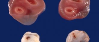
Umbilical cord knots
There are true and false umbilical cord nodes.
False knots
False nodes are an accumulation of Wharton's jelly or local thickening of the umbilical vein due to its varicose veins and have no practical significance (do not affect pregnancy and childbirth).
True nodes
True nodes form during pregnancy (in the early stages), when the embryo is still too small and “floats” freely in the amniotic fluid. In this case, it can slip through the loop of the umbilical cord, resulting in a knot. As long as the knot is not tightened, this pathology does not affect the condition of the fetus, but when the umbilical cord is stretched, which most often occurs during childbirth, the knot is tightened, which leads to hypoxia and the death of the child. If the knot tightens during pregnancy, it ends either in miscarriage or antenatal death of the fetus.
Childbirth with umbilical cord entanglement
4.5 hours have passed since Yulia arrived. The contractions quickly became more frequent, stronger and longer lasting. During a repeated examination on the chair, it was revealed that the cervix was dilated by 7 cm, an amniotomy was performed (instrumental opening of the amniotic sac) - 250 ml of light transparent amniotic fluid was released. According to CTG and regular listening to heart sounds with an obstetric stethoscope, the fetus's condition was satisfactory. Yulia refused the proposed drug pain relief, saying that she felt quite normal.
When the umbilical cord is entangled, the principles of labor management have a number of important points:
- the intrauterine condition of the fetus is carefully monitored using CTG and by listening to the fetal heartbeat through the anterior abdominal wall;
- If signs of fetal hypoxia appear, tactics will depend on the period of labor when these signs appeared. If signs of fetal suffering appear in the first stage of labor (the period of cervical dilatation), when the end of labor is still far away, a cesarean section is performed; if fetal hypoxia is detected in the second period (the period of expulsion of the fetus), then to speedily complete labor when the head erupts, a dissection is performed perineum (episiotomy), at the birth of the head, without waiting for the birth of the whole body of the child, the umbilical cord loops are removed if possible.
Advertising