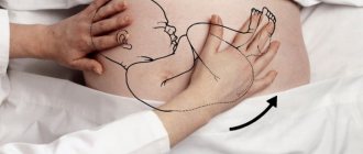
The placenta is an organ that circulates oxygen and nutrition from mother to fetus. This "fleshy patty" adjacent to the wall of the uterus is often called the baby's place. The embryo is connected to it through the umbilical cord.
Placental abruption in late pregnancy is one of the dangerous diseases that causes reasonable concern in the expectant mother. Doctors recommend going to the hospital at the first symptoms of a problem so as not to endanger the child’s life.
Causes of placental abruption
This pathology occurs on average in one woman out of a hundred, and the factors that provoke this process can be very diverse. The main classification of the causes of premature placental abruption divides them into exogenous and endogenous or external and internal.
Endogenous causes
According to numerous medical studies, the main cause of placental abruption is high blood pressure in pregnant women, which leads to increased pressure on the blood vessels and subsequent bleeding. In addition to high blood pressure, the following causes of the development of pathology are identified:
- Problems with blood clotting: If the indicators are poor, the risk of bleeding increases significantly.
- Vascular disorders, thinning of the capillaries of the uterus.
Placenta
- Numerous births, as a result of which there is a thinning and change in the structure of the tissue of the uterine mucosa, to which the placenta is attached.
- Pregnancy after childbirth by caesarean section. Placental abruption can occur when it attaches to the site of a postoperative scar.
- Pathology can occur during spontaneous delivery during multiple pregnancy.
- Diseases of the kidneys and other organs of the genitourinary system are of an infectious nature, especially pyelonephritis, which requires antibacterial treatment and can cause dangerous complications.
- Problems with excessive weight gain during pregnancy.
- Pathologies of the endocrine system, for example, diabetes mellitus or thyroid disease.
- Constant stress and nervous tension.
- Post-term pregnancy.
- Severe toxicosis suffered in the early stages, gestosis in the later stages or preeclampsia.
- Antiphospholipid syndrome.
- Diseases of an autoimmune nature, when the body's own cells are perceived as foreign.
- Pathologies in the development of the uterus, for example, a bicornuate uterus.
- Allergic reactions, including to medications prescribed to maintain pregnancy.
We recommend reading about discharge in late pregnancy. From the article you will learn about what kind of discharge is normal, what kind of discharge should alert the expectant mother, what symptoms should promptly see a doctor, as well as about diagnosis and treatment. And here is more information about how dangerous a hematoma in the uterus is during pregnancy.
Exogenous factors
External causes of placental abruption are largely due to the incorrect lifestyle of the pregnant woman, but there are situations that a woman cannot influence.
Exogenous factors of premature placental abruption include:
- smoking, since nicotine and other harmful substances contained in cigarettes penetrate directly into the blood of the unborn child and into the placenta, which can cause pathologies in its development;
- drug addiction;
- regular consumption of alcoholic beverages during pregnancy;
- injuries sustained during a fall, blow, or car accident; blunt abdominal trauma is especially dangerous.
Symptoms in later stages
At the end of pregnancy, placental abruption occurs most often, and it is almost impossible to compensate for it at this stage.
The main symptoms of the pathology are the same as in the early stages and are as follows:
- bleeding, which in some cases can be very intense, occurs due to damage to blood vessels in the uterus and placenta;
- severe pain in the lower abdomen;
- the uterus is in constant tone, and when palpating the fetus the sensations are very painful;
- upon examination, symptoms of fetal distress are recorded, it hardly moves, and hypoxia increases.
The intensity of symptoms and their severity depends on the degree of placental abruption. In a mild form, they almost do not appear, and the pathology is revealed during a routine ultrasound examination or after childbirth, when, during a mandatory examination of the separated child’s place, hematomas and deformations are found on it.
With moderate severity of detachment, patients complain of a small amount of blood in the discharge and periodic pain in the lower abdomen; sometimes, with a small size of the hematoma, there may be no bleeding at all. The uterus is a little painful and tense.
In severe cases of placental abruption, a woman may sometimes lose consciousness and show signs of severe anemia and tachycardia. In this case, bleeding occurs with significant intensity and is accompanied by pain in the lower abdomen and peritoneum.
A convex area on one side is found on the uterus, and it is very painful on palpation. The fetus is diagnosed with a muffled heartbeat, in some cases it may not be audible at all.
Expert opinion
Daria Shirochina (obstetrician-gynecologist)
When the first signs of abruption appear, it is important to contact the clinic as soon as possible, since it is possible to save the life of the mother and child with an emergency cesarean section.
Symptoms of pathology can appear separately. So, in some cases, despite all the pronounced signs of placental abruption, a woman has no bleeding from the vagina.
This course of the disease suggests that blood, having no way out, enters the wall of the uterus in large quantities, making it impossible for it to contract. In medicine, this pathology is called Cuveller's uterus, and it threatens the most serious consequences for the pregnant woman and the fetus, since it causes severe bleeding. The only way to save a woman’s life in such cases is hysterectomy, that is, complete removal of the uterus.
Since in such situations the child survives extremely rarely, the woman forever loses the opportunity to have children.
Degrees of placental abruption
Pathological changes in the body associated with detachment of the child's place are divided into 3 stages:
- Mild form - slight detachment, marginal. There is a slight vaginal discharge with blood and uterine excitability. At this stage, the child and mother are not affected. It passes without consequences, thanks to the formation of blood clots that stop the bleeding. Detected by ultrasound or after childbirth.
- Detachment of moderate severity - on the part of the woman is accompanied by bleeding, increased heart rate and hypertonicity of the uterus. When one third to half of the placenta is detached, the child experiences a severe lack of oxygen. The functioning of his heart slows down, the fetus is in danger of dying from oxygen starvation. The woman’s painful sensations are periodic and radiate to the perineum and lumbar region.
- Severe degree - tissues peel off by more than half, the process is accompanied by acute pain, intense bleeding or its absence. When the central part is separated and the placenta is filled with blood, the woman’s abdomen swells and the uterus becomes asymmetrical. A noticeable condition is characteristic of significant blood loss, pale skin, cold sweat, low blood pressure. The fetus's heart stops beating and it dies from lack of oxygen.
Determining the degree of separation of the placenta allows doctors to choose treatment tactics and make a prognosis for the further course of pregnancy. The second and third stages of abruption leave no chance of saving the fetus. An initial and non-progressive degree of separation gives hope for a favorable outcome and requires regular monitoring.
Manifestation of partial detachment
When the placenta is not completely separated from the damaged vessels, it begins to accumulate at the site of detachment, forming a hematoma, the size of which depends on the degree of damage. In this case, the separated area of the placenta ceases to function normally, and the gradual growth of the hematoma leads to an increase in the area of exfoliated tissue.
If the pathological process has affected only a small area of the placenta, blood vessels thrombose at the site of its separation and further detachment stops; this course of the disease is called partial non-progressive detachment. If the hematoma stops growing, the pregnancy can end in a normal birth without complications.
The placenta may begin to partially separate centrally or in peripheral areas. If the process begins at its edges, then the woman may begin to bleed, and with the central separation, a retroplacental hematoma forms, in which case blood can penetrate into the abdominal cavity.
With partial placental abruption, the woman’s main complaints are pain in the lower abdomen, which can radiate to the lumbar region, sacral spine and groin area.
Symptoms of PONRP [edit | edit code ]
When a small area of the placenta is detached, a retroplacental hematoma can form. In this case, the vessels of the uterus are thrombosed and the progression of placental abruption will stop. In some cases, blood permeates the uterine wall with significant placental abruption and a large retroplacental hematoma. In these cases, the contractile activity of the myometrium is disrupted. If marginal placental abruption occurs, then blood passes between the membranes and the uterine wall, then symptoms of external bleeding are observed, as blood flows into the vagina. Blood from the genital tract immediately after placental abruption is scarlet. The dark color indicates the amount of time that elapsed from the moment of detachment to the onset of bleeding. Premature placental abruption can be mild or severe.
Mild form [edit | edit code ]
In a mild form of premature placental abruption, there is a slight bloody discharge from the vagina, the tone of the uterus is unchanged, but slight tension is noted, the woman’s condition is satisfactory, the fetal heartbeat is normal.
Severe form [edit | edit code ]
In severe cases of premature placental abruption, pain with severe bleeding is noted. In the case of accumulation of blood between the wall of the placenta and the uterus, bleeding may be absent; a retroplacental hematoma is formed in this place, a local painful swelling occurs with an increase in pain and spread to all parts of the uterus. Local pain may not be expressed in cases where the placenta is located on the posterior wall of the uterus, as well as with external bleeding. In this case, the following signs are noted: tachycardia and tachypnea, arterial hypotension, wetness and pallor of the skin, weakness, dizziness, bloating. Tension and soreness of the uterus are noted. The uterus takes on an asymmetrical shape.
With the onset of placental abruption, signs of fetal hypoxia increase. Fetal death can occur as a result of an increase in retroplacental hematoma up to 500 ml, as well as an increase in the area of placental abruption.
Diagnosis of the condition
The first signs of placental abruption are in many ways similar to the symptoms of other diseases, so identifying the characteristic features of the pathology and assessing the severity of the condition is of particular importance.
When a patient presents with characteristic complaints, the doctor must first perform a gynecological examination and determine the condition of the cervix and vagina. Anamnesis is important for making a correct diagnosis, during which the presence of factors is clarified.
The most informative diagnostic method in this case is ultrasound, which can determine not only the fact of placental abruption itself, but also accurately determine the extent of the lesion, its location and the size of the resulting hematoma.
Classification
I. By area
1) Partial (detachment of a section of the placenta)
— progressive (detachment began from a small area, but with dynamic observation its severity increases)
— non-progressive (the area of detachment does not increase during dynamic observation, an area of retroplacental hematoma gradually forms)
2) Complete (placental abruption over the entire area)
II. By shape
1) Regional (detachment begins from the edge of the placenta, bleeding occurs outward, scarlet blood is released from the genital tract)
2) Central (placental abruption occurs in the center, this is a more unfavorable form of this pathology, since blood accumulates in the “pocket” between the wall of the uterus and the placenta and is not released outside)
3) Placental abruption with combined bleeding (combines both previous forms, some of the blood is released out, this form is dangerous because the amount of blood loss does not correspond to the severity of the condition of the mother and fetus)
III. By severity
1) Mild degree (detachment is a small area, no more than 1/4 - 1/3 of the placental attachment area) 2) Moderate degree (detachment area is 1/3 - 2/3) 3) Severe degree (detachment area is more than 2/3) 3)
How doctors can help
At the end of pregnancy, the main therapeutic measure is to develop tactics for the most optimal method of delivery in order not to harm the health of the mother and the unborn child.
In most cases, childbirth becomes possible only with the help of an emergency cesarean section, but under certain conditions, while a woman is in the clinic, pregnancy can be maintained until the planned date of birth.
Pregnancy management tactics when placental abruption is detected in the last trimester depends on the following factors:
- whether the detachment occurred during pregnancy or during labor;
- how intense was the bleeding, how much blood was lost;
- can the fetal heartbeat be heard, are parts of the body identified during examination, and how does the expectant mother feel.
The advisability of placing a woman in the last trimester of pregnancy with a diagnosis of placental abruption in a hospital is determined by the following conditions:
- the gestational age should not exceed 36 weeks; if it is higher, then delivery is indicated, since the child at this stage is already fully viable, and further preservation can be fraught with significant risks for both the mother and the fetus;
- According to the results of ultrasound, it was determined that the detachment is not progressing, the area of separation from the uterine tissue is small;
- the woman has no complaints of poor health, the condition of the fetus does not cause concern;
- Vaginal bleeding is insignificant and blood loss is minimal.
If doctors nevertheless decide to place a pregnant woman in a hospital, she is prescribed strict bed rest and constant monitoring of the condition of the placenta and the unborn child is carried out. It is necessary to constantly monitor blood clotting indicators and conduct regular examinations using ultrasound and cardiotocography.
Drug therapy is indicated, which includes the following drugs:
- means to relieve uterine tone and vasospasm, for example, No-Shpa, Drotaverine, Magnesia and others;
- drugs to stop bleeding - Vikasol, Ascorutin, sodium etamsylate;
- vitamins, especially A, E and C;
- medicines to combat anemia, in particular preparations containing iron.
It is necessary to constantly monitor sugar levels and blood pressure.
Treatment of diseases that caused the emergence and development of the pathological process during pregnancy should also be carried out.
Expert opinion
Daria Shirochina (obstetrician-gynecologist)
If, despite the measures taken, the condition of the pregnant woman does not improve, and even minimal bleeding appears, urgent measures for delivery are necessary.
After childbirth, it is necessary to take measures to prevent recurrent bleeding, for which drugs that increase uterine contractions, for example, Oxytocin, are used. Transfusion of donor blood components is also indicated to stop bleeding and relieve symptoms of anemia.
Placenta as an organ and its functions
The placenta is a provisional or temporary organ that forms during pregnancy. Another name for the placenta is the baby's place, and after the end of the pushing period the separation of the placenta (placenta) begins, therefore the third stage of labor is called the placenta (see pain relief during childbirth).
The placenta (translated from Latin as flat cake) is necessary for the connection between the body of the mother and the fetus. The formation of this organ begins 10–13 days after fertilization of the egg. The final completion of the development of the child's place occurs by 16–18 weeks, when the transition from histotrophic nutrition of the embryo to hematotrophic nutrition occurs. As a result of this transition, a hematoplacental barrier is formed, due to which the placenta performs its functions. The “responsibilities” of a child’s place include:
Gas exchange
From the mother's blood, oxygen enters the blood of the fetus, and carbon dioxide, formed during the breathing process of the unborn child, enters back into the woman's blood. Thus, the placenta carries out fetal breathing (respiratory function).
Nutritious
The intervillous space, located between the wall of the uterus and the villi of the placenta, receives maternal blood containing nutrients, vitamins and minerals, from where the listed components enter the placental vessels and are delivered to the fetus.
excretory
During the life of the unborn child, metabolic metabolites (urea, creatinine, creatine) are formed, which are removed by the placenta.
Hormonal
The child's place also plays the role of the endocrine gland. The placenta synthesizes a number of hormones that are necessary for the normal course of the gestational period. These include human chorionic gonadotropin, which supports the functions of the placenta and promotes the synthesis of progesterone by the corpus luteum. Placental lactogen is involved in the development of the mammary glands during gestation; in addition, this hormone prepares the mammary glands for milk production. Prolactin, which is responsible for the synthesis of milk, progesterone and estrogens, which stimulate the growth of the uterine lining and prevent new ovulations, serotonin, relaxin and other hormones.
Protective
The baby's place allows maternal antibodies to pass to the fetus, thereby providing immunity to the still unformed child. In most cases, the placenta prevents the development of an immune conflict between the maternal and fetal organisms. Also, the child's place is involved in the formation and regulation of immunity in the woman and the fetus. However, it should be remembered that the placenta is not able to protect the child from the penetration of a number of drugs, drugs, ethyl alcohol, nicotine and viruses into his body.
The normal localization of the placenta is the area of the fundus of the uterus with a transition to the posterior (usually) or anterior wall.
Consequences for mother and baby
The pathology is fraught with the most serious consequences for a woman and her unborn child.
complications. According to medical statistics, every tenth pregnancy with this pathology ends in death.
Death can occur as a result of massive blood loss and loss of uterine contractility in the postpartum period, as well as due to blockage of blood vessels.
Also, placental abruption poses a threat to the life of the child, especially in the period before the onset of labor. With a large area of detachment, the fetus may die due to an insufficient amount of oxygen entering the blood, resulting in severe hypoxia, incompatible with life.
Even if the baby is born, the effects of oxygen deprivation during the fetal period can affect its mental and physical development. A frequent complication in children born with placental abruption is pathology of the cardiovascular and other vital systems.
How to prevent detachment in later stages
Prevention of pathology lies primarily in the correct approach to pregnancy planning. Before conceiving a child, a woman must cure all existing diseases, change her diet to a balanced and rational one, and give up all bad habits.
We recommend reading about gestosis during late pregnancy. From the article you will learn about the causes and forms of gestosis, its signs, the danger of the condition, and management of pregnancy. And here is more information about polyhydramnios during pregnancy.
The key to a successful pregnancy and childbirth is timely registration at the antenatal clinic and strict adherence to all medical recommendations. In no case should you refuse hospitalization if the gynecologist suspects placental abruption, since only timely measures can save the health and life of both mother and child.
Treatment methods
If the detachment has already occurred, then it is impossible to attach it back and make it function as before. The treatment tactics for the pathology depend on the severity of the situation, the condition of the patient and the fetus. The duration of pregnancy is also important.
Conservative treatment is acceptable if, as a result of examination and laboratory tests, the doctor confirms minor detachment and the satisfactory condition of the patient and fetus.
In such cases, the goals of therapy are:
- stopping bleeding;
- regulation of blood pressure and replenishment of blood volume;
- relaxation of the uterus;
- therapy aimed at treating diseases that provoke premature detachment.
A pregnant woman must be provided with medical supervision, rest and bed rest. For moderate and severe forms, intensive therapy is necessary. In cases of significant blood loss, blood transfusions should be given immediately. If necessary, the patient is also given mechanical ventilation.
In case of critical condition, urgent surgical intervention is indicated - caesarean section.
Useful video
About the causes, symptoms, diagnosis and treatment of placental abruption in late stages, watch this video:
Similar articles
- Discharge during late pregnancy: what...
What is normal discharge during late pregnancy. ... Diagnosis of placental abruption usually does not cause difficulties: in addition to bloody discharge from the vagina, the doctor can determine increased tone of the uterus on ... Read more - Preeclampsia during late pregnancy: what is it...
Watch the video about gestosis in late pregnancy... We recommend reading the article about placental abruption during pregnancy. From it you will learn about the symptoms of premature rejection, causes, detection of detachment and... Read more
- Polyhydramnios during pregnancy: why is it detected on...
The danger of this pathology in late pregnancy is the risk of premature birth and... A complication of childbirth with polyhydramnios can be placental abruption, which can occur due to severe distension of the uterus. Read more
- Hematoma in the uterus during pregnancy: causes, sizes...
We recommend reading the article about edema during late pregnancy. ... Placental abruption in the early stages: are there any chances... Why does the uterus hurt after childbirth. Read more
Reasons for PONRP [edit | edit code ]
First group [edit | edit code ]
Factors leading to the development of this complication:
- Prolonged gestosis
- Heart defects
- Diabetes
- Incompatibility of mother and fetus by Rh factor or blood group
- Systemic lupus erythematosus
- Antiphospholipid syndrome
- Previous operations on the uterus
- Uterine malformations
- Post-term pregnancy
Second group [edit | edit code ]
Factors leading to premature placental abruption against the background of existing disorders:
- Overstretching of the uterine walls due to polyhydramnios
- Multiple pregnancy
- Large fruit
- Traumatic injury to the placenta
- Impaired synchrony in uterine contractions
All these factors lead to disruption of the connection between the placenta and the uterine wall, rupture of blood vessels with the formation of a retroplacental hematoma.