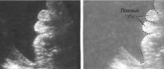
By the third trimester, all the internal systems and organs of the child are fully formed. The fetus develops and grows in the womb. Using ultrasound, it is possible to identify defects and hidden anomalies formed by this time that were not visible before.
As part of the procedure, the size of the fetus is measured, its location in the uterus, respiratory and motor activity, and overall development are assessed in accordance with age standards. Using an auxiliary ultrasound technique - Dopplerography - the nature and speed of blood flow in the vessels of the fetus, umbilical cord and uterus is assessed.
Based on the data obtained, assessment of the functions of the placenta, the woman’s birth canal, the size and presentation of the fetus, and possible entanglements, the doctor makes a conclusion whether the woman can give birth on her own or whether a cesarean section is indicated for her. The final position of the placenta, the quantity and quality of amniotic fluid, and the risk of late toxicosis are also established. Taking these factors into account, the pregnant woman may be recommended to be hospitalized to prevent premature birth.
Monitoring the baby: Ultrasound in the 3rd trimester
Ultrasound examination makes it possible to timely identify pathologies in the fetus and take measures to eliminate them. In the 3rd trimester, situations often arise in which the doctor recommends an ultrasound. Using this procedure, you can solve a number of issues:
- assess the condition of the fetus;
- determine the method of delivery;
- see changes in the organs of the woman’s reproductive system that affect the course of pregnancy.
There is only one planned ultrasound in the 3rd trimester, and additional ones are performed according to the doctor’s decision. Women with pathologies and diseases that put the development of the fetus at risk are required to undergo an ultrasound scan. Identified anomalies in the development of the child also require ultrasound monitoring over time.
Additional ultrasound examinations
If the doctor conducting third trimester screening still has doubts about the health of the woman or baby, he may order additional tests.
CTG or cardiotocography - measurement of fetal contractions. This type of diagnosis allows the doctor to hear the baby’s heartbeat and measure its frequency and strength. This gives an idea of the health of the unborn child.
Dopplerometry is a more serious procedure. This checks the blood flow in the uterus, placenta and umbilical cord. Often this procedure is done several times during pregnancy, as well as in the third trimester, together with an ultrasound scan, but if the fetus develops normally and the ultrasound scan does not reveal pathologies, the doctor may not prescribe it at all.
Ultrasound examination is very informative and absolutely harmless for mother and child. During pregnancy, the mother needs to follow a routine, walk more and eat properly, and if pain or discharge occurs, immediately consult a doctor. Regular visits to the antenatal clinic and scheduled screenings will help maintain the health of mother and child.
How many times can you do it during the third trimester?
The regularity of ultrasound depends on the individual characteristics of the woman. In addition to routine ultrasound, it is recommended to check the condition of the fetus in the following cases:
- slow growth of the abdomen or complete cessation of its expansion;
- weight loss in pregnant women;
- if there are scars on the uterus, to monitor their condition;
- to determine the position of the fetus if breech presentation is suspected;
- when a woman receives any type of injury;
- in the absence of movement for a long time.
If there is no movement for more than 12 hours in a row, you should immediately contact a medical facility for an ultrasound examination. This can often help save the life of the fetus.
Additionally, ultrasound is recommended for women whose work involves hazardous work or high physical or mental stress.
Pathologies that can be identified at this time
In the third trimester, pregnancy is already moving towards its successful completion and this period is characterized by the following possible pathologies, which are detected using ultrasound:
- abnormal position of the fetus;
- deviation in the amount of amniotic fluid;
- entwining the baby with the umbilical cord;
- degree of placental maturity;
- mismatch of fetometric measurement parameters;
- pathology of the heart, lungs and abdominal organs.
How is an ultrasound performed in the 3rd trimester?
In the 3rd trimester, ultrasound is performed only transabdominally. The fetus at this stage is already too large to see in its entirety, so the doctor evaluates individual parts of the body and internal organs. The woman lies on her back, exposing her entire stomach, after which the specialist begins the examination.
How long does it last
The duration of the ultrasound examination depends entirely on the characteristics of the pregnancy. On average, at this time the procedure takes 20-30 minutes, this is due to the large number of areas being examined in the child, as well as the circulatory system. In some cases, the required time may be increased:
- During multiple pregnancy. In this case, the doctor needs to assess the condition of each fetus, check the blood flow, their position relative to each other and the uterus. This is necessary to determine the method of delivery.
- For pathologies in the fetus. In this case, the doctor must examine in detail the abnormally developed organ or system in order to assess the risks and determine how viable the fetus is.
- For problems with the health of the uterus. Scars and other changes on its walls require more careful ultrasound monitoring.
When performing an ultrasound, the doctor may leave the woman with images of the fetus. You should show them to your doctor at your next appointment so that the specialist can assess the course of pregnancy not only by description.
What are they watching?
In the third trimester, the purpose of an ultrasound examination is the same as in the previous ones - to assess the condition of the fetus and identify pathologies, if any. The doctor evaluates the following parameters:
- the maturity of the placenta, on which the supply of nutrients to the baby depends;
- blood supply to the fetus and delivery of the required amount of oxygen to it;
- umbilical cord position, exclusion or confirmation of entanglement;
- the size of the child, compliance with standards, identification of delayed or advanced development;
- the presence of infections that affect the internal organs of the fetus.
Standard measurements of the limbs and body, heart rate are also included in the standard research in the 3rd trimester. At this time, you can see the sex of the baby on an ultrasound, if this was not possible before.
Purposes of ultrasound examination in the 3rd trimester
In the 3rd trimester, an ultrasound scan is done to detect abnormal development of the baby in the womb. It is impossible to do this at earlier dates. No preparation is required for the examination.
During an ultrasound scan, it is checked whether the gestational age corresponds to the development of the baby in the womb. It is determined whether there is a risk of premature birth and signs of oxygen deficiency. The information obtained helps to track and correct many violations. This helps to bring the pregnant woman to a successful delivery.
During an ultrasound, not only the condition of the child, but also his mother is assessed. All reproductive organs are examined. The examination helps doctors prepare if urgent intervention is required. During an ultrasound, the following is examined:
- motor activity in the fetus;
- respiratory system (especially lungs);
- blood flow condition;
- internal organs of the baby;
- fetometric indicators;
- signs of hypoxia;
- umbilical cord (blood flow, vessels);
- heartbeat and fetal position;
- amniotic fluid level;
- fetal presentation;
- length of the cervical tract.
During an ultrasound, the sex of the baby is determined and an approximate due date is set. Particular attention is paid to assessing the placenta. Its improper attachment (low) is a contraindication for childbirth in the usual way.
Norms and interpretation of ultrasound examinations
Only the doctor leading the pregnancy can interpret the ultrasound. Norms and standards are adjusted depending on previous data and other survey methods. An important indicator is the size of the fetus, by which already in the 3rd trimester you can determine how large the child will be. This is important if a woman has a narrow pelvis and other characteristics of her body. During a routine ultrasound, the baby’s size should be within the following limits:
- head circumference from 30 to 32 cm;
- abdominal circumference 28-31 cm;
- femur length 6-6.5 cm;
- shin length 5.5-6 cm;
- humerus length 5.6-6 cm;
- forearm length 4.9-5.2 cm.
Sizes may be larger if parents are tall or the term is determined incorrectly.
A fetus that is too small requires close monitoring, unless the expectant mother is very petite. The normal thickness of the placenta should be from 32 to 44 mm, which means that the baby is adequately fed with the necessary substances. Her maturity at the beginning of the 3rd trimester normally remains 1 degree, and aging begins after 36 weeks. The diameter of amniotic fluid should not exceed 70 mm, as this is a symptom of polyhydramnios, and an amount less than 20 mm indicates oligohydramnios and requires observation in a hospital.
Data from each ultrasound is compared with the previous ones, on the basis of which the doctor makes a conclusion about the course of pregnancy and the condition of the fetus.
What points are paid special attention to during an ultrasound?
At this time, attention is paid to such points as:
- The position in which the fetus is in relation to the mother's uterus. If it is positioned head down, then there is no cause for concern, the child lies normally and takes the correct position. But it often happens that the unborn child is positioned transversely and the doctor gives him a period of 2-3 weeks to assume a normal position. If during this period the revolution does not occur, the mother will be prepared for a caesarean section, so as not to harm either the baby or his parent.
- The amount of amniotic fluid is sufficient, because it is when an ultrasound is done in the third trimester that such a deviation from the norm as oligohydramnios or polyhydramnios can be detected. Both the first and second are very dangerous for expectant mothers, as they signal the presence of any infections in the body.
- The entanglement of a child in the umbilical cord is a fairly common deviation, and at this stage it is even possible to detect double entanglement. If the fact of entanglement with the umbilical cord is confirmed by ultrasound, then experts recommend only a cesarean section - in the process of natural birth, the child may simply be strangled by his own umbilical cord during the passage of the birth canal.
- The degree of maturation of the placenta - if it has matured before the due date corresponding to the stage of development of pregnancy, then the woman should be constantly monitored so that premature contractions and birth do not begin, in addition, with early maturation of the placenta, the child will experience a deficiency of nutrients and oxygen.
- Only an ultrasound in the third trimester allows you to most accurately determine the weight of the unborn baby, which is of great importance if a pregnant woman has a narrow pelvis, when the doctor has doubts whether she will be able to give birth on her own.
- Fetometry. These are parameters for measuring the volume of the fetus - the head, abdomen, length of the hips, because it is by these indicators that the gestational age is determined. Having detected deviations, the doctor is obliged to carry out an extended phytometry procedure - he measures the head circumference in the fronto-occipital part and calculates its percentage with other measurements. Then it takes a second measurement of the tummy and compares it with the measurement of the femur. After the measurements, the doctor examines the brain, considering the state of the vascular plexus, the size of the lobes of the brain and cerebellum, which is required to check for the presence of brain diseases and intrauterine infections that can negatively affect the motor and swallowing abilities of the child. After this, the doctor examines the structure of the nose, lips, eyes and spine.
- The condition of the fetal organs - especially the lungs and heart. If his diaphragm is underdeveloped, then his lungs will not correspond to the norm. To check cardiac activity, the correct functioning of valves, vessels and partitions, a special study is carried out - cardiotocography, which allows you to determine the frequency of heart contractions and check the entire cardiac activity of the system. This procedure can be carried out only after 32 weeks, otherwise the diagnosis will provide unreliable data.
- The condition of the abdominal cavity - the coherence of the intestines, liver, kidneys and bladder is checked. Among the pathologies, abnormalities most often occur in the kidneys.
Can an ultrasound be done if the baby is post-term?
A pregnancy is considered post-term if its duration exceeds 40 obstetric weeks. Doctors recommend undergoing an ultrasound to determine the maturity of the placenta, the condition of the waters and the fetus itself. The specialist checks the readiness of the cervix. The need for urgent delivery depends on the results obtained.
A period of more than 42 weeks requires medical intervention, which is also preceded by an ultrasound. The doctor evaluates all indicators, after which a decision is made to induce labor or perform a cesarean section.
In the 3rd trimester, an ultrasound scan must be performed at least once, at 32 weeks. Timely measures taken in case of any deviations from the norm will help carry the child to the end of the term, and monitoring the condition of the uterus will avoid a threat to the health of the expectant mother.
Features of the third ultrasound in pregnant women
At 32–34 weeks, the main attention is paid to fetometry (measurement of the physical parameters of the fetus), the condition of the placenta, as well as analysis of the circulatory system and cardiac function of the child.
Some vascular malformations (veins and arteries) form in the last trimester, which explains the diagnostic importance of the third screening for the health of the unborn newborn.
Ultrasound in the third trimester of pregnancy is performed transabdominally - with a conventional sensor, the doctor examines through the abdominal wall. At this stage, ultrasound no longer requires special preparation - the location of the uterus, the amount of amniotic fluid and the size of the fetus provide good visibility during the examination.
For a detailed study of the vascular network, blood flow of the placenta and the cardiac function of the child, a standard ultrasound is necessarily performed in combination with Doppler ultrasound.
Additionally: first and second ultrasounds during pregnancy.
What is examined and what is looked at
- In the third trimester, the anatomical and physiological parameters of the child and fetometry are assessed.
- Position of the fetus in the uterus.
- Defects that develop at a later stage are identified.
- They look at the placenta (place of attachment, thickness, maturity).
- The amount of amniotic fluid is determined.
- Some contraindications for natural childbirth are excluded (entanglement of the umbilical cord, low attachment of the placenta).
- Determine the preliminary date of birth.
- The state of blood flow in the vessels of the umbilical cord and uterus is assessed.
Ultrasound diagnostics in the third trimester is carried out transabdominally (through the anterior wall of the abdomen). No special preparation is required.
When deciphering the data during the last ultrasound, specialists determine the characteristics of the upcoming birth. The tactics of labor management are finally chosen (natural birth canal, cesarean section). Eliminates the danger of post-term pregnancy or its premature resolution.
What examinations are done during an ultrasound in the third trimester:
- Two-dimensional or three-dimensional echography;
- Cardiotocography (CTG);
- Doppler.
Features of the fetus at the beginning of the third trimester
When and how many times to carry out echography is decided when monitoring the prenatal development of the fetus. If necessary, doctors prescribe ultrasound in late pregnancy.
Let us list some of the normal developmental features that a fetus has in the 3rd trimester of pregnancy:
- Continues to actively grow and gain subcutaneous fat.
- The respiratory system is improving, but is not yet sufficiently developed.
- The cerebral cortex is actively developing.
- The child moves actively and continuously, can bend and straighten his limbs.
- The development of the reproductive system is completed.
- Vellus hair gradually disappears.
- Sense organs are formed: reacts to sound, light, feels pain.
- The child sneezes, hiccups, may change his facial expression, blink.
- The liver grows and stores iron for hemoglobin.
- The pancreatic cell increases in volume, but enzymes are not yet released.
Video: 3 screening
Goals and timing of the study
At what week is it better to have the third ultrasound? At 30-34 weeks of pregnancy, the final formation of all organs and systems of the child occurs. He experiences the maximum need for nutrients, which are delivered through the placenta and umbilical cord. How the birth will proceed and what vital signs the child will be born with depends on the “health” during this period.
Now it becomes clear why and when the third ultrasound is performed. The main objectives of this procedure are:
- Determination of fetal position. In the second trimester of pregnancy, the baby moves freely in the uterine cavity and can change position at any time. When a woman undergoes her third ultrasound during pregnancy, the presentation of the fetus is already formed (cephalic, pelvic, transverse).
- The study makes it possible to clarify all the dimensions of the fetus: its weight, body proportions, head circumference, and the length of individual parts of the body. Not only the sizes themselves are important, but also what week of gestation they correspond to. “Large fetus” requires special attention during delivery.
- 3rd trimester screening assesses brain development. At the same time, signs of intrauterine infection become noticeable. Sometimes, after an ultrasound at 32 weeks of pregnancy, doctors decide on emergency delivery based on vital indications for the fetus.
- Study of amniotic fluid, which indirectly indicates the normal development of the baby.
- Final confirmation of the readiness of the mother's birth canal for natural childbirth. If, as a result of the 3rd screening, space-occupying formations or incompetence of the cervix are detected, then a decision is made on a Caesarean section.
- At this time, the respiratory movements of the fetus and its motor activity are already well determined. Deviation of indicators from the norm indicates intrauterine hypoxia and the need to look for its cause.
Interpretation of the ultrasound examination allows you to set the date of birth and decide on its method (natural or surgical).


