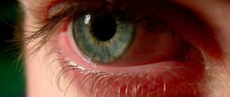
Retinal detachment is a serious disease that requires urgent treatment. This condition is characterized by the separation of the layer with photoreceptor cells from the retinal epithelium (outer layer), which disrupts the nutrition of the outer layers. This phenomenon occurs when intraocular fluid accumulates between these layers. Retinal detachment leads to rapid vision loss. When a detachment occurs, patients turn to the ophthalmologist with complaints of a sharp deterioration in vision, the appearance of sparks and flashes before the eyes.
What is retinal detachment
Adhesions can form in the vitreous body, which attach to the retina and, when moved, provoke its rupture, ingress of ocular moisture and detachment. The risk of detachment increases in the presence of a high degree of myopia, dystrophy, diabetes mellitus, a history of surgical intervention, trauma and vascular pathologies of the eye.
Retinal detachments are divided into types depending on the cause of the detachment. Correct diagnosis and precise determination of the cause help to choose the appropriate treatment.
Types of retinal detachments:
- Rhegmatogenous (primary or idiopathic). It develops when there is a gap through which moisture from the vitreous body enters under the retina. In turn, breaks occur in places where the retina is thinned against the background of its dystrophy (racemosal, ethmoid, retinoschisis, and others). With degenerative changes in the retina, breaks appear from sudden movements, tension, or spontaneously.
- Traction. The focus of pathology occurs as a result of tension during the formation of fibrin strands or vessels that grow into the vitreous body.
- Exudative (serous). Occurs when fluid accumulates under the retina against the background of a pathological process (without the formation of ruptures).
- Traumatic. Begins as a result of injury to the eyeball. Retinal detachment can occur at the time of damage or be a consequence. Damage resulting from surgery is also classified as traumatic.
- Secondary. Such detachments can be the result of diseases: tumors, inflammation, thrombosis and hemorrhages, retinopathy, sickle cell anemia and others.
There are flat, high and bubble-shaped retinal detachments. According to the equality of the process, fresh, stale and old are divided. Classification according to the degree of distribution: local retinal detachment, widespread, total, subtotal.
Symptoms
Note that this disease does not always have pronounced symptoms, like the symptoms of keratitis. Often the first harbingers of retinal detachment are light hallucinations - phosphenes in the form of bright flashes, flying glowing flies, and other phenomena.
If the detachment is accompanied by a rupture of a small blood vessel, which is quite common, black circles will also be observed before the eyes. Painful sensations are also possible.
Retinal detachment in the traditional classical form progresses quickly, so treatment must begin immediately.
If measures are not taken in time, the primary symptoms will be replaced by a feeling of a veil before the eyes, which will greatly impede visibility. And the resulting veil will begin to become denser - and quite quickly. Surgical treatment of eye catarths helps to get rid of the disease forever.
diagram of retinal detachment
If the retinal detachment is rhegmatogenous in nature, the person experiences a hallucination in the form of double objects (diplopia). Treatment of retinal dystrophy at home is effective in combination with traditional treatment.
Note that patients with this disease often observe a temporary improvement in vision in the morning. This fact is associated with the resorption of intraocular fluid overnight. But during the day and, especially, in the evening, vision will decline again.
If a complete retinal detachment occurs, the following symptoms are likely to occur:
- loss of vision (sometimes complete);
- bleeding from the eyes;
- swelling.
In this case, only urgent surgical intervention will help eliminate the problem.
How does retinal detachment manifest?
The onset of detachment can be determined by characteristic symptoms. Patients most often complain of metamorphopsia (curvature of straight lines) and photopsia (light flashes). When a retinal vessel ruptures, a large number of floaters and black dots appear in the field of vision.
During direct retinal detachment, there is a presence of a veil before the eyes, a veil or a dark shadow. Vision rapidly deteriorates, although in the morning it may improve slightly and the field of vision expand. The black curtain obscures part of the field of vision, spreads to the entire retina, and the person goes blind.
Age-related macular degeneration (AMD)
The disease is most often detected in people over 50 years of age and is the main cause of irreversible vision loss. The risk of the disease increases with a genetic predisposition, as well as in patients with diabetes mellitus, hypertension, and atherosclerosis. Symptoms of the disease are associated with damage to the macula area of the retina.
The initial manifestations of AMD do not have any clinical manifestations and can only be detected during an examination by an ophthalmologist. As the disease progresses, symptoms such as distorted outlines of objects, blurred vision, and blurred vision appear.
The dry form of AMD does not require special treatment. To treat the wet form of age-related macular degeneration, intravitreal injections (injections into the vitreous cavity) of drugs that block the growth of defective vessels under the retina (anti-VEGF drugs) are used. In addition, photodynamic therapy and laser photocoagulation of the retina are indicated.
Read more about AMD treatment >>>
Diagnosis of retinal detachment
If retinal detachment is suspected, a comprehensive examination of the patient is performed. Early detection of the disease is the key to maintaining visual function.
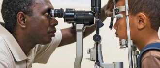
The main method for diagnosing detachment is ophthalmoscopy. This is a procedure for examining the fundus of the eye using various techniques (indirect forehead ophthalmoscope, non-contact and contact lenses). A combination of techniques and examination of the fundus in different positions allows for a comprehensive study of the condition of the retina.
Ophthalmoscopy makes it possible to determine the extent of the process, its shape and localization, as well as to identify areas of dystrophy. During detachment, the doctor sees the disappearance of the red fundus reflex, and in the detachment zone it becomes grayish-white. Small-height detachments can be recognized by changes in the location of blood vessels and a decrease in the clarity of the choroid.
High detachments are diagnosed by white or gray bubbles that sway when the eye moves. Old processes provoke the appearance of rough folds and star-shaped scars. The detached retina is rigid and immobile.
When examined, the tears are red in color and vary in shape. The characteristics of the ruptures will determine the rate at which the detachment process spreads and the prospects for treatment. When ruptures are localized in the upper part of the eye, detachments progress faster. In the lower regions the process will be slow and the flow will be favorable.
In cases where examination of the fundus is impossible or difficult, ultrasound diagnostic methods are used. Electrophysiological studies are designed to assess the functionality of the retina in the presence of an old detachment. Additionally, intraocular pressure is measured. With detachment, there may be a decrease in pressure in the affected eye.
Perimetry (examination of visual fields) also turns out to be informative. Detachment is characterized by loss of vision. Their characteristics will depend on the extent of the detachment and its location. It is also important to take into account the fact that the macular region is involved in the pathological process. Typically, loss occurs opposite to detachment.
Proper nutrition for dystrophy
An important part of the treatment of retinal dystrophy of the visual organs is the normalization of nutrient metabolism. To do this, the patient needs to reconsider his diet.
It is recommended to eat as many fresh vegetables and fruits and seafood as possible. Honey and cinnamon have a beneficial effect on the visual apparatus.
A diet for degenerative processes in the eyeball involves avoiding the following foods:
- alcohol;
- flour products containing baking powder;
- sausages;
- canned food;
- semi-finished products;
- fast food.
If you have vision problems, you should not eat foods that are too salty, fatty or sweet. Instead, you should eat low-fat dairy products, fish, and various cereals. Meals should be fractional.
You can improve the condition of your visual system if you regularly add spices such as cumin, coriander and cumin to your dishes.
Surgical methods for treating retinal detachment
Retinal detachment requires immediate treatment. A long-term pathological process will provoke persistent hypotension, cataracts, iridocyclitis, eye subatrophy and blindness. The main goal of therapy is to bring the layers of the retina together and block breaks. When treating detachment, it is important to bring the photoreceptor layer closer to the pigment epithelium and limit the rupture to foci of chorioretinal inflammation. This is a local sterile inflammation that glues the retina to the choroid and stops the progression of the disease.
At-risk groups:
- a high degree of myopia and astigmatism, which provoke thinning and rupture of the retina;
- age from 45 years;
- heavy loads and high risk of injury in athletes;
- diabetes mellitus, causing diabetic retinopathy and hemorrhages;
- heredity.
Surgical intervention for detachment can be extrascleral (on the surface of the sclera) and endovitreal (from inside the eye). The best treatment option is vitrectomy. This is a procedure to remove the vitreous and replace it with silicone or gas to ensure that the detachment adheres tightly to adjacent layers.
Possible surgeries for detachment
- Extrascleral filling. The operation is performed in the presence of ruptures that do not require exposure from inside the eye. The seal is installed externally.
- Vitreoretinal surgery. It is used for old detachments, when careful cleansing and straightening of the retina is required. Special silicone is injected through pinhole punctures using long instruments.
- Cryocoagulation of ruptures, as well as subclinical detachments.
The goal of treatment is to block the retinal tear. The sooner the operation is performed, the more reliable the result will be and the better vision will be restored. Doctors give the most favorable prognosis for detachments that do not affect the central zone. If the pathology has managed to close the center of the retina, even after a successful operation it is not possible to completely restore vision.
Since retinal detachment is a consequence of a tear, it is necessary to undergo regular preventive examinations and detect them on time. Laser coagulation techniques are used to treat ruptures.
Detachments that were treated incorrectly or unsuccessfully must be operated on within a year, while the eye still perceives light. Surgical treatment of retinal detachments is carried out painlessly, safely and quickly. The operation is carried out using the latest equipment and only highly qualified specialists. Outpatient procedures take from 40 minutes to 1.5 hours, taking into account the complexity of the operation and complications.
Retinal detachment can be corrected through surgery, but not in every case it is possible to restore the integrity of the retina and full vision. Even after successful treatment of severe detachment, vision rarely returns completely. Only in some cases is it restored to its original level.
After surgical treatment there are no restrictions on visual stress, but for a month the patient is prohibited from visiting the bathhouse, sauna and swimming pool. Physical activity should be minimized for a period of one month to a year, depending on the severity of the condition.
After surgery to eliminate detachment, refractive errors (myopia, astigmatism) often increase. Sometimes relapses occur and repeated surgery is required, which often turns out to be ineffective. The success of surgery for detachment is determined by the timeliness of treatment. A long-term pathological process, as a rule, ends with irreversible changes in the retina and the death of visual neurons.
Endovitreal treatment of retinal detachment
Endovitreal surgery involves intervention from the cavity of the eyeball. The doctor makes three incisions in the sclera (approximately 1 mm each), through which he gains access to the vitreous body and retina. This procedure is called sclerotomy. Instruments, a light source, and a solution are introduced through the incisions to maintain the tone of the eye. The most commonly used is a vitreotome - a 1 mm cylinder in which a knife is hidden that cuts the intraocular tissues. If necessary, the doctor may use other instruments.
Expanding gases, silicone oil or perfluoroorganic compounds are used to straighten and press the retina to the membranes. After the introduction of a special substance, laser coagulation of the retina can be performed.
Indications for vitrectomy for detachment:
- big sizes;
- long retinal detachments along the dentate line;
- proliferative vitreoretinopathy, presence of folds;
- posterior retinal tear;
- combination of rupture with hemophthalmos.
During endovetral intervention, the vitreous body is removed (transciliary vitrectomy). Sometimes prolonged tamponade of the cavity with silicone oil or gas is required. The gas bubble will resolve within 2-4 weeks, shrinking and being replaced by intraocular fluid. Silicone oil takes a little longer to remove (2-3 months).
For rhegmatogenous detachment, the doctor removes the vitreous body and the posterior hyaloid membrane. To relieve traction, cords and membranes are removed. During surgery, a bubble of “heavy water” is created in the fundus of the eye, which presses on the retina. Excess fluid is removed through the gap, and laser coagulation of the affected areas is performed. After this, the “heavy water” is replaced with saline solution, and the incisions are sutured. When proliferative vitreoretinopathy of the tissue occurs with an old detachment and it is impossible to smooth it out, loosening peripheral incisions (retinotomy) are required.
Extrascleral treatment of retinal detachment
With endovitreal access, the operation is performed from inside the eye, and with extrasleral intervention, the rapprochement of the retina and pigment epithelium is achieved by indenting the sclera (filling). During the operation, the doctor creates an indentation shaft that blocks the rupture, and the accumulated fluid is gradually absorbed into the epithelium and choroid.
Before such an operation, bed rest is required so that the detachment bubbles decrease as the subretinal fluid is absorbed. This makes it easier to detect the break. After the operation, bed rest is also prescribed, at least for a day.
Scleral filling
When filling the sclera, the layers of the retina are brought together by pressing the sclera from the outside. In the projection of the rupture, a silicone strip or filling of the required size is attached to the sclera. The strip is literally sewn on. Under its pressure, the sclera is pressed inward, pressing the choroid to the retina. In this position, the accumulated fluid begins to dissolve.
Filling stages:
- Determining the location of the rupture, marking this zone on the sclera. For these purposes, a diathermocautery is used, with the tip of which the doctor applies pressure, creates a shaft and marks the location of the projection of the rupture on the sclera.
- Cutting out the filling and suturing it to the sclera in the projection area. The position of the filling will depend on the type of pathology, location and number of breaks. There are radial, sectoral and circular fillings.
- If there is a large amount of fluid, it must be removed through an opening to the sclera (drainage).
- Additionally, air or gas can be introduced into the vitreous body. While the bubble dissolves (several days), vision will remain poor.
- Applying sutures to the conjunctiva.
If there is a large accumulation of subretinal fluid, it is drained through a puncture in the sclera. When filling, soft silicone sponges are used. It is easy to cut out a filling from silicone according to the required parameters.
The type of filling is determined by the doctor, taking into account the type and location of the rupture. There are radial, sectoral and circular fillings. In some cases, they resort to cerclage (circular indentation with silicone thread or braid). The circlage is created in the equator area of the eye.
Possible complications after filling:
- Early: infection of the eyeball and orbit, vascular detachment, glaucoma, dysfunction of the extraocular muscles, ptosis, strabismus.
- Late: exposure of the filling, changes in the central area, refractive errors, cataracts.
- Consequences: lack of detachment adhesion, relapse.
Restoration of vision after filling occurs gradually. This process usually takes several months.
Ballooning of the sclera
The operation involves temporarily placing a catheter with a balloon to the sclera (in the area of projection of the rupture). Liquid is pumped into the balloon, its volume increases, creating the effect of indentation of the sclera, similar to a filling operation.
Ballooning creates conditions for the resorption of subretinal fluid and successful delineation laser coagulation of the retina. The balloon is removed after the formation of adhesions between the retina and adjacent tissues. Ballooning is low-traumatic and is used for various pathologies of the visual system.
Laser coagulation for detachment
After extrascleral operations, diathermal, laser or photographic coagulation is possible. The effect can be consolidated by cryopexy along the boundaries of the detachment from the side of the cavity through the pupil (transpupillary) or the sclera (transscleral). Additional exposure provokes the formation of adhesions around the break and reliable fixation of the retina.
Laser treatment for detachment allows you to create adhesions between the retina and the lining of the blood vessels. The doctor uses laser coagulators to create microburns. This operation is effective for preventing detachment, limiting existing lesions (flat detachments) and additional coagulation after surgery.
Laser coagulation of the retina is performed under local anesthesia. A Goldmann lens is placed on the eye, which focuses laser radiation on a specific area of the fundus of the eye. Adhesions form within two weeks.
Possible complications of laser coagulation:
- exudative detachment;
- vascular detachment;
- degenerative changes in the central region.
Risk factors
Despite the variety of causes that can cause retinal diseases, it is possible to identify factors that significantly increase the risk of developing pathology. The most important ones include:
- smoking;
- increased cholesterol levels;
- obesity;
- arterial hypertension;
- prolonged exposure to bright sunlight, artificial light;
- working on a computer.
Factors contributing to hypertension
The risk of thrombosis of arteries or veins increases in people who take combined oral contraceptives. Inflammatory pathologies more often develop in people with reduced immunity. This may be due to taking corticosteroids, HIV infection or tuberculosis.
Prevention of retinal detachment
The main measure to prevent retinal detachment is timely contact with a specialist if characteristic symptoms occur. Therefore, you need to be attentive to your health and respond to any discomfort in a timely manner. It is very important to undergo regular preventive examinations, even if there are no risk factors.
After a head or eye injury, you need to undergo a full examination. Pregnant women are recommended to undergo examinations and undergo preventive laser coagulation to prevent abruption during childbirth. Patients with a high degree of myopia, retinal dystrophy and a history of surgery should exclude certain sports and heavy loads to avoid detachment.
In case of detachment, the prognosis will depend on the duration of the process, localization and condition of the vitreous body. For the result to be reliable and effective, the operation must be performed within two months from the onset of detachment. After treatment, patients should be observed by an ophthalmologist and limit physical activity.
Similar articles
Inflammatory diseases
In the occurrence of inflammatory diseases of the retina, the main role is played by the hematological route of infection. It is through the bloodstream that pathogens from foci of infection in other organs enter here. For this reason, infectious retinitis is often combined with inflammation of the vascular layer of the eyeball - choreoretinitis, inflammation of the optic nerve head - neuroretinitis, inflammation of the vessels of the retina itself - retinal periphlebitis.
The causative agents may be:
- staphylococci, streptococci, pneumococci;
- pathogens of tuberculosis, syphilis, toxoplasmosis;
- herpes viruses, measles, adenoviruses.
Inflammation of the retina is characterized by visual disturbances (the appearance of scotomas above the lesion at the bottom of the eye), metamorphopsia (distortion of shape), photopsia (flashes of light). Unfortunately, after purulent retinitis, complete restoration of visual acuity is almost impossible to achieve.
Therapy is etiologically directed against the underlying disease (antibiotics, antiviral or antifungal drugs). Additionally, trophic, vasodilating and anti-inflammatory drugs are used.
If the vitreous body is damaged, its removal is indicated as a dangerous source of infection; if retinal detachment occurs due to inflammatory edema or pus, it is coagulated with a laser.
Possible complications
As noted earlier, improper or untimely treatment of retinal detachment can lead to serious complications. Also, health problems may arise immediately after the operation. As a rule, such complications manifest themselves in the form of glaucoma, cataracts or general weakness of the patient’s body.
The most common complications of retinal detachment include the following:
- development of endophthalmitis (purulent inflammation of the membranes of the eyeball caused by infection);
- proliferative vitreoretinopathy - a pathology accompanied by the proliferation of fibrous tissue (scarring);
- relapse of the disease (repeated detachment), requiring repeated surgery.
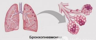
If you notice symptoms such as chest pain, frequent cough, difficulty breathing, redness and swelling of the skin, chills, fever or purulent discharge from the eyes after surgery, you should immediately contact a specialist. The appearance of such symptoms may indicate the development of serious diseases, so ignoring them can lead to disastrous consequences.
Postoperative period
After the operation, the patient remains unable to work for a month. At this time, physical activity, lifting weights, bending the body, and visiting the bathhouse are prohibited. In the future, increasing physical activity is possible after consulting a doctor. In the postoperative period, drug therapy is carried out with the mandatory use of anti-inflammatory, antibacterial, and reparative agents.
In case of clouding or decreased vision in the operated eye, you should contact an ophthalmologist. It is recommended to periodically examine the second eye for early diagnosis of possible retinal detachment.
Grigorova Valeria, medical observer
7, total, today
( 178 votes, average: 4.51 out of 5)
Strabismus in children: causes, symptoms, treatment
Allergic conjunctivitis: symptoms and treatment