With the onset of pregnancy, a restructuring of the functions of all organs and systems occurs in a woman’s body. According to Russian statistics, 70–80% of all pregnant women experience pathological disorders called gestosis, better known as toxicosis.
Preeclampsia during pregnancy can occur at any time and manifest itself in mild and severe forms. About 18% of pregnant women are registered with antenatal clinics for this matter. From 8 to 16% suffer from toxicosis in the second half of pregnancy, and among pregnant women in specialized hospitals - up to 30%.
The proportion of mortality as a result of eclampsia (severe form of gestosis) is 20–25%; the mortality rate of children due to gestosis in the period from the 22nd week of pregnancy to the 7th day after birth (perinatal mortality) is 3–4 times higher than the average mortality rate.
What is gestosis
Statistics vary widely from source to source. This is mainly due to three reasons:
- Incomplete coverage of all pregnant women with dispensary observation.
- Late referral of pregnant women to antenatal clinics, when mild forms of gestosis become more severe (preeclampsia and eclampsia).
- Change in the general name and classification of pathological disorders.
In connection with the last point, some clarity should be provided. This will avoid substitution of concepts and confusion in ideas.
Previously, all pathological symptoms throughout pregnancy and deviations from the norm in the results of laboratory and instrumental studies were called toxicosis of pregnant women. Depending on the duration and form, it was divided into toxicosis of the first and second half of pregnancy.
In accordance with this classification, the term “toxicosis” is replaced by the term “preeclampsia”. Some experts believe that it can be early (in the first trimester of pregnancy) and late (in the second half). Others recognize only gestosis in the second half of pregnancy, and those disorders that appear in the first half are considered either physiological or not related to the etiology (cause) and pathogenesis (mechanism of development) of gestosis. The first option seems more convenient, allowing one to get an idea of any pathological deviations from the normal course of pregnancy.
They are associated with pregnancy and disappear after delivery. This is the basis for the assumption by all scientists that the cause of gestosis lies in:
- the negative role of the fertilized egg and fetus;
- a disorder of a woman’s adaptive mechanisms designed to provide the fetus with the opportunity for normal development.
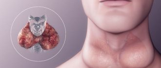
There are many factors contributing to the occurrence of gestosis during pregnancy. However, among them, the greatest attention is paid to concomitant overt or hidden endocrine diseases, impaired renal and liver function, high blood pressure, multiple pregnancies, hydatidiform moles and some others.
There are many versions and theories about the initial and subsequent mechanisms of the development of pathology, but all of them, except the autoimmune one, raise great doubts. Rather, they reflect individual links in the cascade development of a single mechanism.
Complications
important Preeclampsia is a truly dangerous pathology, in which sometimes doctors have to forget about the child and do everything to save the life of at least the mother. Preeclampsia in the second half of pregnancy is not a condition with which you should joke: severe forms (preeclampsia, eclampsia) can develop in a matter of hours and lead to extremely serious complications.
Possible complications of gestosis:
- Premature termination of pregnancy;
- Intrauterine growth retardation;
- Fetoplacental insufficiency;
- Premature abruption of a normally located placenta;
- Kidney, heart, liver failure;
- Heart attack;
- Retinal detachment;
- Hemorrhage in internal organs, including the brain (stroke);
- Thrombosis of large vessels;
- Cerebral edema followed by coma;
- Fetal death;
- Death of a woman
.
Never refuse hospitalization if your doctor strongly recommends it: remember that your life and the life of your child are most valuable!
Preeclampsia in early pregnancy
It occurs in the first three months of pregnancy and goes away completely by the beginning of the second trimester. The leading role in its pathogenesis is given to:
- Functional disorders of regulation of the central nervous system according to the type of neuroses.
- Disadaptation of vegetative centers. Pregnancy first of all requires a restructuring of the function of the digestive system, which is connected through nerve receptors and trunks with the autonomic centers of the hypothalamic region of the brain. Impulses arriving at these same centers from the altered uterus or nerve pathways against the background of nervous system disorders can be perverted. They cause response signals from the centers, but already to the digestive tract, which leads to nausea and vomiting.
- Neuroendocrine (neurohormonal) disorders and metabolic disorders. They also contribute to the emergence of perverted pathological impulses and the development of early gestosis. For example, the timing of the onset of vomiting was observed to coincide with a decrease in the secretion of corticosteroids by the adrenal cortex and a peak in the content of human chorionic gonadotropin in the blood.
What forms of gestosis can occur in the first trimester of pregnancy?
In medicine, it is customary to distinguish rare forms of gestosis:
- fatty liver (due to cholelithiasis in the period of exacerbation, intoxication)
- dermatitis (it should be differentiated from atypical forms of syphilis, because the RV reaction can be weakly positive)
- osteomalacia (due to impaired calcium phosphorus metabolism, decreased function of calcitriol in the parathyroid glands, prolonged leaching of calcium from the bones). A pregnant woman is prescribed parathyroidin, calcium supplements and vitamin D2. Children are prescribed a solution of ergocalciferol in drops - 1 drop throughout the year.
- tetany (characterized by the appearance of clonic convulsions). To eliminate convulsive syndrome, bolus therapy with magnesium is used, and vitamin therapy (calcemin, ergocalciferol) is also indicated.
Clinical manifestations and treatment
Mild gestosis is most often manifested by salivation, nausea and vomiting (50 - 60%), less often - dermatoses in the form of skin itching and rash, eczema, very rarely - bronchial asthma, osteomalacia (softening of the bones), acute liver dystrophy, tonic spasms of the limbs or muscles of the neck and face.
The earlier vomiting occurs, the more severe it is and is accompanied by fluid loss and disturbances in water and electrolyte balance. There are 3 degrees of severity:
- I degree is a mild form. Vomiting can be on an empty stomach or due to food intake or an unpleasant odor. Its frequency is no more than 5 times a day, and body weight loss is no more than 2–3 kg per week.
- II degree - moderate severity. Vomiting is repeated 6–10 times, and weight loss is 3 kg over 7–10 days. Weakness occurs, the pulse rate increases to 90–100 beats/min at rest, a slight decrease in blood pressure, and a positive urine reaction to the presence of acetone.
- III degree - indomitable vomiting of pregnant women. It occurs with any movement, food or water intake up to 20 - 25 times a day. Weight loss - 8 - 10 kg or more. Severe signs of dehydration, weakness, increased body temperature, dry skin and mucous membranes appear. The pulse exceeds 120 beats/min, blood pressure decreases, the daily volume of urine decreases, urine tests reveal a pronounced positive reaction to acetone. Blood tests show disturbances in protein, salt and carbohydrate metabolism.
If symptoms of gestosis occur in the form of vomiting of pregnant women, correction is required only in 8–12%. Treatment of mild forms is carried out on an outpatient basis in the form of recommendations regarding nutrition and regimen, and the prescription of sedatives in the form of tinctures of medicinal herbs. Frequent monitoring of weight dynamics, blood and urine tests, and blood pressure (normal) is necessary.
In grades II and III, rest, sedative (calming) drugs, corrective therapy using intravenous drips of water-salt solutions, vitamins, and replacement of protein loss are prescribed. With adequate therapy, the condition quickly recovers.
In severe cases, treatment is carried out only in a hospital. If symptoms progress during treatment, artificial termination of pregnancy is indicated.
Therapy
If necessary, the list of diagnostic measures is expanded. Often consultations with related specialists are included - neurologist, ophthalmologist, surgeon. Treatment depends on how long ago the first signs of the disease appeared.
Early
Correction of early gestosis (toxicosis) is as follows.
- Mode. Mild and moderate forms of pathology can be treated at home, severe forms can be treated in a hospital. You should adhere to a diet limiting fatty, salty, and smoked foods. It is recommended to consume acidic foods and drinks to help reduce the frequency of vomiting.
- Anti-vomiting medications. The most common antiemetic drug is Metoclopramide. Available in the form of injections and tablets.
- Restoring water balance. It is carried out using plenty of fluids or intravenous injections of glucose-saline solutions.
- Vitamins and minerals. They help replenish the supply of nutrients lost through vomiting and support the body.
Artificial termination of pregnancy is used as a last resort if early toxicosis is associated with dangerous complications and worsens the mother's condition.
Late
Correction of late gestosis involves the following measures.
- Mode. A woman may be offered hospitalization as she needs constant medical supervision and complete rest.
- Nutrition and weight. The expectant mother is prescribed a diet that excludes the consumption of sweet, salty and fatty foods. At the same time, weight is monitored daily.
- Magnesia. This is the main remedy that helps cope with the main symptoms of the condition. Intravenous infusions can be prescribed around the clock - for this, slow administration of the drug is used using special devices, lineomatov.
- Correction of edema. It is carried out using solutions based on starch compounds, which help restore water balance in the body by redirecting fluid into the bloodstream.
- Normalization of pressure. Metoprolol, Diltiazem, Methyldopa are allowed. Infusion of magnesium also reduces blood pressure.
- Liver protection. Almost always, gestosis affects the functioning of the liver, so drugs are prescribed that normalize its function. Preference is given to natural-based products. For example, "Heptral".
- Prevention of thrombosis. Cardiomagnyl is prescribed, as well as aspirin-based drugs. Less commonly, anticoagulants Fragmin and Fraxiparin.
To protect the fetus and maintain the functions of the placenta, special medications are used individually for each case. These can be metabolic agents (for example, Actovegin) or those that improve blood circulation (Pentoxifylline). In severe forms of pathology, deterioration of the condition of the woman or fetus, the option of delivery by cesarean section at any stage of gestation is considered.
Late gestosis
Preeclampsia in late pregnancy is accompanied by a violation of all types of metabolism, total damage to all systems (see calculator for calculating the gestational age). At the same time, the properties of the blood and vascular walls change, and degeneration of organs and tissues occurs.
There are neurogenic, hormonal, and renal theories of the mechanisms of pathology development. Currently, preference is given to immunological. It has several varieties, differing in some elements. Its general meaning is tissue incompatibility at the cellular level. As a result, the vascular changes in the uterus necessary for the formation of placental blood flow do not occur.
This confirms the fact that gestosis occurs from the very beginning of pregnancy. Therefore, applying the term “late” to it does not reflect reality.
The resulting disturbances lead to a decrease in blood flow in the chorionic villi and a deterioration in the delivery of oxygen to the tissues (hypoxia). This causes local damage to the inner lining of blood vessels (endothelium), which becomes widespread due to the release of biologically active substances that are toxic to them.
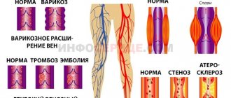
The consequence is an increase in vascular permeability and the occurrence of edema, increased sensitivity to vasoconstrictor hormones. A widespread spasm of blood vessels occurs with an increase in blood pressure. The ability of the endothelial cells of the inner wall of blood vessels to resist thrombus formation is reduced, and the physicochemical properties of the blood (thickening, fluidity, etc.) are disrupted.
As a result of these processes, microthrombi are formed, microcirculation and tissue nutrition are disrupted, the structure of the liver, lungs, and kidneys is damaged, which is manifested in corresponding changes in the general condition and the results of laboratory diagnostic and instrumental studies.
Classification by period of occurrence
Depending on the period, early and late gestosis are distinguished.
Early gestosis in pregnant women
Early gestosis in pregnant women begins before 12 weeks and can continue until the middle of the second trimester
Most often, this condition manifests itself in the expectant mother in the form of symptoms of body intoxication (toxicosis):
- nausea, vomiting and other unpleasant symptoms from the digestive system;
- intolerance to certain smells and foods;
- loss of appetite;
- dizziness;
- salivation (sometimes more than 1 liter per day).
More or less pronounced manifestations of toxicosis in the first weeks of pregnancy worry about 60% of expectant mothers. By the 20th week, by the time the placenta forms, the unpleasant manifestations of toxicosis, as a rule, disappear.
However, there are also special, extremely rare forms of early gestosis:
- "Jaundice" of pregnant women
“Jaundice” of pregnant women (cholestasis) - usually develops after the 12th week from the moment of conception and is manifested in the coloring of the skin and mucous membranes in a yellow color, often accompanied by itching throughout the body.
In most cases, it is benign, but can mask serious damage to the liver and kidneys, gallbladder, and pancreas. Test results and medical supervision will help you figure it out.
- Dermatosis.
Dermatosis during pregnancy causes a lot of unpleasant sensations due to incessant itching of a local nature (usually in the external genitalia) or general.
Dermatoses can appear both as a result of the process of adaptation of a woman’s body to the state of pregnancy, and as a result of exacerbation of diseases that cause itching (from allergic to infectious).
Manifestations of dermatoses occur in the form of urticaria, eczema, herpes and other skin lesions.
- Tetany of pregnant women.
It manifests itself in the form of convulsions that occur due to calcium metabolism disorders, as well as due to certain mental disorders and a tendency to convulsive syndromes.
- Osteomalacia.
It occurs due to calcium-phosphorus deficiency, means softening of bone tissue and manifests itself in the form of pain in the joints and limbs, aggravated by movement.
In most cases, early gestosis can be dealt with using modern methods of influence and absolutely avoiding unpleasant consequences for the course of pregnancy and fetal development.
Late gestosis
Particularly dangerous is gestosis in the second half of pregnancy (late gestosis), in which the layer of endothelial cells lining the internal walls of blood vessels is damaged, resulting in vascular spasm.
This leads to difficulty in blood circulation in the body, which leads to degenerative changes in organs and tissues, including their death.
The kidneys, liver, brain and placenta are most affected by hypoxia.
Dysfunction of the placenta leads to oxygen starvation of the fetus and its retardation in intrauterine development.
Most often, late gestosis occurs against the background of an existing disease, which worsens during pregnancy.
This form of pathological pregnancy is called “combined” gestosis.
Complications of pregnancy are much less common in mothers who are not at risk for health reasons, but, nevertheless, in approximately 10-30% of cases this happens, it is called “pure” preeclampsia and has a more favorable prognosis in treatment.
Symptoms
The classification distinguishes between “pure” gestosis and those arising against the background of concomitant diseases. The main clinical signs of late gestosis:
- High blood pressure. It is more convenient to navigate by average pressure indicators. It is determined using a machine or formula by dividing by “3” the sum of systolic and double diastolic pressure. Normally it should not exceed 100 mm. Hg Art. The onset of the disease is indicated by exceeding this figure by 15 mm.
- Presence of edema.
- Increased protein content in urine.
Some women may only have two of the three main symptoms. Depending on their severity and taking into account subjective symptoms and other indicators, the following forms of gestosis in late pregnancy are distinguished:
- Dropsy of pregnancy.
- Nephropathy.
- Preeclampsia.
- Eclampsia.
Dropsy of pregnancy
This is the mildest form of late gestosis. Its main symptom is swelling of varying degrees:
- 1st - only shins;
- 2nd - anterior abdominal wall and legs;
- 3rd - swelling of the face also occurs;
- 4th - anasarca (total swelling).
Edema is accompanied by a decrease in the amount of daily urine (up to 30–60% or more) and a weight increase of more than 350 grams within a week.
Nephropathy
It can develop independently or as a result of ineffective treatment of dropsy, as evidenced by the addition of high blood pressure and/or protein in the urine. Depending on the symptoms, there are 4 degrees of nephropathy, which are conveniently determined using the presented 8-point Wittlinger scale. Based on the calculated sum of points, the severity of nephropathy is determined:
- Easy – 2 – 10 points.
- Average – 11 – 20 points.
- Severe - more than 21 points.
| Number of points | Blood pressure indicators (mm Hg) | Amount of protein in urine g/day | Edema | Daily urine output (diuresis) in liters | Increase in body weight (kg) from the beginning of pregnancy. | Subjective symptoms |
| 0 | 120 and 80 | No | No | More than 1.0 | Up to 12 | No |
| 2 | 140 and 90 | up to 1.0 | limited | — | 13 — 15 | weakness, increased thirst, slight shortness of breath when walking, etc. |
| 4 | 160 and 100 | 2 – 3 | common | 0,9 – 0,6 | From 16 and over | — |
| 6 | — | 4 or more | — | Less than 0.5 | — | — |
| 8 | 180 and 110 | — | — | No urine for more than 6 hours | — | — |
Risk groups for the occurrence of gestosis
Special observation and more detailed and thorough examination during pregnancy are required by women who are at increased risk of gestosis:
- by age: under 18 years old,
- over 35 years old;
- according to social and living conditions: working in hazardous industries,
- living in conditions of increased stress,
- having bad habits;
- lack of weight due to poor nutrition;
- by the presence of extragenital diseases: having kidney, liver diseases;
- having endocrine disorders;
- having cardiovascular diseases, etc.;
- according to obstetric and gynecological predisposition: multiple pregnancy;
- polyhydramnios;
- genetic predisposition (severe gestosis in the patient’s mother);
- previously experienced gestosis (regardless of the outcome of pregnancy);
- infantilism of the genital organs, etc.
- infectious diseases.
If, at an initial appointment with a gynecologist, the expectant mother finds out that for some reason she falls into a group at increased risk of gestosis, then she should be prepared for increased attention from medical specialists throughout pregnancy.
Special observation must be taken without irritation and especially without panic, remembering that all the actions of doctors are aimed at the benefit of her health and the future baby.
Preeclampsia
With it, in addition to the symptoms of nephropathy, there are also signs of impaired blood circulation in the vessels of the brain, swelling of its membranes and increased intracranial pressure:
- headache;
- lethargy, drowsiness, sometimes unmotivated good mood (euphoria) and excitement;
- nausea, vomiting, pain in the epigastric region;
- increased or, conversely, decreased reaction to external stimuli (bright light, loud sound, air movement);
- blurred vision, photopsia - flashing “spots”, dark or multi-colored circles and spots before the eyes.
The duration of preeclampsia can range from minutes to several hours.
Case Study - Preeclampsia
A primigravida woman was admitted to the pathology ward with a diagnosis of 36 weeks of pregnancy. Nephropathy 2 – 3 degrees. The classic Zangheimester triad is evident: swelling of the legs, hands and anterior abdominal wall, protein in the urine up to 0.5 g/l, pressure 150/100. There are no complaints upon admission. I, (obstetrician-gynecologist Anna Sozinova), prescribed her full treatment and careful monitoring, in particular, monitoring her blood pressure every 2 hours until she falls asleep. Late in the evening I am urgently called to work. The pressure jumped to 190/120.
I began to actively ask her about complaints. The woman describes the classic symptoms of preeclampsia: a headache, spots in front of her eyes, and a stuffy nose. According to the rules, you need to treat for 2 hours, after which the question of surgical delivery is decided. We are trying to create a protective treatment regime as best we can: the patient is alone in the room, the overhead light is extinguished and only the table lamp is on, we speak almost in a whisper. At the same time, we call a resuscitator-anesthesiologist, since a caesarean section is inevitable in this case. And, of course, there was a caesarean section, fortunately, both mother and child felt great after everything.
Preeclampsia and how to treat it?
Treat preeclampsia or prevent the development of eclampsia as follows.
At the first stage, it is necessary to use these drugs (for moderate and severe severity):
- bolus therapy with magnesium sulfate (16.0-25% magnesium sulfate - bolus, 20.0-25% MgSO4 on physical solution);
- antihypertensive drugs: (nifedipine (adalat 10 mg 2-3 times, maximum daily dose - 100 mg), Labetalol (normodine IV 10 mg), methyldopa (aldomed - 0.25 g 3-4 times a day, maximum dose – 3 g); aminophylline, clonidine (0.5–1 ml of 0.01% solution intravenously (i.m.) papaverine, drotoverine, Hydralazine (1 ml) + 20 ml of 0.9% saline solution solution to administer intravenously - a peripheral vasodilator, Diazepam (10 mg (2 ml) intravenously + 10 ml of 0.9% sodium chloride solution.
- administration of crystalloids during intoxication (Crystalloids - 10 - 15 ml/kg (0.9% NaCl solution, Ringer's solution, etc.) Synthetic colloids - 10 ml/kg (refortan, etc.).
- in case of impaired renal function, the use of calcium gluconate (3 ml intravenous bolus), furosemide (100 mg 1 time in the morning) is indicated
Important!
Any degree of eclampsia requires the supervision of a doctor in the department of pathology of pregnant women, since this condition threatens the life of the mother and fetus. The antenatal clinic deals with the prevention of gestosis
Eclampsia
Eclampsia is the result of severe damage to all systems and manifests itself as a syndrome of multiple organ failure. More often it develops following the previous form, but can occur suddenly with any severity of nephropathy.
Eclampsia occurs with convulsions, which may be preceded by increased headaches, blurred vision, twitching of the eyelids and facial muscles, anxiety, and mental disorders are possible. Seizures can be triggered by bright light, minor pain or a sharp sound, but often occur on their own either once or in a series of seizures.
Their duration is 1 - 2 minutes, after which a loss of consciousness occurs, which returns slowly, followed by amnesia. Sometimes loss of consciousness occurs without convulsions.
Diagnostics
The diagnosis of “preeclampsia” is made based on the results of a number of medical examinations:
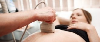
- During a routine ultrasound, when insufficient fetal growth is noticed. This indicates a lack of necessary microelements in the child’s diet.
- During a visual examination, the gynecologist will notice changes in the mother’s skin, namely, he will detect extensive swelling and bruising.
It is impossible to diagnose the disease yourself with one hundred percent certainty. But systematic edema is a reason to consult your doctor.
Prevention of pathology
No one is immune from the development of this complication. However, it is possible to prevent the development of a more complex form of the disease.
- The expectant mother's diet should contain foods with a high protein content - cottage cheese, lean meats, eggs, fish. You should limit or completely eliminate the consumption of salty foods and salt. This also applies to confectionery products (ice cream, candies, cakes, pastries, etc.). Sweets can be replaced with fruits. You need to include fiber in your diet, which is found in dried fruits, seaweed, vegetables, and herbs.
- You should not drink a lot of liquid. The maximum amount of liquid you drink per day is no more than 1.5 liters, including first courses.
- Watch your weight. Normally, its increase during pregnancy is 12 kg. If you eat properly and nutritiously, you will not gain excess weight.
- It is recommended that a pregnant woman take a daily walk in the fresh air, which helps improve blood circulation.
- You should not skip visits to the gynecologist and tests, because this will allow you to identify gestosis in pregnant women at an early stage and prevent the development of serious consequences for the mother and child.