Sacroiliitis is an inflammation of the sacroiliac joint. This disease occurs due to infections and injuries. Treatment of sacroiliitis is urgent, because this disease can lead to the development of severe complications and even disability. The inflammatory process in the joint of the sacrum and iliac region can be either an independent pathology or one of the symptoms of other diseases. To understand how to treat sacroiliitis, you need to understand the causes, forms and signs of this disease.
Main types of sacroiliitis and causes of the disease
Depending on the nature of the inflammatory process, there are several main types of sacroiliitis:
| View | Cause of occurrence |
| Nonspecific or purulent sacroiliitis | The disease is the result of infection of tissues by microorganisms (streptococci, staphylococci) |
| Specific or infectious | The result of specific infections entering the joint - brucellosis, syphilis, tuberculosis |
| Non-infectious sacroiliitis | Develops after injuries, against the background of excessive stress, metabolic pathologies due to tissue wear |
| Aseptic or infectious-allergic | Consequence of immune dysfunction |
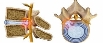
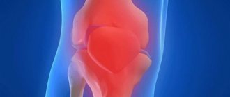
Osteoarthritis of the sacroiliac joint
Pathology can result from:
- Violations of tissue integrity - ruptures of the tendon-ligament apparatus, cracks, fractures.
- Congenital developmental defects - incomplete fusion of the sacrum, dislocations and subluxations of the joint.
- Oncological and benign neoplasms – cysts, tumors, metastases.
- Infectious processes in the body - abscesses (ulcers), soft tissue phlegmon (purulent inflammation without clear boundaries), osteomyelitis (a purulent process developing in the bone and bone marrow), syphilis.
- Metabolic disorders – calcium deficiency.
- Constant and heavy loads - physical labor, sports training.
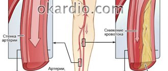
- Lack of blood supply - due to sedentary, sedentary work.
More often, sacroiliitis is a symptom of other diseases, and not an independent pathology.
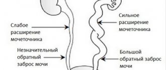
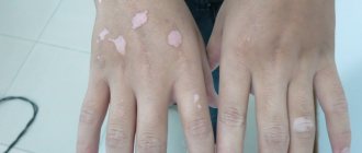
Possible causes of sacroiliitis: 1 – pelvic trauma; 2 – infectious agents (for example, Treponema pallidum – the causative agent of syphilis); 3 – heavy physical activity; 4 – sedentary lifestyle
Prevention and prognosis
Simple recommendations:
- daily physical education classes;
- strengthening the immune system;
- timely treatment of infectious pathologies so that pathogenic organisms do not penetrate the joints;
- reducing the risk of stagnation during sedentary work: periodic warm-up, change of body position;
- refusal to overload when joint pain occurs;
- timely visit to a vertebrologist, discipline during treatment.
If you suspect sacroiliitis or pain in the iliosacral, gluteal, or femoral region, you should promptly contact a rheumatologist or vertebrologist to find out the cause of the discomfort. Treatment of the underlying disease and elimination of the consequences of injuries reduces the likelihood of relapses and improves the condition of the problem joint.
Any inflammatory spinal disease is dangerous, so if you have characteristic symptoms, you should not self-medicate. Timely diagnosis and treatment of the problem will mean a favorable outcome and an improved prognosis for full recovery.
As a preventive measure, it is recommended to lead an active, healthy lifestyle, eat right, avoid hypothermia, do exercises daily, and if your health worsens, immediately consult a doctor.
Characteristic symptoms (common to the disease)
The severity and strength of the manifestations of the pathology depend on the nature of the inflammatory process.
Purulent sacroiliitis is acute:
- signs of poisoning of the body with inflammatory products increase;
- body temperature rises (up to 40 degrees);
- Intense pain occurs below the lower back and in the abdomen.
The patient's condition quickly deteriorates, it is difficult for him to bend, straighten and bend the leg (located on the side of the diseased joint), any movement causes acute pain.
Aseptic and non-infectious processes are not so rapid, pathology develops gradually.
Therefore, at the beginning of the disease, the patient is bothered by mild symptoms of sacroiliitis:
- unexpected pain;
- stiffness in the back that occurs after exercise or in a static position (when a person stands or sits for a long time).
As the pathology develops, the gait may change (swinging from side to side, “duck” gait, lameness), and difficulties may arise with performing certain movements (difficulty raising a straight leg).
The later stages are characterized by limited mobility of the joint, accompanied by cramps and constant cutting pain.
| General symptoms inherent in the pathology | Complications of advanced sacroiliitis |
| Pain below the lower back that radiates to the abdomen, gluteal muscle, hip or knee (from the inflamed joint) | Breakthrough of pus into other cavities (pelvis) and adjacent tissues |
| Pain occurs and intensifies with pressure on the sacroiliac joint | Limitations of joint function and disability |
| Lack of mobility in the lower back and lower back |
General symptoms of sacroiliitis: 1.2 – pain below the lower back, which can radiate to the buttock and thigh; 3 – when palpating the sacroiliac joint, the pain intensifies
Signs of different types of sacroiliitis
In addition to the general symptoms inherent in all types of sacroiliitis, there are signs and features characteristic of certain types (purulent, aseptic, non-purulent).
Nonspecific purulent
The cause of purulent inflammation of the left or right sacroiliac joint can be:
- Pathogenic microorganisms that have entered an open wound.
- A focus of infection adjacent to the joint (for example, an abscess or phlegmon - purulent inflammation of soft tissues).
Signs of inflammation quickly increase. The patient tucks his legs under him, trying to relieve the intense pain in the lower back and abdomen.
Unpleasant sensations increase when trying to put pressure on the sacroiliac joint and when extending the limb from the side of the inflamed joint.
In the case when pus breaks out of the joint (flows):
- in the buttock - painful, pulsating swelling occurs in the thickness of the muscle;
- into the pelvic cavity - a dense, elastic formation with vibrations of the fluid inside when pressed is palpated through the rectum.
| Purulent sacroiliitis is characterized by | A complication of the process may be |
| Sharp, cutting, twitching pain in the affected joint, weakening in a forced position (with legs tucked in) and intensifying with movement and pressure | Dissolution of adjacent tissues by pus accumulated in the joint |
| Increase in general body temperature (up to 40 degrees) | Damage to the spinal cord (due to leakage into the spinal canal) |
| Weakness | Sepsis (general inflammation) |
| Signs of general intoxication (sweating, chills, dry mouth, increased heart rate) |
Tuberculous sacroiliitis
What is it - sacroiliitis in tuberculosis? The cause of infectious inflammation of the joint is a focus of tuberculosis in the area of the iliac or sacral bones.
The process develops slowly, without any pronounced manifestations. It is accompanied by:
- dull, weak pain of unclear localization or along the sciatic nerve, in the pelvic area, radiating to the knee and thigh;
- stiffness, limited mobility;
- an increase in general (up to 37.2–37.5 degrees) and local temperature;
- swelling and discoloration of the skin over the source of inflammation (leakage of lymph and blood through the walls of blood vessels, infiltration).
How difficult is it to move when bone tissue is damaged? The patient is forced to protect the limb so as not to provoke pain and discomfort; it is difficult for him to bend and straighten his back.
In 75% of cases, complications of the process include:
- Dissolution of adjacent tissues by accumulated pus.
- Formation of abscesses (purulent inflammation of soft tissues) and fistulas (formation of a channel through which pus flows into the tissues).
- Destruction and deformation (change in shape) of bones.
- Disability.
X-rays reveal destruction of the sacral and iliac bones of the joint and a clearly visible narrowing of the joint space.
X-ray diagnosis of tuberculous sacroiliitis. Arrows indicate affected areas of bones
Syphilitic sacroiliitis
Syphilitic sacroiliitis is a rare pathology that occurs against the background of sluggish chronic syphilis (the causative agent is Treponema pallidum).
Click on photo to enlarge
The inflammatory process is accompanied by:
- weak night pain;
- stiffness of the back below the waist.
With secondary and tertiary syphilis (at the stage of spread of the process, with extensive damage to the skin and internal organs) it is possible:
- the appearance of gummous tumors of connective tissue or bones (formations in the form of nodes of different sizes);
- complete destruction, immobility of the joint.
At the beginning of the disease, small changes are noted on radiographs, which over time turn into partial or complete destruction of the bone tissue of the joint.
Brucellosis form
Brucellosis sacroiliitis occurs in different ways. More often, a distinctive feature of the pathological process is periodically occurring and quickly passing pain (sometimes on both sides).
Less commonly, a persistent inflammatory process that involves the synovium (synovitis) and other tissues of the sacroiliac joint (arthritis) and is difficult to treat.
Pathology occurs with:
- Pain in the lower back and joints, which intensifies with movements of the spine.
- Painful sensations that appear when trying to lift a straightened limb (in this case they are localized on the back of the thigh).
- Muscle tension.
- Stiffness of the spine.
A characteristic feature of brucellosis sacroiliitis is the absence of radiological signs (bone destruction, narrowing of the joint space) even with obvious signs of the disease.
Normal radiograph of the pelvis. Click on photo to enlarge
Aseptic sacroiliitis
What is aseptic sacroiliitis (that is, not purulent)? The pathology develops against the background of autoimmune diseases, which are a consequence of deficiencies in the body’s immune system (for example, ankylosing spondylitis, which affects the joints).
The inflammatory process in these cases:
- double-sided;
- progresses over decades;
- manifested by stiffness of the spine and mild pain in the buttocks, radiating to the hips.
A characteristic feature of aseptic sacroiliitis is a weakening of symptoms during movement or during exercise (the back seems to be “developed”).
Non-infectious sacroiliitis
This sacroiliitis occurs as a result of trauma, metabolic disorders or regular physical activity.
Characteristic signs are attacks of pain, aggravated in static (stationary) postures or when there is a load on the spine.
Other symptoms:
- unilateral discomfort in the thigh, buttock, lower back;
- stiffness, impaired joint mobility;
- deformation of posture (scoliosis);
- change in gait (“duck-like”, limping).
Pain occurs when pressure is applied to the area of the affected joint.
Degree of development of sacroiliitis
In addition to the fact that the process can be primary and secondary, specific and nonspecific, it can also be aseptic (again, autoimmune), and non-infectious (for example, due to overload).
There are three degrees, or stages, of sacroiliitis, which vary depending on the clinical picture:
1) At the first stage, clinical manifestations do not dominate. Occasionally, pain may appear in the lower back or lower back, radiating to the legs. Basically, signs of inflammation are present on X-ray images: the joint spaces lose their clear edges and become somewhat “blurred”.
2) The second stage is characterized by both the progression of the X-ray picture (narrowing of the gaps, disruption of their congruence), and the appearance of stiffness, persistent pain, and the possible appearance of general reactions (fever, malaise).
3) At the third degree of sacroiliitis, ankylosis develops - complete healing of the joint spaces. This leads to deformation of the muscles of the lower back and pelvic floor, the occurrence of neurological disorders and other symptoms.
Why is sacroiliitis dangerous?
A simple aseptic post-traumatic process, for example, right-sided sacroiliitis, may not be considered dangerous. As time passes, as with any sprain, the inflammatory response will subside. But in the case of secondary, bilateral sacroiliitis, a long course is possible, which ultimately can lead to disability.
In the case of ankylosing spondylitis, this will be “ossification” of the spine. Even more dangerous is the secondary tumor process, which after some time can lead to the death of the patient.
It is important to understand that in the case of tumor growth, initially there may be a one-sided lesion that does not bother the patient, and a year later he will die from an inoperable stage of cancer with metastases to the ilium. Therefore, we can talk about the danger of sacroiliitis only by specifying its cause.
In some cases it is harmless, sometimes it leads to disability, and sometimes to death. But, in the latter case, we repeat, sacroiliitis is not to blame here, since it “turned up” to the oncological growth.
Features of pathology in children
How does sacroiliitis occur in children? The onset of pathology is often mistaken for infection or acute appendicitis.
The process occurs with severe or mild symptoms, the following manifestations are characteristic:
- Periodic, acute, cramping pain and bloating with stool disturbances.
- Intense back pain radiating to the buttock, knee, lower leg and hip joint.
- Spasm (painful contraction) of the back muscles.
- Increase in general temperature (from 37 to 39 degrees).
Symptoms of general intoxication are very widely represented:
- chills, fever, sweating;
- weakness, dehydration;
- nausea, vomiting;
- lack of appetite;
- dizziness and headaches;
- confusion, lethargy.
The child develops a spinal deformity (scoliosis). He “drags” his leg on the side of the diseased joint, limps or refuses to move, taking forced positions (on his back with straightened legs).
Some of the signs of sacroiliitis in children: 1 – scoliosis; 2 – lameness; 3 – forced position in bed
Marfan syndrome
In recent years, there has been a significant increase in hereditary diseases affecting connective tissue. Some experts believe that the reason for this phenomenon lies in the accumulation of new genetic changes (mutations) during human evolution, which is caused by internal and external environmental factors. Others believe that, in fact, diagnostic capabilities have simply increased due to the improvement of medical genetic knowledge, which makes it possible to recognize genetic pathology and make the correct diagnosis.
A large group of hereditary diseases consists of connective tissue damage. Since it is an integral part of all organs and systems in the body, such pathologies are distinguished by many disorders and clinical symptoms. One of the most famous genetic diseases affecting connective tissue is Marfan syndrome.
This pathology is characterized by a wide variability of symptoms (from latent forms to life-incompatible course options) and most often includes lesions of the cardiovascular system, eyes, central nervous system and musculoskeletal system.
The disease is quite rare - 1 case per 10,000 people. But if we look at the statistics of European countries, the frequency is much higher - 1-3 cases per 5000 people, which is due to the greater availability of specific diagnostics of latent forms of pathology.
Causes and genetics of pathology
The disease was first described in detail in 1896 by the French pediatrician A. Marfan, after whom the pathology was named. He observed a 5-year-old girl of asthenic build with disproportionately long limbs and congenital arachnodactyly (long fingers). By the mid-20s of the last century, there were already many similar clinical cases described in children and adults. American geneticist McCusick conducted a detailed study of chromosome mutations and discovered a new group of hereditary connective tissue diseases, which included Marfan syndrome.
According to its etiology, Marfan syndrome is a genetic disease with an autosomal dominant type of inheritance, with varying degrees of expressivity (clinical manifestations of genetic changes). Approximately 85% of cases of the disease are hereditary, that is, inherited from parents. The remaining cases are new, that is, they arise due to new spontaneous mutations and are not inherited.
The direct cause of the pathology in 95% of cases is a mutation in the gene that encodes the structure of fibrillin-1 and/or fibrillin-2. The mutation of the FBN1 and FBN2 genes is localized on chromosomes 15 and 3.
Fibrillin is the basis of elastic fibers of connective tissue of a glycoprotein nature. It forms the framework of the intercellular substance, vascular walls, cartilage, the lens of the eye and many other organs and tissues. If the patient has the described mutation, the connective tissue has an increased ability to stretch, becomes less durable and resistant to mechanical stress, which becomes the cause of the clinical manifestations of the syndrome.
In approximately 5% of cases, the direct cause of Marfan syndrome (atypical forms of pathology) is a point mutation of the gene that encodes the structure of the α2 chain of type 1 collagen.
Classification
According to ICD-10, Marfan syndrome is included in the class of congenital developmental defects and chromosomal pathologies, here the pathology can be found under the code Q87.4.
A detailed clinical classification of the disease does not exist today, but several forms of the disease are distinguished, depending on certain criteria.
Depending on the severity of symptoms:
- erased form - signs of pathology are mild and may remain unnoticed throughout life; as a rule, changes affect no more than 2 organ systems;
- clinically pronounced form - the symptoms of the pathology are clearly visible and occur in more than 2 organ systems.
Depending on the genetic factor:
- the familial form is diagnosed in cases where the disease is inherited;
- The sporadic form is defined when the pathology is caused by a new spontaneous mutation in an individual and does not occur in his relatives.
Symptoms of Marfan syndrome
The signs of Marfan syndrome are very varied. Therefore, they are considered from the point of view of damage to individual organs and systems:
- musculoskeletal system;
- organs of vision;
- of cardio-vascular system;
- nervous system;
- respiratory system;
- skin and soft tissues;
- other organs and systems.
Musculoskeletal system
Pathological connective tissue causes the development of a number of specific phenotypic signs and skeletal deformations in patients with this pathology.
As a rule, externally, patients with Marfan syndrome look quite specific. They are characterized by:
- asthenic physique;
- high growth;
- poor development of subcutaneous fat tissue, which makes people look thin;
- very long upper and lower limbs with a relatively short body;
- elongated skull (dolichocephalic);
- elongated fingers – arachnid (arachnodactyly);
- the face is narrow, elongated vertically;
- Gothic upper sky;
- underdevelopment of cheekbones;
- protruding lower jaw (prognathism);
- abnormal growth (crowding) of teeth and pathological bite;
- hypermobility of joints, their “looseness”;
- eyes set deep in the skull.
As the child grows, various skeletal deformities may appear. Most often, curvatures of the spinal column appear. Patients are diagnosed with scoliosis, pathological kyphosis and lordosis, and “straight back” posture (smoothing of physiological lumbar lordosis). Subluxations and dislocations also appear in the cervical spine; approximately 20% of patients are diagnosed with lumbar spondylolisthesis.
Other skeletal deformities include:
- protrusion of the acetabulum of the hip joint of 2-3 degrees, which becomes the cause of dysplastic coxarthrosis and disability of many patients if endoprosthetics is not performed;
- keeled and depressed deformity of the chest;
- flat feet (longitudinal and transverse).
Patients with SM are also characterized by osteopenia (decreased bone mineral density) and frequent pathological bone fractures against its background, as well as a tendency to habitual dislocations, for example, of the shoulder.
The cardiovascular system
Among the lesions of the cardiovascular system in SM, the most common are:
- mitral valve prolapse with or without regurgitation
- cardiac myxamatosis;
- dilated cardiomyopathy with the development of heart failure;
- aneurysms of the aorta and other vessels (cerebral, renal, etc.);
- expansion of the pulmonary artery and various parts of the aorta.
It is cardiovascular pathological changes in SM that determine the prognosis and life expectancy of patients. Approximately 90% of all patients with this genetic pathology die at the age of 40-50 years due to complications such as dissection and rupture of aortic aneurysm, other vessels, progressive heart failure due to dilatation of its chambers and changes in the valve apparatus.
In the presence of SM, the presence of congenital heart defects in children is possible. The most common are coarctation of the aorta, stenosis (narrowing) of the pulmonary artery, ventricular and atrial septal defect.
Also, such patients are prone to various cardiac arrhythmias, including life-threatening ones (atrial fibrillation, ventricular tachycardia and extrasystole), and to infective endocarditis.
Organ of vision
Pathological changes in the eyes are very characteristic of this disease. Approximately 60-80% of patients are diagnosed with lens dislocation due to weakness of its ligamentous apparatus, even in infancy. Other characteristic features include:
- flattening of the cornea;
- increase in the size of the eyeball in length;
- myopia or hypermetropia;
- disruption of the accommodation process due to underdevelopment of the ciliary muscle.
If the described lesions of the organ of vision are detected in a newborn child, one should think about a possible genetic pathology.
Nervous system
Due to the pathological structure of the vessel walls, patients with SM have an increased risk of hemorrhagic strokes, as well as cerebral hemorrhages due to rupture of vascular aneurysms and subarachnoid hemorrhages.
Developmental anomalies include dural ectasia. Most often you have to deal with lumbosacral ectasia of the meninges (protrusion of the dura mater beyond the spinal canal through a defect in the structure of the vertebrae). This is a big criterion for SM, which occurs in 40% of cases of the disease.
Some patients have deviations in intellectual development, but most people with SM are characterized by high IQ scores.
Respiratory system organs
In most cases, changes in the bronchopulmonary apparatus are diagnosed accidentally. The development of bullae in the upper parts of the lungs is characteristic, which can sometimes rupture with the development of spontaneous pneumothorax.
Also, due to chest deformations, patients are prone to developing emphysema, frequent respiratory infections and respiratory failure.
Diagnostics, 4 degrees of illness
A preliminary diagnosis is made based on a survey, examination of the patient, and diagnostic tests:
- Lasègue test. The patient is placed on his back on a flat surface. In this position the patient does not experience pain. The doctor slowly bends the leg at the hip joint. The patient's knee should be flexed. Next, the doctor smoothly lifts the patient’s leg and straightens it.
Carrying out the Lasegue test The test is considered negative if the patient does not feel pain during its execution. With a positive test, pain occurs along the back of the thigh when raising the leg and disappears when it bends at the knee - a sign of sacroiliitis - inflammation of the sacroiliac joint. - Ferpson test. The patient sits on a chair and tries to lower his leg down. The test is negative if the patient does not experience pain at this time. A test during which pain occurs in the sacroiliac joint is considered positive.
Movements of a straightened leg up and down cause characteristic pain at a certain point below the lower back and muscle contraction - this indicates that a person has inflammation of the sacroiliac joint.
The main method for diagnosing sacroiliitis is radiography; there are 4 degrees of changes in the articulation with characteristic radiographic signs:
| Degree of pathology | X-ray signs |
| 1st degree | The images show changes in the size of the joint space |
| 2nd degree | The outlines of the joint are unclear, blurry Bone compaction, erosion (destruction of the joint surface) |
| 3rd degree | The joint space noticeably narrows, signs of ankylosis appear (fusion of the joint, leading to its immobility) With purulent sacroiliitis, the bone expands, loses density, and osteoporosis develops. |
| 4th degree | Complete fusion of the joint space, destruction (destruction) of bone tissue Uneven, “eaten” outlines of the joint |
If the X-ray is insufficiently informative, the patient is prescribed a CT or MRI.
MRI procedure of the sacroiliac joints
Laboratory diagnostics:
- serological, enzyme immunoassay or PCR analysis (determination of a specific pathogen);
- general and biochemical tests (signs of inflammation - increased ESR (erythrocyte sedimentation rate), leukocytes, increased C-reactive protein).
Diagnosis of inflammation of the sacroiliac joints
There are several methods for diagnosing the disease. First of all, this is radiography. With its help, the specialist analyzes the existing changes in the sacroiliac joint.
If there are complaints of pain on both sides, and there is a suspicion of bilateral sacroiliitis, the patient is prescribed a laboratory blood test. It helps to determine whether there is a causative agent of infectious diseases in the body. When these are identified, the diagnostic and therapeutic measures of the vertebrologist are joined by the measures of an infectious disease specialist, a pulmonologist and a dermatovenerologist.
Treatment methods
How successful is the treatment of sacroiliitis? At the beginning (at stage 1), sacroiliitis can be eliminated by eliminating the cause of the disease (infection).
Later, the goal of treatment is to relieve severe symptoms and prevent or stop joint destruction.
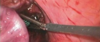
Therapy is carried out using conservative methods. Surgical intervention is indicated for purulent sacroiliitis - opening the lesion, removing tissue, drainage, prescribing medications.
Drug therapy
Drug therapy consists of a set of drugs that are used to eliminate the cause and severe symptoms of the disease (pain, inflammation, stiffness).
The doctor prescribes:
- Painkillers, non-hormonal anti-inflammatory drugs - Naproxen, Ibuprofen, Piroxicam.
- Hormonal anti-inflammatory drugs - Prednisolone, Methylprednisolone, Hydrocortisone.
- Immunosuppressors that can suppress the activity of autoimmune inflammatory reactions - Azathioprine, Infliximab.
- Broad-spectrum antibiotics that inhibit the development and reproduction of pathogenic microorganisms - Clarithromycin, Kanamycin.
- Anti-tuberculosis drugs – Isoniazid.
Medicines for the treatment of sacroiliitis
In case of unbearable pain and obvious impairment of mobility, injections of Lidocaine or Diprospan are given into the muscles or capsule of the iliac joint.
Until complete recovery, it is recommended to wear support bandages or corsets during treatment and during the rehabilitation period. They help relieve stress on your back.
Physiotherapy
Physiotherapeutic recovery methods are combined with drug treatment. This complex effectively delays functional changes in the joint and helps maintain its mobility.
In the acute period, only electrophoresis or ultraphonophoresis with drugs is used.
Other physical procedures - magnetic therapy, mud therapy, massage, paraffin therapy - are prescribed after the acute manifestations of the disease have subsided, during the rehabilitation period.
Mobility is restored through physical therapy exercises.
Examples of physical therapy exercises for sacroiliitis
Treatment of arthrosis of the acromioclavicular joint: medications and physiotherapy
Have you been trying to heal your JOINTS for many years?
Head of the Institute for Joint Treatment: “You will be amazed at how easy it is to heal your joints by taking every day...
Read more "
In the human body there are many hidden joints, the functions of which are invisible. So, the scapula is connected to the collarbone by a movable joint. The main role of this joint is to help lift the arms up and increase the range of motion in the shoulder. Sometimes age-related or traumatic degeneration of the articular surfaces of the acromioclavicular joint occurs, which leads to an unpleasant disease - arthrosis. As a result of the disease, the function of the upper limb is sharply impaired, which seriously impairs the quality of life of the sick person.
Causes, anatomy and biomechanics of the problem
Movements associated with lifting the upper limbs are carried out by the shoulder joint, scapula and collarbone. These bone formations form joints with different levels of activity. Just above the humerus is the acromion, a semicircular process of the scapula. Its distal end is connected to the proximal part of the clavicle. This place is usually called the acromioclavicular joint. In addition to bone structures, ligaments and cartilage tissue are present in the junction area. The latter forms a capsule around the joint, but inside there is practically no synovial fluid, since the range of movements of the joint is limited.
For the joint to function fully, it is sufficient to contain fairly dense cartilage tissue inside the cavity. However, for various reasons, bone structures grow, growths (osteophytes) appear, which leads to the appearance of clinical symptoms of the disease. The exact factor that leads to arthrosis of the acromioclavicular joint has not been identified, but there are situations that provoke the appearance of the disease. These include:
- lifting weights;
- prolonged work with arms raised;
- hereditary characteristics of the osteochondral junction (weakness of the ligamentous apparatus, underdevelopment of chondrocytes);
- injuries of the acromioclavicular region;
- systemic connective tissue diseases (lupus, rheumatoid arthritis);
- previous operations in the same area;
- habit of sleeping with a bent arm under your head.
The greatest importance in the development of arthrosis of the acromioclavicular joint is the professional high load on the shoulder region. Therefore, the disease mainly affects people over 35 years of age who have been employed for a long period in the following professions:
- weightlifters;
- bodybuilders;
- welders;
- track and field athletes training with parallel bars or horizontal bars;
- miners;
- other professions in which the arms are subject to maximum load when raised (electricians, trainers, boxers, etc.).
The main immediate cause of the appearance of clinical symptoms is the replacement of cartilaginous mobile tissue with bone static structures. Osteophytes appear, which limit the already meager activity of the joint. Acromioclavicular arthrosis develops over a long period, so when painful sensations begin, the pathology is already far advanced.
Main symptoms and clinical manifestations
The disease develops slowly. The first symptoms appear gradually, usually at the height of serious physical exertion. For a long time the patient does not pay any attention to them, but the unpleasant signs of trouble in the joint progress. The main symptoms characterizing arthrosis of the acromioclavicular joint include:
- pain in the shoulder area;
- irradiation of painful sensations in the arm, neck and shoulder blade;
- the ability to raise your arms up is sharply limited;
- sleep is disturbed, as the pain intensifies in a supine position;
- irritability and depression appear;
- The entire upper shoulder girdle suffers: stiffness of movements and limited function of the limb are formed. The quality of life deteriorates sharply.
The main symptom of acromioclavicular arthrosis is pain. It initially appears only at the height of physical activity or when raising your arms up. Then it becomes constant, intensifying with the slightest movements. A person cannot cope with the simplest everyday situations:
- cross your arms;
- raise them as high as possible;
- sleep on the affected side;
- lift a bag even with a small weight;
- put the baby on your shoulders;
- rotate the affected half of the body to the side.
The biggest problem is the problem of falling asleep. Painful sensations increase sharply, which requires constant use of analgesics. There are no fatal complications from arthrosis of the acromioclavicular joint. However, severe, constant pain and restrictions on daily movements cause serious trouble and impair the quality of life. The highest degree of problem that the disease can cause is the complete inability to lift the affected arm.
If we compare the characteristics of the course of the disease due to various causes, a pattern appears. Symptoms progress most favorably and slowly during degenerative-dystrophic processes in the joint due to involutional changes. In athletes, especially those with increased body weight, pain is significantly less pronounced than limitations in limb function. The most unfavorable course of pathology is associated with traumatic injuries and previous operations. Equally difficult, but effectively treatable, symptoms occur with systemic connective tissue diseases.
Difficulties in diagnosis
With typical symptoms, it is not too difficult to suspect a problem; it is more difficult to act from the standpoint of evidence-based medicine, that is, to identify the anatomical substrate of the disease. The bone fragments of the acromion and clavicle are directly affected in the most advanced stages, when treatment is aimed at relieving symptoms. Initially, the changes concern cartilage tissue and the appearance of small osteophytes. Therefore, the full range of diagnostic measures should include:
- blood test to exclude a systemic process;
- x-ray of the shoulder - an experienced doctor will be able to notice a decrease in the distance between the distal end of the scapula and the collarbone;
- computed tomography – all the subtleties of changes in bone tissue are visible;
- MRI – makes it possible to evaluate the pathology of tendons, cartilage and bones;
- densitometry – to identify age-related osteoporosis.
All questions can be resolved only by magnetic resonance examination. The destruction of the articular capsule, a decrease in the amount of cartilage tissue in the articulation cavity, and marginal bone growths are clearly visible.
For differential diagnosis, the involvement of doctors of the following specialties is indicated:
- therapist - primary care, organization of the diagnostic process;
- rheumatologist - exclusion of systemic nature of the lesion;
- neurologist – assessing the condition of the upper limb and prescribing conservative treatment;
- traumatologist – determining indications for surgical correction and performing surgical procedures;
- physical therapy doctor (instructor) – development of individual exercises to activate recovery processes in the joint.
Only the joint work of specialists will allow us to establish effective activities for the full diagnosis and treatment of arthrosis of the acromioclavicular joint. In some cases, there is an underestimation of the significance of damage in this joint, which leads to delayed diagnosis and a protracted period of painful symptoms for the patient.
The same banned issue for which Ernst fired Malakhov!
Joints and cartilage will be cured in 14 days with the help of ordinary...
Treatment and rehabilitation
It is impossible to completely cure the disease with conservative methods of therapy. However, this method of helping the patient is effective in terms of controlling the symptoms of the disease. It is possible to relieve the manifestations of acromioclavicular arthrosis and stabilize the progression of osteochondral changes. The basic principles of treatment can be presented as follows:
- effective pain relief - NSAIDs and simple analgesics, intra-articular blockades are used;
- improving blood flow in the joint area - peripheral vasodilators are used;
- anti-inflammatory treatment - hormones are used parenterally for a short course and intra-articular administration;
- chondroprotector therapy – restoration of cartilage tissue;
- preparations for external use – enhance the effectiveness of systemic agents;
- Exercise therapy, massage, acupuncture.
If the entire complex of conservative methods is ineffective and clinical symptoms increase, surgical correction of arthrosis is performed.
The table below presents the main drugs, course of treatment and main dosages for various types of drug delivery to the affected area.
| Drug – type of therapy | Externally | Systemically | Inside the joint |
| NSAIDs | In the form of ointment and gel 2 times a day. Ketoprofen, Ortofen and Nimesulide are commonly used | Intramuscularly for no more than 5 days - Ketorolac, Diclofenac (up to 150 mg per day), then oral tablets in half the dose. For problems with the gastrointestinal tract - Nimesulide 200 mg per day or rectal administration of the drug. General course of therapy - up to 1 month | Not entered |
| Local anesthetics | In combination with NSAIDs (usually lidocaine) | Not applicable | For pain relief, a single dose of 1-2 ml of a 10% solution. It is a diagnostic criterion - pain completely stops when an anesthetic is injected into the joint cavity |
| Hormones | Not applicable | Intravenously in a short course, usually prednisolone at a dose of 90-120 mg. Duration - no more than 5 days | To relieve the inflammatory reaction - hydrocortisone or betamethasone. Usually a single injection is sufficient, which can be repeated after 1 month. |
| Chondroprotectors | In parallel with NSAIDs or in isolation. Chondroitin or glucosamine is used | Long term inside. The effect of therapy for arthrosis of the acromioclavicular joint has not been proven. Average dose - 1000 mg of chondroitin sulfate per day, course - up to 12 months or more | Only in experimental medicine, not used in routine practice |
| Simple analgesics | Not applicable | For rapid pain relief, parenterally or orally. Drugs of choice in the presence of ulcerative defects of the gastric mucosa. Metamizole or Paracetamol is used. The average dose is up to 1500 mg per day for Analgin and 3000 mg for Paracetamol | Not applicable |
In clinical practice, several drugs from different groups are usually combined with different routes of administration. Intra-articular agents are actively used together with oral medications. Chondroprotectors are often prescribed for arthrosis of the acromioclavicular joint. However, no studies have been conducted to prove the effectiveness of drugs from the chondroitin and glucosamine group for diseases of this joint.
Surgical correction is indicated in the presence of large numbers of osteophytes and destruction of bone tissue at the distal end of the acromion and the proximal portion of the clavicle. Usually, grinding of the bone structures is performed to increase the distance between them. A false joint filled with connective tissue is formed. Its function is sufficient for shoulder movements without pain. The operation is performed both openly and using endoscopic techniques.
Forecast
Since the pathology is associated with the performance of work duties, preventing arthrosis means changing daily work. If professional reorientation is not possible, the main task becomes early detection of pathology. Each episode of pain when raising your arms up, especially during physical activity (picking up a child, carrying a bag, lifting gymnastic equipment), should not go unnoticed. If the pain has become regular, but still tolerable, an MRI should be performed when discomfort first appears. This study will allow a detailed study of the condition of bone and cartilage structures to assess the situation.
Timely treatment will stop and significantly slow down tissue destruction, which makes the disease easier to tolerate and has little effect on the quality of life. Pathology of the acromioclavicular joint does not lead to severe complications. The prognosis will always be favorable.
IT IS IMPORTANT TO KNOW! The only remedy for treating joints recommended by doctors! Read more…
MRI images clearly show defects in the anatomy of joints, damage to tendons and ligaments, degenerative processes of soft tissues, foci of inflammation, fluid accumulations and internal bleeding, bone growths, as well as swelling and tumors. In a word, this diagnosis is more suitable for studying soft tissues, including blood vessels and lymph flows.
Indications:
- Diagnosis of injuries (including sports), fractures.
- Detection of tumor processes and metastases.
- Inflammatory joint damage.
- Dislocations.
- Congenital abnormalities and developmental pathologies.
- Inflammation of the synovial bursa.
- Intervertebral hernia, osteochondrosis.
- Study of the ligaments and muscles surrounding the joint.
It is possible to perform MRI not only according to the doctor’s indications, but also at the patient’s initiative. This is possible with complaints of pain in the joint area, limited mobility, swelling and redness in the joint area. This study is also carried out to monitor the progress of treatment in preparation for surgery.
Advantages:
- The ability to evaluate not only bone, but also soft tissue.
- No harmful exposure to health.
- Suitable for frequent examination of the body.
- Safe for children, used to examine infants and, if necessary, even for pregnant women.
- High research accuracy.
- The ability to see metastases at the stage of their inception, when they have not yet led to structural changes.
Flaws
The main disadvantage of MRI is the inability to examine patients who have metal or ferromagnetic prostheses installed in the area of interest or have a pacemaker. The hemostatic vessels of the brain, as well as the Ilizarov apparatus, also serve as an obstacle to scanning. The patient must inform his doctor about the presence of such implants in his body, otherwise the tomography may have negative consequences. In addition, metal elements in the body obscure the surrounding tissues in the image, so the examination becomes meaningless - in this case, MRI will not provide any information about the condition of the joints. For the same reason, examination is difficult if the patient has a tattoo with metal-containing drugs.
The disadvantages of MRI include the fact that the examination lasts a long time (up to 40 minutes) and requires absolute immobility from the patient, otherwise the images will be blurry and uninformative.
Also worth noting is the cost. The price of MRI is quite high, and the more accurate the device, the more expensive the study will be.
Risks
If the patient does not have metal or other implants in the body, then this examination does not pose any danger. However, we should not forget that the examination is necessarily carried out with contrast, which in itself is safe for humans. But if the patient has problems with kidney function, then additional consultation with a therapist will be required, who will determine whether the procedure can be performed.
CT or MRI of joints: which is better?
Each of these techniques has its own advantages and features. The choice depends on the goals set for the study, as well as on the symptoms. If it is assumed that the disease is associated with the condition of soft tissues, blood vessels, tendons, then an MRI is prescribed. If all the signs point to bone pathologies, their damage and growth, then they are often referred to a CT scan. Examination of joints in this sense stands apart because they are complex structures consisting of both soft and bone structures. It is impossible to choose just one type of study, as this will greatly limit the capabilities of specialists and reduce the accuracy of diagnosis.
The patient's age also plays a role. Thus, they prefer to examine children and older people using MRI, since it is a safer diagnostic method. In some cases, after an MRI, the patient is immediately sent to a CT scan, and vice versa. This is possible if one diagnostic method does not answer all questions and the results need clarification. Having in hand images from two scans, the doctor gets a complete picture of the structure of the joints and their condition, which will allow him to choose the most effective treatment.
A specialist’s opinion about two examination methods - CT and MRI, as well as about the diseases for which they are prescribed, is in this video.
Conducting CT and MRI at the same time is impossible, since the examination is carried out on different devices that differ in the principle of operation and the contrast agents used. But carrying out one study after another is possible if it is necessary for diagnosis.
Prognosis for recovery
Most often, sacroiliitis is not an independent pathology, but a symptom that appears against the background of other diseases (infections, metabolic disorders, injuries, immune disorders).
With timely elimination of the causes and proper therapy, the prognosis is usually favorable. Treatment times vary.
| Disease | Forecast |
| Sacroiliitis resulting from ankylosing spondylitis | Incurable At stage 1, it is possible to stop the pathology and prevent complete fusion of the joint At stage 3 – complete immobility of the sacroiliac joint, disability |
| Purulent, nonspecific sacroiliitis without complications - tissue melting, purulent leaks, abscesses, fistulas | After opening the lesion, it is treated with a course of antibiotics - from 2 to 4 weeks (in 70% of cases) |
| Tuberculous sacroiliitis | The duration of treatment depends on the degree of bone tissue damage - usually at least 6 months The prognosis is unfavorable - 50% of patients become disabled, the rest experience joint deformation, posture and gait disorders The mortality rate with the modern approach to the treatment of bone tuberculosis has been greatly reduced (3–5%) |
| Sacroiliitis against the background of incurable pathologies (cancer) | Quite quickly leads to partial or complete immobility, disability |
Severe symptoms of syphilitic sacroiliitis are treated with a course of antibiotics for 7–10 days (for primary syphilis - successful in 90%).
Causes
In medicine, there is a list of factors that provoke the process of inflammation of the sacroiliac joint and the development of the disease. Causes of sacroiliitis:
- autoimmune diseases,
- sedentary lifestyle (sedentary work),
- joint overload (for example, during pregnancy, lifting heavy objects),
- congenital developmental abnormalities,
- musculoskeletal injuries,
- infections,
- the presence of tumors,
- metabolic disorders.