Hygromas (synovial cysts, ganglia) are benign formations that never become malignant. They are capsules consisting of connective tissue, inside of which there is a transparent or yellowish secretion of a viscous consistency. Localized near joints or tendon sheaths, they are round or irregular in shape.
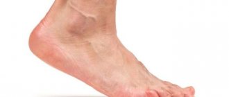
In the photo of the foot hygroma you can see how unattractive it looks. In addition, as the size of the tumor increases, a person begins to feel discomfort when walking, and difficulties arise with choosing shoes.
What is hygroma and what causes it?
Any hygroma is a benign neoplasm that is not life-threatening. It does not degenerate into a cancerous tumor or other types of malignant formations; its only danger is the possibility of developing secondary inflammation, which, in turn, will cause pain and limited mobility. A hygroma is essentially a fluid-filled cyst that has a clear shell and is localized near the joint. The neoplasm is formed from the articular membranes and has a pedicle with which it is attached to the articular capsule, which explains its stable position.
Where is it located?
Hygroma of the foot occurs on the instep, in the area of the heel bone or ankle, depending on which joint it grew from. The most unpleasant location is the sole: such cysts especially interfere with walking and cause severe discomfort.
Why does it appear?
The reasons for the appearance of this neoplasm are still not fully understood. It is known that hygroma is more often formed in women than in men (sometimes several times), and also that this disease affects mainly young people, from 20 to 30 years old. It very rarely occurs in children and the elderly; after forty, its formation is considered rather an exception.
Provoking factors.
These reasons are considered:
- hereditary predisposition. In families where older relatives have had cases of synovial neoplasms, they are likely to appear in the younger generation. It is believed that this is due to the characteristics of tissues that are inherited;
- intensive work of joints. The risk of developing a cyst is higher if a person actively loads a particular joint. In the case of hygroma of the foot, those at risk include hikers, athletes whose activities involve footwork, or representatives of professions that involve walking or standing in one place for long periods of time. Hygroma can be caused by wearing heels, uncomfortable, too tight shoes;
- injuries and illnesses. Suffered joint injuries can lead to the appearance of a synovial cyst; tendonitis, synovitis, bursitis and other inflammatory diseases of the joint and surrounding tissues are also considered risk factors.
Causes of pathology
In medicine, there is not yet a clear explanation of what causes the formation of a pathological cystic formation. The connective tissue that forms the sheath of tendons and joints undergoes degenerative processes, resulting in a characteristic protrusion that continues to grow. Among the factors that can provoke the development of a pathological process, the following stand out:
- hereditary predisposition;
- tight shoes;
- prolonged stress on the legs;
- traumatic joint injuries;
- joint diseases;
- overweight.
In childhood, the formation of hygroma is associated with injuries received during games. A child can damage a joint even with a slight blow to the limb, which can provoke the development of the disease.
How to detect hygroma
First signs
Secondary symptoms usually do not appear immediately. In the early stages, hygroma looks like a nodule or ball under the skin, soft, but nevertheless dense, with clearly defined boundaries. The shape of the neoplasm is usually regular, close to spherical, there is no sensation of germination into other tissues, or blurring of the boundaries. The seal can move freely, but with a very small amplitude. Inside the hygroma is synovial fluid, a substance that is usually found in the joint, sometimes interspersed with mucus or fibrin.
Development of the disease
If at first a hygroma is just a lump that is not painful in itself and does not cause discomfort, then over time secondary symptoms appear. It is associated with the growth of the tumor, which can be quite rapid: synovial cysts can reach impressive sizes. Secondary symptoms are associated with the fact that the hygroma begins to compress the surrounding tissues, pinching them and thereby interfering with their normal functioning.
Secondary signs
These include:
- pain that manifests itself when pressing on the hygroma or when trying to move the affected joint;
- discomfort when moving (especially pronounced in the case of hygroma on the foot in places that experience stress while walking);
- redness, swelling, other symptoms of inflammation resulting from tissue compression;
- blood stagnation, which is accompanied by numbness, a tingling sensation, and a change in the temperature of the foot.
The color of the skin over the hygroma is no different from normal; in some cases, the skin becomes rougher and rougher than before the cyst appeared. The tumor can grow for several years, remaining unnoticed until it begins to cause discomfort, or it can rapidly develop in a short time - up to several days.
Important
A swelling found on the foot is not always a hygroma. The lump can be a symptom of other diseases, so you should not diagnose yourself. It is best to consult a competent doctor and find out for sure what kind of disease manifests itself this way.
Symptoms
The symptoms of the disease are quite typical, which in most cases allows an accurate diagnosis to be made based on complaints and examination of the patient. At the very beginning of the development of hygroma on the leg, it appears as a small bulge with clearly defined edges.
Other characteristic signs of a synovial cyst:
- the tumor is inactive because it is tightly attached to the tendon;
- the neoplasm can be of different sizes - from 2 mm to 6 cm in diameter;
- the skin over the lesion looks normal, in rare cases it can be rough and peel off;
- The structure of the cyst is often soft and elastic.
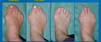
Hygroma on the toe is painful because it experiences frequent stress. There is no restriction of movement, but tight shoes can be a problem.
In advanced cases, as the tumor grows, it begins to compress nearby tissues and nerve roots, causing noticeable discomfort, tingling or numbness. Untreated hygroma often worsens joint mobility. It can take several months or years for a tumor to develop to this state.
Why the disease is unpleasant
Hygroma has no risk of malignancy and, as a rule, is not capable of causing death. Often a person does not notice it at all until it grows to a state in which it becomes visible to the naked eye. The cyst begins to cause discomfort when it grows to a certain size: as a rule, doctors talk about a diameter of over 10 mm. Much depends on its location: the most unpleasant are hygromas on the sole of the foot; neoplasms on the instep, heel or ankle are not so uncomfortable, but can cause problems when choosing shoes, walking, or changing shoes. If the cyst begins to compress the surrounding surfaces, secondary aseptic inflammation may occur, which is characterized by pain and limited mobility.
It is better to remove the hygroma of the foot immediately - it will not resolve
Hygroma is a non-dangerous benign formation, which resembles a synovial cyst in type, filled with transparent, colorless, sometimes cloudy and thick contents. It most often forms near the joints connecting the tubular bones of the joints (wrist, knee, ankle, elbow), although occasionally it is possible to form a hygroma in the area of the lymph nodes of the cervical region and even between the membranes of the brain. The most common are hygroma of the wrist and hygroma of the foot (see photo).
What will happen if left untreated?
Depending on the specific case, hygroma on the foot can behave very differently without treatment. Sometimes growth stops at a certain point; in some situations, the tumor continues to grow until it reaches a substantial size. The rate of growth also varies from person to person, with the development of a synovial cyst taking anywhere from a matter of days to several years. You can predict something only after reading a specific case, but there are certain observations that apply to all patients:
- Overloading the joint increases the likelihood of hygroma growth, so it is recommended to reduce physical activity during treatment;
- physiotherapeutic and drug treatment affects concomitant inflammation and other secondary symptoms, but does not in any way reduce the size of the tumor and is not able to influence its development;
- It is extremely rare for hygroma to resolve on its own, but this happens infrequently, and you should not count on such a possibility.
Prevention
Hygroma on the foot is a preventable disease. To do this, you must adhere to the following recommendations:
- during physical exercise, distribute the load on both legs without overworking a certain group of joints;
- fix the joints with an elastic bandage or bandage;
- avoid injury, and if trouble has already happened, see a doctor immediately.
High-quality shoes will also help to avoid the appearance of hygroma on the foot. Properly selected mid-heeled shoes with orthopedic instep support will protect your feet not only from synovial cysts, but also from many other joint pathologies.
Hygroma of the foot should not be ignored. Despite the benign nature of the tumor, its contents can become infected, leading to serious complications.
Author: Elena Medvedeva, doctor, especially for Ortopediya.pro
Accidental hygroma injuries
If a synovial cyst is damaged, it can burst and quickly disappear, but it is highly not recommended to injure it yourself in the hope of such a possibility. In this way, the surrounding healthy tissue can be damaged, in addition, the contents of the cyst will remain around the joint and in the future will lead to the formation of new hygromas. Crushing the tumor often causes relapses, including multiple ones, in which several hygromas form at once near one joint. External damage that disrupts the integrity of the skin (for example, puncture) can lead to infection and the development of a secondary infectious process with the formation of pus. In the case of a synovial cyst of the foot, the risk of injury is much higher than in other cases, so it is not recommended to delay contacting a doctor.
Content
Hygroma of the foot - what is it? Hygroma is a hollow, round-shaped neoplasm filled from the inside with a viscous jelly-like mass, either transparent or with a yellow tint. On the outside, the formation is surrounded by a compacted connective tissue capsule. The hygroma is located near the joint, because anatomically has a connection with the synovial capsule and the sheath of the adjacent tendon.
Hygroma has a round shape
Important! The localization of hygroma can be different, but always in the vicinity of one or another joint. Hygromas of the hands are most common; The second place in the frequency of formation is occupied by foot hygromas.
The formation of hygroma is associated with pathological degeneration of the connective tissue membranes of joints and tendons. The cells of normal connective tissue take on a different appearance and form the capsule of the hygroma.
Pathological cells of the capsule are divided into two types:
- Spindle cells take part in the formation of the capsule.
- Spherical cells produce fluid that fills the cavity of the neoplasm.
Diagnostics
The importance of visiting a doctor
Diagnosis and treatment of hygroma of the instep and other areas of the foot begin with a visit to a specialist. A traumatologist and rheumatologist deal with cysts near the joints: these doctors are the first ones to start examining if you suspect a hygroma. Despite the fact that the neoplasm is diagnosed quite easily and is usually visible to the naked eye or manifested by palpation, the doctor’s goal is to exclude other diseases. Such symptoms are caused not only by hygroma: the cause may be an epithelial cyst, lipoma, vascular formation and other types of benign tumors. Also, swelling and growths near the joints are a common symptom of arthritis and other inflammatory diseases of the musculoskeletal system.
Diagnostic methods
To make an accurate diagnosis, the doctor usually prescribes:
- general and biochemical blood test, urine test (they are needed to accurately exclude diseases that one way or another cause changes in the composition of urine and blood);
- Ultrasound or radiography (allows you to see what is happening under the skin and inside the joint).
Often these measures are enough to make an accurate diagnosis, but if doubts remain, a specialist may recommend a puncture or MRI. Conversely, in cases where the diagnosis is clear without additional measures, the doctor can report hygroma immediately after visual examination, palpation and history taking.
Why does hygroma appear on the foot and how to deal with it
Conservative methods of treating hygroma are often prescribed to patients who either do not want to undergo surgery, or whose tumor allows this option. They include a wide variety of medical techniques that have one thing in common – minimal invasiveness. The most common option is puncture - removing spherical cells from the ganglion capsule using a syringe.
Folk remedies for the treatment of leg tendon hygroma can be effective only if they are used as auxiliary techniques. At the same time, to minimize the risk, you should definitely consult with your doctor about the relevance of using a particular prescription.
Traditional methods, as a rule, involve the use of medicinal mixtures and ointments based on celandine, aloe, red clay, physalis, cabbage and other medicinal plants. They are able to activate and accelerate regenerative processes in the body. But remember that hygroma, despite its safety, can become a real problem if it is constantly disturbed.
Hygroma of the foot is a disease that does not pose a threat to health and life, because the tumor never progresses to the malignant stage. But, despite its harmlessness, the disease must be treated, because the presence of such a tumor increases the risk of foot injury and can serve as an impetus for the development of more serious diseases.
Treatment of hygroma with copper. To do this, you need a copper coin and an elastic bandage. The coin should be applied to the site of the tumor, and then a bandage should be applied tightly. And you need to walk like this for at least 3 days.
The simplest and most painless treatment option is a compress. You need to take a handful of ficus leaves and fill them with kerosene. Soak gauze or cotton wool with the resulting liquid and apply it to the tumor. Within 2 weeks you can get rid of the tumor forever.
There is another effective recipe. You need to dissolve 250 g of iodine (10%) in warm water. Grind 20-30 analgesic tablets. Pour the resulting powder into the solution and mix thoroughly. This solution must be used to treat the tumor until it goes away completely.
Conservative medicine offers hope for a chance to avoid unwanted surgical interventions when treating a tumor. There is no guarantee that folk remedies for combating hygroma will be more effective than traditional medicine. If the disease is already advanced, home recipes will not help and you cannot do without a doctor. Folk remedies are based on plants and natural products. Here are the most common ones.
Compresses
Lotions are made from raw eggs and wine vinegar.
A compress of 60% alcohol is also used at night. Gauze cloth soaked in the solution is applied to the tumor, and a layer of cotton wool and polyethylene is placed on top. The compress is fixed with a bandage.
Crushed cranberry fruits are applied to the neoplasm in the form of a compress.
Ointment "folk"
Powder from 30 analgin tablets is mixed well with 200 g of iodine. The ointment is used until the tumor disappears. When using this method, you need to follow safety precautions. The mixture may cause allergies or “corrode” the skin. Be careful with folk remedies.
Treatment
For foot hygroma, conservative or surgical treatment is used. It is important to understand that the tumor will not disappear or decrease by taking medications and undergoing physiotherapeutic procedures: they are needed to relieve inflammation and increase joint mobility, but none of these measures will get rid of the cyst itself. There is no evidence that any medications or procedures can shrink or eliminate a hygroma - only surgery is used for this.
Medication measures
Nonsteroidal anti-inflammatory drugs and, in rare cases, glucocorticosteroids are used as medications. They are designed to reduce swelling around the cyst, relieve secondary inflammation that occurs due to compression or concomitant diseases of the musculoskeletal system. Antihistamines are sometimes added to anti-inflammatory drugs. If an infection has been introduced into the hygroma (this happens if you try to puncture the cyst yourself), the doctor prescribes antibiotics that should overcome the infectious process.
Physiotherapeutic measures
They are selected based on the specific case and may include mud therapy, ultrasound or shock resonance therapy, paraffin therapy and other methods. Some evidence suggests that these treatments may shrink a hygroma, but this does not always happen, and even when this happens, the risk of the tumor growing again is extremely high.
Characteristics of tendon cyst
Bump on the foot
Hygroma of the foot is a neoplasm that has a hollow, rounded structure; it is filled with a viscous yellowish or transparent mass. The connective capsule of the cyst is connected to the sheath of the tendon tissue or joint of the leg. Therefore, most often the appearance of a neoplasm is observed in the mentioned area. Do not forget that the hands are also susceptible to a similar ailment, although the back of the foot suffers much more often.
In addition to the name “tendon cyst,” the hygroma capsule also received a third definition that reveals its essence – ganglion. Translated from Greek, this word means a single “nodule” of a benign tumor filled with fluid. For a more precise understanding of what it is, it should be clarified that in medicine, the term ganglion usually means a degenerative type of synovial cyst.
Cyst rupture occurs due to fluid filling or trauma. Immediately wash, treat the wound and consult a doctor. Based on the condition of the cyst, the doctor will recommend surgical intervention with a scalpel or laser. Previously, doctors specially opened the hygroma for treatment. The method turned out to be ineffective in practice. The tissues of the cyst grew together, and a new tumor formed in other parts of the leg. Removal is considered the most effective.
Surgical treatment
This is the only method that today allows you to completely get rid of a synovial cyst. In the past, crushing was used as a therapeutic measure, but today it is not used due to the very high likelihood of relapse and the possibility of damaging surrounding tissue. Surgery for hygroma is easy and does not require general anesthesia or a long hospital stay. It is usually prescribed if the cyst is growing rapidly, causing discomfort or pain in the patient, or interfering with his normal life activities - in other cases, the operation is performed only at the request of the person himself.
How it goes
Under local anesthesia, a skin incision is made, after which the surgeon performs resection of the cyst. Usually it is removed directly in the capsule; damage to the walls of the hygroma is not allowed. The wound is sutured, it is treated with antiseptic drugs, after which the patient can go home. For a certain period of time, you must follow the recommendations (do not disturb the limb, treat the incision site), and then come back to remove the sutures.
Modern method
This is an operation using a laser that burns out the cyst. Its use is justified if the hygroma is located near blood vessels or nerve endings that can be damaged during standard surgery. After laser surgery, tissue recovers faster.
The Doctors Online service will help you find a good doctor to diagnose hygroma. With its help, you can make an appointment with a specialist and find out what the symptoms mean right now.
Choose treatment
Clinical manifestations
The pathological process on the fingers looks like a tumor-like formation that is dense to the touch. It is localized in the joint area, but can grow throughout the entire phalanx. The cyst does not cause pain and does not limit the mobility of the fingers.
Pain and discomfort occurs only when nerve endings, blood vessels are compressed, and if the hygroma grows under the tendons.
Read also: Hygroma of the shoulder joint
Tumors often occur on the right arm, since this limb bears a greater load. A benign formation can originate from the tendon sheath or joint. In the first case, the hygroma is mobile, of a dense consistency, and in the second - immobile, hard. Its base is fixed to the lower tissues; the capsule is not fused to the subcutaneous tissue.
The tumors are single, but sometimes several nodes appear in the area of the ring, index and little fingers. When pressed, a sharp pain occurs that can radiate to the elbow. The intensity of the pain depends on the location and size of the hygroma. After physical exertion, the tumor increases, the skin over it turns red, and at rest the size decreases. The formation grows slowly or rapidly, and can reach 6 cm in diameter.
The most painful hygromas are located on the fingers on the palm side. They grow from the vaginal flexor tendons and are larger in size than when localized on the dorsal side. Seals are observed on several phalanges; as they grow, the nerve fibers are compressed, causing pain resembling neuralgia.
Recovery after surgery
After surgery, a special rubber band is inserted into the wound and a suture is applied. The elastic band is removed and the stitch is removed seven days after surgery. To avoid the formation of adhesions, special exercises are prescribed. Recovery is important, as is surgery. Proper return to form minimizes the risk of disease recurrence.
Without indications for removal, it is permissible not to get rid of the hygroma and live with it all your life. The patient decides to leave the lump or remove it for aesthetic purposes.