Your body and breasts continue to grow. If you haven’t started preventing stretch marks and dry skin yet, now is the time! Italians rub olive oil into their skin, Indians rub coconut milk. You will probably hear a lot of advice from friends or advertisers. The choice is great. Start using special creams for pregnant women, the smell of which does not cause you discomfort, moisturize your body skin every time after taking a shower or bath. To improve blood microcirculation, rub your legs, arms, back, and shoulders with a soft towel. Apply nourishing masks to your hair and face, treat yourself to spa treatments that are allowed for pregnant women: as a rule, these are procedures that do not cause heating of the body and skin.
When is the second ultrasound performed?
The timing of the second ultrasound is 20-24 weeks of pregnancy. They were chosen deliberately.
Firstly, the sex of the fetus is clearly determined. This makes it possible not only to come up with a name or choose children's things, but in the case of some genetic diseases, it makes it possible to predict the chance of their development.
Secondly, it is by this age that the main structures of the central nervous system finish forming and the heart and large vessels take their “final form”. If these organs are not developed correctly, this will already be visible, and measures can be taken to treat these problems (for example, some heart defects can be operated on - then the child will be born healthy).
Thirdly, the main bones and soft tissues are already fully formed. Therefore, a second planned ultrasound scan during pregnancy can accurately refute or confirm the diagnosis of a malformation if it was suspected during the first study. If at 20-24 weeks, when performing sonography on an expert-class device, the expert did not find any defects, then, even if they form later, there will be no danger to the baby’s life.
Fourthly, according to the law, it is only possible to terminate a pregnancy up to 22 weeks. This procedure is performed when there are medical indications associated with formed developmental anomalies:
- it is impossible to perform an intrauterine operation that would correct the situation;
- the defect will lead to severe disability or death soon after birth;
- vital functions after birth will need to be supported with the help of equipment (for example, a ventilator) or medications.
If the detected malformation is compatible with life, but causes significant defects in appearance (for example, a continuous cleft palate, or the absence of a limb/several limbs), or if there are social contraindications to childbirth, abortion can also be performed up to 22 weeks. This issue is decided by a special commission consisting of several medical experts.
Based on the above, the timing of this planned ultrasound must be strictly observed.
Also in the section: What will the first planned ultrasound tell you about?
Recommendations before the study
At 20 weeks of pregnancy, you need to undergo another scheduled ultrasound, during which the doctor will be able to see how the fetus is developing. If any problems are discovered or there are suspicions of pathology and deviation from the norm, you will have to do another examination a week later. It is for this reason that an ultrasound must be done on time; after 24 weeks, some problems are extremely difficult or impossible to correct.
The procedure does not require special preparation. About a day before the ultrasound, you should not eat foods that cause gas, as they may interfere with the examination. There is also no need to be afraid of the negative impact of waves on the fetus, because it is minimal, and diagnostics is aimed at identifying pathologies and abnormalities of the fetus, which is more important and can save the life and health of you and your baby.
6 minutes Author: Irina Bredikhina 166
The birth of a baby is a real miracle, and many expectant parents want to know the gender of the long-awaited child in advance in order to have time to properly prepare for changes in their lives depending on who will be born. With the help of modern methods of examining pregnant women, this is not particularly difficult - ultrasound methods make it possible to find out the sex of the fetus as early as 20 obstetric weeks.
To date, this diagnostic technique is still the safest and most informative. In our article we will provide information about the development of the fetus from the moment of conception to the appearance of sexual characteristics, the differences between the ultrasound of a boy and a girl at 20 weeks and how they look in the photo, as well as possible errors in the diagnostic procedure.
Baby at 20 weeks
The main organs have already been formed by this time and most of them are in their final places. So, the heart already consists of the required number of chambers (normally there are 4, but with developmental defects there may be fewer). Vessels depart from it and flow into it, which will no longer change their relative positions on their own.
The child's brain is fully developed, but the development of neural connections is still ongoing. Therefore, he no longer moves so “randomly”, but as if he is testing his capabilities: he bends his limbs in all joints, moves his fingers, can grab the umbilical cord or taste a finger. At this time, he can already hear sounds, understand whether it is light or dark. A sleep-wake schedule is established. In addition, fetal activity can be provoked by certain sounds, stroking the stomach, or eating sweet foods.
The fetus already has facial muscles working, and he can frown, yawn or attempt to smile. A feeling of sweetness is also formed. The digestive tract is already absorbing this liquid, but feces are not yet formed.
The integumentary tissue is already the same as that of adults in structure, but wrinkled and looser. It has a developed coat of vellus hair; it holds a special cheese-like lubricant that protects the skin from the action of water. Fat cells are formed under the covering tissue, which provide protection from the cold. Hair begins to appear on the head, and nails begin to appear on the fingers.
The reproductive system also matures: in girls, about 10 million oocytes appear in the ovaries, and in boys, the testicles begin to move towards the exit from the abdominal cavity.
The immune system completes its “training” necessary to distinguish “strangers” from “our own”.
No matter how developed the central nervous system of the fetus is, its lungs are not yet differentiated, and they will not be able to deliver oxygen to the blood (this will become minimally possible by week 24), which makes it not ready for birth.
Determination of gender
At 20 weeks, an ultrasound can most accurately show the sex of the unborn baby. It is not difficult to determine. In boys, the genitals are immediately visible, while in girls there are no bulges.
Difficulties in determining sex may arise due to the location of the fetus. In this case, you can re-test after 15 minutes. Pregnant women are advised to eat some chocolate to make the baby active.
When the baby is on his stomach, it is also difficult to determine his gender. Some babies turn away or hide during an ultrasound scan, so the sex of the child remains a mystery to parents until birth.
Even with the use of modern ultrasound equipment, errors in determining gender are possible; everyone should be prepared for this.
Baby at 21 weeks
The second planned ultrasound during pregnancy, performed at this time, will show the further development of the fetus. In just 1 week, his skin becomes less wrinkled, eyebrows and eyelashes appear, and his eyelids make blinking movements. The tongue begins to perceive not only sweets, but “embryos” of baby teeth appear in the depths of the gums. The subcutaneous tissue thickens.
This week the urinary system “starts up”, but the intestines are still poorly able to peristalt. The endocrine organs are already working, and they can partially take over the provision of hormones to the mother’s body if there are any malfunctions in it (for example, diabetes).
A fetus at 21 weeks of gestation should sleep about 22 hours a day, occasionally being awake and actively moving. Periods of prolonged or frequent (more than 5 times a day) activity, as well as excessively “sluggish” behavior of the child, require additional ultrasound examinations for hypoxia.
Possible problems
BPR and LZR of the head are less than normal
A slight deviation from the average tabulated values of the baby’s head size does not indicate anything alarming. All children in the middle of pregnancy grow at different rates, and no one excludes a hereditary feature of appearance - a small head. A deviation from the lower threshold of normal for 2 weeks is considered alarming (if the fetal BPD, for example, corresponds not to the twentieth, but to the eighteenth week of pregnancy).
A shrinkage of the head can be caused by microcephaly , as well as by malnutrition. In this case, doctors will talk about intrauterine developmental delay of the baby. Additional research will help identify the exact cause; if development is delayed, the prognosis is favorable - vitamin preparations, as well as medications that improve uteroplacental blood flow, help correct the situation.
The baby's head is larger than the age norm
Exceeding the normal size of the fetal head this week is sometimes a sign of hydrocephalus, swelling due to infectious processes or developmental defects. A slight excess of the indicators (by less than 2 obstetric weeks) may indicate that during this pregnancy there is a tendency towards a large (about 4 kg) or gigantic (more than 5 kg) fetus.
Again, the doctor needs to look at mom and dad, perhaps they have big heads - a family trait, and then no correction will be required.
The baby's abdominal circumference is less than the gestational norm
This indicator may be a feature of the child’s physique, because thin mothers and fathers usually give birth to equally slender children. As is the case with the size of the fetal head, a lag from the norm of more than 2 weeks is considered critical.
This situation requires a careful study of the uteroplacental blood flow, especially the placenta, in order to promptly identify possible causes of malnutrition. Thinness of the fetus in itself is not dangerous. The task of doctors is to find its true cause, which may pose a risk for the further development of the child.
Bone length does not correspond to age
An indirect marker of genetic pathologies during the second ultrasound is the shortening of the shin bones; the length of the remaining bones is measured only in order to imagine the proportions of the baby, as well as to track its development.
Shorter or longer forearms or femurs in most cases turn out to be features of the child’s appearance, because he may have genetically long legs or short arms. Such changes are considered physiological.
Pathological abnormalities and shortening of the limbs cannot go unnoticed, and the woman will immediately be referred to a geneticist for consultation.
Low placenta
The uterus is growing, the placenta still has every chance to rise to its normal height, which most often happens if the pregnant woman follows all the prescriptions of her doctor.
What does ultrasound show?
Ultrasound scan at 20 weeks of pregnancy
According to ultrasound in the second trimester of pregnancy, an assessment should be made of:
- number of fruits;
- fetal presentation;
- the circumferences of the head, chest and abdominal cavity of the fetus, the lengths of its bones and the spaces between them (the norm of these measurements is discussed below);
- brain structures, width of the ventricles in it;
- the fetal face, especially the nasal bone. Its shortening (less than 5.6 mm) or absence indicates a high probability of Down syndrome, and an increase of more than 6.1 mm indicates other genetic abnormalities;
- the size and number of chambers in the heart, the vessels emanating from it, the heart rate;
- condition of the spine: it should not be split, the spinal cord should not exit through it under the skin;
- the condition of the anterior abdominal wall, which should not be split;
- the abdominal cavity, which should not contain free fluid;
- kidney and intestinal development;
- fetal gender;
- condition of the placenta: normally there should be zero maturity, absence of areas of calcification or infarction;
- location of the placenta relative to the exit to the cervix (internal os): norm - not lower than 70 mm;
- amount of amniotic fluid (oligohydramnios or polyhydramnios);
- condition of the cervix: length - at least 25 mm, pharynx, both internal and external, must be closed;
- the number of vessels in the umbilical cord (normally 3), the nature and speed of blood flow through them;
- uterine muscle tone: normally it is described as “normotonus”. “Hypertonicity,” especially accompanied by nagging, aching or more acute pain below the navel or in the lower back, requires urgent hospitalization in order to save the pregnancy from spontaneous abortion.
A sonologist conducting a study shows a woman her baby on the monitor. To better understand how these contours make up the image, you can sign up for a 3D or 4D study. In this case, a three-dimensional image of the fetus completed by the program is displayed on the monitor; with four-dimensional ultrasound, it moves in real time. If desired, you can obtain a photograph of the fetus, as well as a recording of its behavior during the study, but these wishes must be expressed before the start of the study so that the doctor prepares the equipment.
Three- and four-dimensional ultrasounds are performed over a longer period of time (about 1-1.3 hours, as opposed to 20-30 minutes with the 2D method). Some scientists believe that such prolonged exposure to high-frequency sound on fetal fluids and amniotic fluid causes the phenomenon of cavitation, and has a detrimental effect on the condition of the child.
What will the ultrasound show?
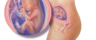
During the first half of pregnancy, the baby has made a huge evolutionary journey. Almost all his organs are formed, the functioning of his systems is being improved - the nervous system, in particular. The baby's height this week is about 26 centimeters. The child weighs only about 300 grams.
This week the baby has a big event - his eyes are acquiring photoreceptors, and now the baby can distinguish between light and dark. If you shine a flashlight on your belly, your baby's movements may increase. At the end of the fifth month, the baby learned to “make faces”, and now on an ultrasound, if you’re lucky, you can see how he squints, blinks, and grimaces.
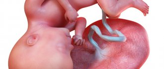
The child looks like a small, but already fully formed, man. He already has excellent control over his arms, plays with the umbilical cord, and sucks his fists. His intestines are working, the baby is swallowing amniotic fluid, his small stomach has begun to function.
At week 20, the doctor carefully examines the fetus (or fetuses, if there are several of them in the uterus), assesses the size of the baby, and compares the results with tables of normal values for the gestational age. This allows you to set not only the exact date and adjust the date of birth, if necessary, but also to understand whether the baby is getting enough nutrition and whether he has any diseases that could interfere with his normal development.
The doctor will be able to examine the baby’s internal organs, assess the condition of the placenta, umbilical cord, and amniotic fluid. If the mother’s health leaves much to be desired, a scan will allow her to assess the condition of her reproductive organs, identify inflammation, cysts, and possible problems with the cervix and cervical canal, which may be signs of an impending miscarriage.
An ultrasound examination at the twentieth week clearly shows the sex of the baby ; there should be no problems determining it. At the first ultrasound, it was difficult to accurately see the sexual characteristics because they were too small. In the third, which occurs at the end of pregnancy, gender diagnosis can also be difficult. The child will become large, “curl up” in a compact position, and it will be very difficult to see a boy or girl in the baby.
Now is the right time to find out who to expect - a son or a daughter.
Preparing for the study
An ultrasound at 20-21 weeks of pregnancy is performed with a sensor that is moved across the abdomen (transabdominal). Amniotic fluid serves as a “water layer”. Therefore there is no need to fill the bladder.
Since it is not the woman’s digestive organs that are being examined, but the condition of the fetus, there is no need to follow any specific diet.
Preparation and performance of ultrasound
At 20 weeks, ultrasound can be performed without special preparation. A pregnant woman does not need to stick to a diet and wait for her bladder to fill.
Ultrasound is based on the transabdominal method (the sensor is located on the anterior wall of the abdomen). If you perform an ultrasound with a vaginal probe at 20 weeks, there is a risk of premature miscarriage.
A pregnant woman can lie on her left side or on her back. To allow the ultrasound wave to penetrate the uterus, a special water-soluble gel is applied to the abdomen. It can be easily removed with a regular paper napkin and does not cause any inconvenience.
Who can skip a routine second trimester ultrasound?
Routine ultrasound examinations are an important measure that helps doctors properly manage and maintain this pregnancy. So, they see, for example, the presence of isthmic-cervical insufficiency, and offer the woman a pessary. When determining low placentation, she is advised to lie down more, not lift heavy objects, and if bleeding appears, do not think that it is “menstruation”, but urgently contact the gynecological (up to 23 weeks) or obstetric (after this period) department of the hospital.
But ultrasound is a voluntary study that a woman can refuse. Then she must be prepared for the birth of a child with anomalies, as well as premature birth.
“Officially” those women who underwent a second ultrasound screening during pregnancy at 16-20 weeks can not undergo an ultrasound examination at 20-24 weeks. This is a study that is carried out according to indications for those women who have a high risk of having a child with severe anomalies. It includes a blood test for 3 or 4 hormones at once, as well as an ultrasound performed on an expert-class device by a sonologist - a specialist in prenatal diagnostics. With such sonography, slightly more indicators are assessed than with “conventional” ones, and the examination itself is considered more reliable
Pregnancy screening is a paid procedure; its results are interpreted by a medical geneticist.
Probability of errors in ultrasound diagnostics
The risk of obtaining unreliable results in determining the sex of the unborn child is possible only in the first trimester of pregnancy. The longer the period, the higher the visualization of the genital organs and the less error in the final ultrasound data. In some cases, diagnosis can be difficult not only due to the early stage of pregnancy, but also due to the location of the internal organs of the expectant mother (intestines and bladder) and the unfortunate position of the fetus - it can close or turn away.
The doctor may make a mistake due to bends in the umbilical cord, swelling of the girl’s fingers or labia. The receipt of inaccurate data is not always due to the fault of a medical specialist - not all factors depend on his qualifications and attentiveness. And if in the early stages of the baby’s gestation period inaccuracy is associated with a short period of time, then in the second trimester errors occur due to the quality of diagnostic equipment and from the fetus itself, which can turn “the wrong way.”
In the third trimester, there is also a possibility of receiving inaccurate final ultrasound data - the baby becomes large and cannot move freely in the womb. This phenomenon affects the resulting image; the doctor always warns about the remaining probability of error regarding determining the sex of the child.
Experienced ultrasound diagnostic specialists and obstetrician-gynecologists who have been working with expectant mothers for a long time can determine with great certainty at an early stage who will be born to a married couple.
In conclusion of all the above information, I would like to emphasize once again - today, examination of a pregnant woman and unborn child using ultrasound waves is the safest, high-quality, informative and reliable method. Despite the fact that every parent wants to know who will be born - a boy or a girl, it is much more important that the child is born healthy.
“Who am I having, a girl or a boy?” - this question is asked by almost every expectant mother during a routine ultrasound. Some undergo this procedure specifically to find out the gender of their unborn child. At what time will gender determination be most accurate? Why does ultrasound give an incorrect result in some cases, and how often do mistakes occur in identifying the sex of the fetus?
Indicators are normal
Interpretation of ultrasound during pregnancy 20 weeks includes indicators obtained by measuring the size of the fetus (fetometry), as well as characteristics of the condition of maternal and temporary (placenta, membranes, amniotic fluid) organs. The results obtained should be interpreted by the obstetrician at the antenatal clinic in charge of the pregnancy.
In the table below we present the average norms for this period:
At week 20, if a routine ultrasound examination performed at a antenatal clinic or medical center reveals controversial data, then the woman can still be sent for ultrasound screening at a medical genetic center.
Features of pregnancy
At the twentieth week, exactly half of the pregnancy occurs - expectant mothers are already beginning to fully feel the new life inside themselves.
Many people tolerate this period of pregnancy more easily.
From 19 to 20 weeks, when the second trimester of pregnancy comes to an end, all the negative symptoms that prevented expectant mothers from enjoying their new position at the beginning of pregnancy go away.
Photo:
During this period, doctors begin to insist on conducting a second ultrasound, which will show in detail how the fetus develops.
In addition, young parents have the opportunity to reliably find out the gender of their unborn baby. At week 20, a routine ultrasound can already give a clear idea of the development of the future boy or girl.
Ultrasound in mid-pregnancy makes it possible to compare the growth and development of the fetus with the norm. At this stage, the expectant mother already clearly feels the first movements of the fetus inside her.
The baby can already independently push away from the walls of the uterus, spin and react to unpleasant sharp sounds.
Many women in the period from 19 to 21 weeks begin to experience cramps and severe pain in the joints, which sometimes cause tears to appear.
The occurrence of cramps during this period can be explained by the constantly growing weight of the fetus, as a result of which the pressure on the joints of the legs increases.
In the period from 19 to 21 weeks, the expectant mother should give her legs more rest. Also in this case, a light relaxing massage that can be done by your spouse can help get rid of discomfort in the joints.
From the outside, at this stage of pregnancy you can observe a greatly increased size of the tummy, which is already taking on a streamlined shape.
At week 20, the expectant mother can already know the sex of her baby and prepare for the arrival of a boy or girl.
During this period, a second planned ultrasound is performed, the decoding of which allows you to find out how the fetus is developing and what parameters differ from the norm.
The second planned ultrasound at week 20 can be done in multidimensional 3D or 4D format. This will allow the expectant mother to have a good look at her baby and see all his organs that are completing their formation.
READ Ultrasound indicators at 17 weeks of pregnancy
Video:
During this period of pregnancy, the woman’s hair condition significantly improves and her nails become stronger.
From 19 to 21 weeks, the general hormonal levels in her body stabilize, but there is a lack of calcium. This is due to the fact that most of it is consumed by the child to form his teeth and bones.
During this period of pregnancy, food preference should be given to low-fat cottage cheese, various types of cheeses, and dairy products.
During the period from 19 to 21 weeks, it is extremely important to balance your daily diet and eat right.
During this period, the doctor prescribes a second planned ultrasound for the expectant mother. It is better to opt for a multidimensional 4D study, which will show the exact gender and developmental features of the baby.
At this stage of pregnancy, the baby begins to grow more actively. The expectant mother may notice how her belly protrudes, her hips increase in volume, and her waist begins to disappear.
In the period from 19 to 21 weeks, the uterus also increases in volume. It begins to put constant pressure on the lungs, kidneys and stomach, which leads to severe heartburn, constant shortness of breath and constipation in the expectant mother. Frequent urge to urinate begins, especially at night.
Photo:
During this period, the expectant mother’s body works at its limit and gives most of its energy to the baby, which can provoke various health problems.
You can improve your general condition somewhat by eating in small quantities. At week 20, many expectant mothers develop stretch marks, which special underwear will help get rid of.
In addition, varicose veins may also appear. In this case, the woman is recommended to wear support stockings.