Nature gives a woman a whole nine months to prepare for the miracle of the birth of her baby, and with the help of modern technologies you can find out his gender, weight and height in advance. All this becomes available with ultrasound. An ultrasound at the 13th week of pregnancy is very important, at this time all parts of the body are clearly visible and it becomes possible to examine the baby’s face. Such an examination, in addition, will help identify problems, pathologies or abnormalities in the development of the embryo. An ultrasound machine generates ultrasound waves and uses a transducer to display an image of the fetus. When the sensor comes into contact with the body, a special gel is applied. The ultrasound scanning procedure itself does not cause the patient any pain or discomfort and lasts no more than ten minutes.
What changes are observed in the female body?
A woman's uterus increases in size. The location of the enlarged uterus is felt above the bone of the womb, the placenta continues its development. When walking frequently, you feel tired and weak in the muscles. Therefore, it is so important to comply with the norms of physical activity, but leading a passive lifestyle is also not recommended. Along with some stabilization of the general condition of a pregnant woman, other difficulties may arise: the process of digesting food may be difficult, and constipation is likely.
Often, pain occurs in the tailbone area and lower abdomen, which can bother you throughout the entire pregnancy. These indicators do not indicate a pathological abscess. At the 13th week of pregnancy, you should take a vitamin composition specially created for pregnant women. However, you should consult your doctor in advance about the advisability of their use.
A woman’s belly at 13 weeks of pregnancy is not yet noticeable, but it’s worth thinking about comfortable shoes and clothes. Some women begin to develop age spots on their skin, which will disappear after childbirth. The strip leading from the navel to the forehead becomes darker. The mammary glands swell, the nipples and the area around them become dark. During this period, you should think about cotton underwear without pits.
A woman should listen to her body. So, if your stomach often hurts, there is unacceptable bloody discharge, you need to urgently seek medical help - self-medication in this case is prohibited, and ignoring the symptoms is extremely undesirable.
Preparatory stage
Ultrasound during pregnancy does not require special preparation. A couple of days before the study, doctors recommend that expectant mothers reconsider their diet. The menu should not contain products that contribute to increased gas formation. Everything you need to give up is listed in the table.
| Products | Exclude from the diet: • fried foods; • foods high in fat; • sauces; • pickled products and marinades; • canned food; • home canned preparations; • smoking; • sausages, frankfurters and sausages; • cabbage; • legumes; • confectionery products. |
| Beverages | The following drinks should be avoided: • soda; • kvass. |
Eating and drinking before the procedure is not prohibited. Immediately before the ultrasound performed at the 13th week, the woman should empty her bladder. Otherwise, the diagnosis will not be reliable.
Sometimes an ultrasound at the 13th week, on the contrary, requires a full bladder. That is why before the procedure you need to consult a doctor and follow all the recommendations of the treating doctor. A few days before the examination, it is better to give preference to lean varieties of meat and fish, weak teas, and water-based cereals. Products must be fresh and of high quality. They should not contain chemical additives.
As a rule, doctors recommend drinking more at this time before the procedure.
On the day of the ultrasound, it is better not to overeat. It is recommended to give preference to a light breakfast. For example, a very good option would be oatmeal seasoned with a little honey and unsweetened tea.
A woman can take a snack with her that she can eat immediately after the ultrasound. A good option is an apple. It is strictly forbidden to go hungry.
A woman must have a disposable diaper with her. It is laid on the couch for hygiene. In addition, you need to take dry wipes. This is necessary in order to remove any remaining ultrasound gel. Before conducting a transvaginal examination, a condom is purchased that will be placed on the medical device.
It is advisable to wear comfortable shoes and clothes that are easy to remove for the ultrasound. This will speed up the research process and eliminate unnecessary discomfort. A pregnant woman should not worry before the examination. The procedure is safe and painless.
Read also: is ultrasound harmful to health?
This video will tell you how prenatal screening is carried out at 13 weeks:
Embryo development at 13 weeks
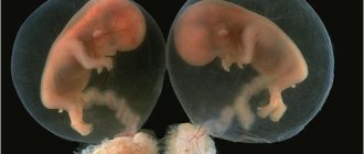
The development of the fetus at the 13th week of pregnancy is due to vigorous activity. Bone cartilage is formed and baby teeth are formed. The expectant mother should add calcium-rich foods to her diet.
At the 13th obstetric week of pregnancy, the baby’s pancreas actively produces insulin and other secretory glands form. During this period, the body still has disproportionate shapes, since the head exceeds the size of the body. The fetus is actively moving, but the woman does not feel this due to its small size.
Progress of the procedure
Ultrasound is the safest and most effective examination prescribed for pregnant women. At the time of pregnancy, a woman must attend 3 ultrasound examinations.
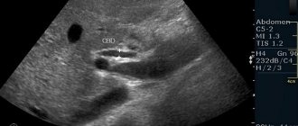
Ultrasound examination is based on the principle of echolocation. A special device emits ultrasonic waves that pass through all internal organs. The method allows you to visualize the entire abdominal cavity.
Transvaginal ultrasound at 13 weeks gives more accurate results
Each organ has a certain echogenicity and reflects ultrasound waves differently. In the first trimester, which means at the 13th week, transvaginal ultrasound is used. In this case, a special sensor is inserted into the woman’s vagina.
Transvaginal ultrasound is more accurate in the early stages. The fetus is rarely examined through the abdominal cavity at the 13th week. The abdominal method is less effective in the first trimester.
Norms of intrauterine development of the fetus by the end of the 13th week of pregnancy
Determination of CTP (coccygeal-parietal size) at 13 weeks is the measurement of the length of the fetus from the crown to the coccyx. The norms for the CTE of a 13-week fetus are 65 mm, and the indicators of minor deviations are 53-77 mm. At this stage of pregnancy, the size of the nasal bone is examined. The normal fetal nasal bone at 13 weeks should be 2.0-4.2 mm. If, as a result of the study, any deviations from these norms are determined, we can talk about an abnormality of fetal development.
The diagnostician also examines the joint bones of the skull and butterfly, vertebral cartilage, limbs and their bone tissue, the anterior abdominal wall, bladder, stomach, cardiovascular system and motor activity. The normal fetal heart rate should be 140-160 beats/min.
Is it possible to determine the pathological process in advance? If the embryo has indicators characterizing Down syndrome, an ultrasound sensor at week 13 will be able to detect signs of this disease. An important indicator of pathologies of this nature is the thickness of the nuchal translucency space (TN). The nuchal translucency index at 13 weeks should not exceed 30 mm, then the risk of Down syndrome is minimal. Thanks to the sensor of the ultrasound machine, the specialist will be able to determine the size and structure of the fetus, confirm or deny the presence of anomalies. In general, ultrasound research shows:
- KTR - about 66 mm;
- biparietal size (BPR) - 20-28 mm;
- length of the nasal bone - 2-4.2 mm;
- fetal head circumference - 73-96 mm;
- TVP - no more than 30 mm;
- abdominal circumference - 58-80 mm;
- length of the femur - 7-11.8 mm.
Ultrasound examination at this stage of pregnancy makes it possible to identify pathologies in the central nervous system, cardiovascular, genitourinary and musculoskeletal systems of the fetus, as well as developmental anomalies in the gastrointestinal tract.
Purposes of ultrasound
At the 13th week of pregnancy, the first ultrasound is performed. Such research is mandatory. Diagnostics is necessary to ensure that there are no serious abnormalities in the fetus.
The doctor studies the anatomical structure of the baby and determines the distance from the tailbone to the head of the fetus.
An ultrasound during pregnancy at the 13th week is necessary in order to understand whether the fetus is fully developing. The doctor compares the obtained numbers with normal values. The doctor makes sure there are no chromosomal abnormalities.
First of all, at the 13th week, Down syndrome may be present in the fetus. If this diagnosis is confirmed, the mother is referred for further research.
It is at this time that doctors prescribe a routine ultrasound
By the 13th week, all internal organs are formed in the baby’s body. That is why this is the best period to exclude possible pathologies:
- hearts;
- brain;
- possible neoplasms.
In the presence of severe pathologies, a woman may be referred for an artificial termination of pregnancy.
Ultrasound methods
The research methods used to obtain information about fetal development are as follows:
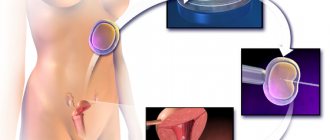
- Transvaginal. How is ultrasound performed using the transvaginal method? This diagnosis is carried out in the early stages of pregnancy, as it provides the most reliable information about the condition of the fetus and its intrauterine development. Transvaginal equipment can also be used to determine the sex of the child. A sensor equipped with a disposable condom is inserted into the pregnant woman’s vagina, and data is displayed on the screen indicating how the systems and organs will develop. The cost of the procedure will be on average 1200 rubles.
- Abdominal. To study using this method, it is necessary to fill the bladder with fluid before the procedure. This study is not prescribed in the early stages due to the possible misdiagnosis associated with the small size of the fetus. Approximate cost: 1500 rubles.
- 3D ultrasound. Using a three-dimensional sensor, a clear photo of the embryo is displayed on the screen, as it looks at the current moment. This ultrasound allows you to determine the sex of the child with complete accuracy. The cost of the study is on average 3,000 rubles.
An unplanned ultrasound may be prescribed in the following cases:
- suspicion of a frozen pregnancy;
- likelihood of miscarriage;
- poor heredity regarding chronic diseases;
- congenital anomalies.
If, as a result of diagnosis, there is a significant difference between the norm and the indications, you should visit a gynecologist-obstetrician. In such cases, a biochemical analysis is prescribed followed by therapy.
The difference between 3D/4D ultrasound and 2D in early pregnancy, is volumetric ultrasound better or worse?
| Differences between 2D, 3D and 4D ultrasound images |
Ultrasound is the optimal method for diagnosing the health status of the pregnant woman and baby throughout the entire gestation period. In addition to the usual two-dimensional black and white image on a monitor screen, new methods of volumetric examination (three-dimensional 3D and four-dimensional 4D ultrasound) have recently become increasingly widespread.
The main differences between volumetric methods and conventional, two-dimensional, ultrasound
:
- With 2D ultrasound, the image on the screen is visible in two projections - length and width. At 3B, another dimension is added - height, so the image becomes three-dimensional, but static. With 4D, another parameter (time factor) allows you to see the object of study (fetus) in real time in dynamics.
- With a two-dimensional ultrasound, the image is displayed in black and white; with 3D/4 D, a golden background is added.
- 3D/4 D ultrasound can only determine the structure of external organs. Pathology of internal structures can be seen in the first trimester only with the help of 2D ultrasound.
- The intensity of the impact of ultrasonic waves during volumetric types of ultrasound is higher than with two-dimensional ultrasound, and the duration of exposure increases almost 2 times. And although there is no convincing evidence of the negative effects of ultrasound under conditions of increased intensity and duration of exposure, there is still reason to fear the occurrence of delayed negative consequences.
- Therefore, one of the main differences between two-dimensional echography and 3D/4 D ultrasound is the recommended examination time. 2D ultrasound, recognized by the World Health Organization as harmless and safe for the pregnant woman and the fetus, is used in the early stages of pregnancy, especially when the need arises, even at 4-5 weeks. The recommended period for volumetric ultrasound is no earlier than the 20th week of gestation, when the main period of organ formation ends.
- There are rare cases when a 3D procedure is required for medical reasons, despite the potential danger. These are the cases when it is necessary to resolve the issue of the possibility of continuing pregnancy or the need to terminate the pregnancy: suspicion of chromosomal abnormalities or severe malformations of the baby that are incompatible with viability. In these cases, a more thorough examination is possible by obtaining a three-dimensional image.
- The availability of two-dimensional ultrasound is much higher, because All medical institutions are equipped with such devices. The opportunity to undergo examination in 3D/4D mode is not available in all clinics or cities. For this, special extra-class devices are needed, which combine three emitters and three receivers of reflected ultrasonic waves. In this case, a special algorithm is used to decipher the received signals using computer programs that form a three-dimensional image.
- The advantage of 3D and 4D ultrasound is the ability to rotate the image at any angle, as required for a better view. And with 4D you can see all the movements in real time.
- Three-dimensional images that are displayed on the screen are easily understood by parents, compared to 2D images. Therefore, expectant mothers and fathers want to see their child’s face as soon as possible, take a photo or record a video of his behavior during pregnancy. Although in the early stages of pregnancy, and in subsequent periods, the child does not share their delight, he does not like it.
- Volumetric types of ultrasound, which are most often done at the request of the parents, and not as prescribed by the doctor, are a paid procedure; it costs 1.5-2 times more than a regular paid ultrasound of a pregnant woman.
| Difference in images of a baby at 18 weeks with 2D and 3D (in the center) ultrasound |
Of course, the woman decides whether or not to do a 3D/4D ultrasound in the first trimester of pregnancy. There is no direct evidence of a direct negative effect on the fetus of these methods. But it’s not for nothing that doctors around the world are guided by WHO recommendations and do not prescribe any optional examination methods in the early stages of pregnancy, for fear of harming the fragile body of the fetus. All methods have advantages and disadvantages, indications and contraindications. Only specialists can compare the pros and cons, so when deciding which ultrasound method to use, you need to trust your doctors.
Preparing for the ultrasound procedure
Research at thirteen weeks is carried out transabdominally, which does not require special preparation. A few days before the procedure, you should not eat foods that cause gas formation in the intestines. You must have with you a diaper to cover the couch, dry wipes to remove residual acoustic gel from the stomach, and an account card. For examination using transvaginal ultrasound, you need to take a medical condom.
Ultrasound examination is carried out transabdominally or intravaginally. During the procedure, the woman does not experience any bodily discomfort; the procedure is painless. The interpretation of ultrasound indicators is carried out by a diagnostician or a gynecologist.
Contraindications
Ultrasound is safe, but, like any other method, it has contraindications. At week 13, the study cannot be carried out if:
- extensive inflammatory lesions, burns and skin diseases on the examined area of the skin;
- undergoing gynecological surgical interventions (transvaginal ultrasound);
- urinary incontinence, if transabdominal ultrasound with bladder filling is recommended.
The doctor first rules out possible contraindications. Ultrasound is not performed when there is increased gas formation or on a full stomach.
What does a screening test show?
Screening consists of a number of research procedures that will help to determine in advance the development of chromosomal diseases of the embryo. Screening is carried out at 10-14 weeks. This is the most appropriate time for timely detection of fetal abnormalities.
The screening study consists of a biochemical blood test and ultrasound diagnostics. Biochemistry is done after an ultrasound, and based on the results of these studies, they talk about the possibility of carrying a healthy/sick baby. The risk group often includes women whose screening test results are 1:350 or lower. These results indicate a high risk of developing abnormalities, but do not guarantee them. Normally, the level of progesterone in a woman’s blood should be:
- in the first trimester - 8.9-468.4 nmol/l;
- in the second trimester - 71.5-303.1 nmol/l.
In addition, biochemistry allows you to find out hCG, PAPP-A and the indicator of the protein component of plasma.
Advice for expectant mothers
To ensure a smooth pregnancy, expectant mothers are advised to follow these recommendations:
- Choose comfortable clothes and shoes without heels for everyday wear.
- Limit your consumption of fatty and floury foods.
- Diversify your menu as much as possible with fresh food, vegetables, and fruits.
- Gymnastics will help avoid uterine tone.
- Don't ignore daily hygiene procedures.
- Don't forget about walks in the fresh air.
During this period, it is important to establish a strong emotional connection with the child. When communicating with him, you need to convey exclusively positive emotions, get more rest and eat right. Ultrasound at this stage must be completed without fail, as it allows early