How does pregnancy develop in the first weeks after conception?
After the fusion of the sperm with the female reproductive cell, the resulting zygote moves towards the uterus.
After 24 hours, the fragmentation process begins, leading to the formation of a blastocyst consisting of approximately 100 cells on the 5th day. On the 8th day, the blastocyst penetrates the uterine endometrium. The fetal tissues begin to produce human chorionic gonadotropin (hCG), and the formation of the fertilized egg begins. 14 days after ovulation, when menstruation should begin, there is no discharge - their delay indicates pregnancy (see also: what kind of discharge can there be in early pregnancy before menstruation is delayed?).
Signs of pregnancy at 6 weeks
Already at such an early stage of pregnancy, a woman begins to notice changes occurring in her body. They are due to the fact that the body begins to actively prepare for bearing a child and produces a large amount of hormones that rebuild all processes in a woman’s body.
Already at this time, severe toxicosis appears, characterized by a feeling of nausea, vomiting, a change in reaction to odors and food, and increased salivation.
Also, due to the influence of a large amount of progesterone, which is necessary to strengthen the uterus and its increased blood supply, digestive disorders may occur, which also affects the manifestation of toxicosis.
Doctors in this condition advise pregnant women to drink more water to cope with the feeling of nausea. A large amount of fluid helps remove salts from the body and helps cope with unpleasant symptoms. Also, to unload the digestive system, which cannot cope with the food entering it, it is recommended to switch to frequent meals in small portions. This can also reduce the feeling of nausea and help cope with discomfort and vomiting.
Also, under the influence of hormones, pregnant women become irritable, they experience a loss of strength and even dizziness.
There may also be noticeable flaccidity in the muscles, affecting all movements of the woman. At the same time, severe stress and emotional unrest negatively affect the unborn child, so it is extremely important during this period of time to try to avoid mental stress and excessive worry.
In some cases, abdominal pain may begin in the sixth week of pregnancy. In this case, be sure to consult a doctor in a timely manner so that he can conduct a full diagnosis and eliminate possible risks for the mother and child. In most cases, abdominal pain is a direct signal that something is going wrong in a woman’s body, so you should not ignore these sensations.
Another obvious sign indicating pregnancy is breast enlargement and a change in its sensitivity. Quite often there is pain and tingling in the nipples, darkening halos and hypertrophied tactile perception. For most pregnant women, even minor touches of the breast become too painful and unpleasant. Many women also note a feeling of heaviness in their breasts that appears with the onset of pregnancy.
In addition to these main manifestations, there may also be a decrease in blood pressure, skin problems, constipation, heartburn and other symptoms.
However, for some women, pregnancy may actually not manifest itself at all and even pass without the toxicosis that is familiar to many. This largely depends on the individual characteristics of the woman’s body and how it reacts to pregnancy and excess hormones. The only symptom that is present in almost everyone is frequent urination. This phenomenon is due to the fact that it is caused by the physiological characteristics of a woman’s body, and not her hormonal background. Already from the sixth week of pregnancy, pressure from the uterus on the bladder may occur, which leads to frequent urge to go to the toilet. Despite the fact that at this stage this reproductive organ is still quite small and does not exceed the size of a plum, due to its location it affects all organs in the abdomen, including the urinary system.
At what stage and at what level of hCG is the fertilized egg visible during an ultrasound?
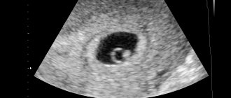
During an ultrasound, the fertilized egg is detected as a small black dot. A blurry white spot is clearly visible in it - an embryo. The study is carried out transvaginally. The fertilized egg is an anechoic (not reflecting ultrasonic waves) formation of a round or oval shape. The microscopic size of the embryo at the very beginning of its development does not make it possible to see it using ultrasound. In the early stages, a sonologist detects pregnancy and determines its duration by detecting the fertilized egg and measuring it.
When is the egg visualized well? In the first weeks, the fertilized egg grows by about 1 mm per day: at 4 weeks its diameter is 3 mm, at 5 weeks – 6 mm. Until 8–10 weeks of pregnancy, its duration is determined by the diameter of the fetal egg, then by the coccygeal-parietal size (CPR) of the fetus.
The doctor will be able to see the fertilized egg and embryo no earlier than the 5th obstetric week. A convincing sign of pregnancy is the hCG level detected through a blood test from 1500 to 5000 IU/l. As a rule, if the test result is normal, you can go for an ultrasound - the doctor will be able to see the fertilized egg.
Visiting the doctor at 6 weeks pregnant
Many people are interested in the question: when should I register for pregnancy and childbirth? Everyone decides for themselves when to visit their gynecologist for the first time. If nothing worries you, you can postpone the visit. But still, the sixth week of pregnancy is the right time for your first visit to the gynecologist.
Typically at this visit, your doctor will confirm your unique condition. Pregnancy management should begin with a pregnancy chart, a detailed medical history and a medical and gynecological examination, as well as palpation of the mammary glands.
The gynecologist determines possible risks associated with pregnancy and potential threats. Do not hide any information about chronic or infectious diseases that you have previously been exposed to.
You should inform your appointment about illnesses in your family or your partner’s family. If you have the opportunity, write down what medications you take regularly, write down their names and doses.
Include reasons for long hospital stays, previous procedures and surgeries. Take all documentation of your treatment, such as medical records, with you. If this is not your first pregnancy, write down the dates of previous pregnancies, miscarriages and births, as well as the weight at which your children were born.
During the examination, the doctor will check the condition of the body and cervix to see if their weakening indicates pregnancy. The doctor will evaluate your health and examine your chest, lungs, and heart. Checks body weight and blood pressure.
When to go to the doctor for the first time if you are pregnant
The results will form the basis for comparison in subsequent studies. He will order laboratory tests - detailed tests will allow the doctor to monitor the pregnancy.
- Blood test including blood type and Rh factor
- General urine analysis
- Fasting blood glucose test
- Tests for syphilis (VDRL). HIV testing recommended
- Viral and virological tests for HCV
- Test for toxoplasmosis (IgG, IgM), cytomegalovirus and rubella
- Cytological and bacteriological studies. Cytology involves a microscopic examination of a smear of the cervix to look for changes that may develop into cancer. The smear must also be bacteriological.
Don't forget to visit your dentist too! Assessing oral health and determining preventative and therapeutic needs during pregnancy is extremely important.
Research shows that nearly 18% of preterm and low birth weight births are associated with periodontal disease and oral inflammation.
When does ultrasound determine the presence of an embryo in the fertilized egg and what does it look like?
If conception occurred in a woman with a regular menstrual cycle, the diagnostician will detect the embryo at 6–7 weeks. By this time, the size of the fertilized egg increases to 7 mm. In addition, during the ultrasound, the fetal heartbeat is heard. On the monitor, the embryo looks like the letter “C”. Towards the end of the 8th week of pregnancy, clearly visible outlines of the head, legs and arms appear. From next week, the embryo is already considered a fetus.
If during the examination the doctor does not detect a fertilized egg in the uterus, and all tests indicate pregnancy, you can undergo a second examination 6-7 days after the first one. In a situation where the fertilized egg is visible but the embryo is not identified, the procedure can be repeated after 2 weeks. Listening to a heartbeat outside the uterine wall suggests an ectopic pregnancy.
What happens to the fetus, how the embryo develops
The child now looks like a tadpole. The size of the baby is from 4 to 6 mm, and its length, measured from the top of the head, can be compared to the width of a grain of rice or wheat. Did you know that you already carry 1 gram of happiness under your heart?
Around the 6th week of pregnancy, the gestational sac fills with fluid, which at this stage is formed from the plasma and body fluids of the woman.
Now amniotic fluid will not only reduce the risk of possible injuries, but will also provide warmth and protection for the baby from bacteria.
The 6th week of pregnancy is also the time when the neural tube closes (this process usually ends 28-30 days after fertilization).
This means that there is no longer a risk of developing defects that could lead to spina bifida or hydrocephalus. On the 25th day after fertilization, the anterior opening of the neural tube closes, and 2 days later the posterior opening closes.
But this does not mean that the threat of developing other defects has also ceased to exist. Unfortunately, the most dangerous period is the period of organogenesis, that is, the formation of all organs and systems of the body, which lasts from 4 to 12 weeks of pregnancy.
During this time, the newly formed organs are exposed to external factors such as drugs, chemicals and others.
Table. Estimated embryo size at 6 weeks
| Pregnancy period by day | Fertilized egg size | Embryo size |
| 6 weeks and 1 day | 12 mm | 3 mm |
| 6 weeks and 2 days | 13 mm | 4 mm |
| 6 weeks and 3 days | 14 mm | 4 mm |
| 6 weeks and 4 days | 15 mm | 5 mm |
| 6 weeks and 5 days | 16 mm | 5 mm |
| 6 weeks and 6 days | 17 mm | 6 mm |
This is why it is so important to avoid all medications and other hazardous activities during this critical time of development.
Baby development in the sixth week of pregnancy:
- A child's brain develops at an insane rate.
- The spinal cord begins to develop from the existing neural tube.
- During the 6th week of pregnancy, the baby's limbs begin to form from the main body.
- A cellular mass is formed, which will give impetus to the formation and development of the eyeballs of the future baby.
- Folds of tissue form that will soon become the cheeks, chin and jaw.
- A bulge will form where the nose will soon appear.
- A hole appears from which the mouth and canals that form the middle ear will later form.
- The heart, which just a few days ago (at 5 weeks of pregnancy) was shaped like a tube, now turns into four chambers and almost beats outside the body of the embryo.
- During the 6th week of pregnancy, your baby's liver, pancreas, lungs and stomach are slowly forming.
- Bronchi appear - the primary respiratory tract
The heart begins to beat around the 23rd day of pregnancy from the day of conception, that is, in the 6th week.
Remember that fertilization age is different from the actual length of pregnancy determined by doctors based on the date of your last menstrual period.
Why is the embryo not visualized at 6–7 weeks?
The woman saw two lines on the test, was delighted, came for an ultrasound, but the doctor did not find an embryo. Why is the embryo not visualized during a positive pregnancy test during an ultrasound? Possible reasons:
- The gestational age was incorrectly calculated. The embryo becomes clearly visible at 6–7 weeks.
- The examination was carried out using outdated equipment, or the doctor is not sufficiently qualified.
- The study was carried out by the transabdominal route, which increases the error of the result.
- A spontaneous miscarriage occurred, which the woman mistook for normal menstruation. The hCG concentration did not have time to decrease by the time of the examination, and ultrasound gives a negative result.
If the doctor said during an ultrasound examination that there is no embryo, the woman should not worry too much. Erroneous detection of anembryony (absence of an embryo) may be due to various factors. An argument in favor of successful conception is an increase in hCG levels over time and the absence of bleeding and other signs of early miscarriage. To get the final result, you need to undergo an ultrasound again after 7-14 days.
Difference between obstetric and actual gestational age
The gestational age can be obstetric or embryonic. Obstetrical is counted from the 1st day of the last menstruation; in the second case, the count is from the day of conception, which is taken as the day of ovulation. As a rule, the obstetric period is 14 days longer than the embryonic period.
When performing an ultrasound, the obstetric period is taken into account. During the procedure, the doctor correlates the identified indicators of embryo development with the obstetric period. The accuracy of the final result depends on the gestational age:
- when examining a pregnant woman up to the 12th week, the permissible deviation is 1–2 days;
- within 12–28 weeks – 1 week;
- after the 28th week the permissible error increases.
All subsequent examinations and tests are carried out taking into account the obstetric period. Its duration is 280 days, or 40 weeks. In practice, the difference from the embryonic gestational age of 2 weeks may vary for individual reasons. According to statistics, in 20% of pregnant women it is less than two weeks, in 45% it can reach 3 weeks, and in 15% it can exceed 3.
External factors: quality of equipment and qualifications of the doctor
Misdiagnosis during ultrasound occurs in rare cases. If the doctor does not detect the embryo, it is necessary to re-examine a week later or undergo the procedure using other equipment. It is possible that the specialist conducting the ultrasound examination does not have sufficient qualifications. It is advisable to clarify information about the equipment and specialist before the ultrasound.
Absence of embryo
The absence of an embryo in the fertilized egg can be caused by various reasons:
- acute viral or bacterial infection in a woman in the first weeks of pregnancy;
- chromosomal and anatomical defects of the embryo;
- blood clotting disorder in a pregnant woman.
There are no pronounced physiological signs of the absence of an embryo in the fertilized egg. Even a gynecologist examining a woman cannot detect anembryonia. Only an ultrasound diagnostic specialist can reliably determine the pathology, and then at least 5-6 obstetric weeks (1-2 weeks after a missed period).
On the monitor screen, an empty fertilized egg is visualized as a black spot of irregular shape, usually smaller in size than it should be according to the stage of pregnancy. Sometimes a specialist sees a normal round-shaped fertilized egg, but there is no yolk sac in it. In the first weeks of pregnancy, an ultrasound examination has low information content, so it is recommended to undergo the first ultrasound at 10–12 weeks - at this time the fetus is already clearly visible.
Symptoms and sensations of the expectant mother
This week of pregnancy is special for the expectant mother. Let us consider in detail how a woman feels during this period. The embryo closes the opening of the neural tube, from which the baby's brain and spinal cord will form. For this you need folic acid. So don't forget to take it every day.
Since you're still in the early stages of pregnancy, you may not have experienced pregnancy symptoms - during this period, some women are constantly hungry, while others feel almost nothing. During the sixth week of pregnancy, the most common symptoms are:
- Sensitive and sore breasts
The chest probably hurts due to increased blood flow. It's hard to believe that a woman's body is so complex that she prepares for breastfeeding at the very beginning of pregnancy.
Photo of an embryo at 6 weeks of pregnancy
- Fatigue
You feel tired all the time because your body is still getting used to hormonal changes, this is normal. Try to rest whenever you have the opportunity.
- Nausea
We often hear that morning sickness is typical during pregnancy. But very often this nausea lasts all day, especially if you are carrying twins.
The most important thing is to find foods that suit you and calm your stomach. Keep them on hand and consume only them regularly, because if your stomach is empty, your nausea will only become more intense.
- Mood changes
The 6th week of pregnancy can be difficult emotionally for the expectant mother. Mood changes are also normal because hormones are still active and determine mood. Mood swings and nervousness during pregnancy are associated with an increase in the level of the beta hormone hCG and your individual sensitivity to changes in its concentration.
This is a temporary condition and everything will return to normal when the hormones restore balance.
So, if you still haven't felt any hormonal changes caused by premenstrual pressure, then pregnancy symptoms may not appear until 6 weeks.
- Frequent urination
This is normal during this period. It is important to pay attention to whether urination is normal or if you have discomfort accompanied by a burning sensation.
Which may be a urinary infection that gets worse during this period. If in doubt, you should consult your doctor, who will refer you for further testing.
- Gas and flatulence
Progesterone, a pregnancy hormone, can cause gas and bloating in the abdomen. Drink plenty of water and eat fiber-rich foods to avoid constipation, which contributes to bloating.
- Cramps and spots
In the first weeks of pregnancy, cramps and light bleeding are common. During this period, intercourse may also cause bleeding because the cervix is more sensitive during pregnancy. If you feel uncomfortable and worried, it is best to consult your doctor.
What to do if the fetus is not visible on ultrasound and what could this mean?
There are situations that during an ultrasound, the embryo is not visualized inside the fertilized egg, and sometimes the fertilized egg itself is not visualized. First of all, you need to try not to panic. There may be no pregnancy at all, or there was an error in calculating its duration, so it is still difficult to diagnose. If a frozen pregnancy is not definitely confirmed, there is no need to rush into cleaning. First, it is better to undergo an ultrasound again, in another clinic. It may be necessary to conduct one or more studies. The best option is when the level of hCG in the blood is monitored in parallel with the diagnosis. If pregnancy develops without deviations, then its level increases. This helps specialists exclude a possible frozen pregnancy.
What to do if pregnancy is not detected during an ultrasound scan?
If a situation arises when the ultrasound doctor cannot see the outline of the embryo, and sometimes even the fertilized egg itself, you must try to remain calm and not succumb to false beliefs! This is possible in the absence of gestation or its period is too short to be noticed on the monitor. Without absolute evidence that the pregnancy has been interrupted, curettage of the uterine cavity cannot be performed!
You should go to another clinic and undergo the examination again - it is better to do this using expert-class equipment with high resolution. It is also necessary that the ultrasound be accompanied by blood tests for hCG levels. The examination may need to be completed several times. Future parents should make every effort to ensure that diagnostic errors do not cost the child’s life!
Timing of the examination
The first ultrasound is prescribed when a woman comes to the antenatal clinic to register. Usually this is a period of 10-12 weeks, if there is a positive hCG test. Then it is already possible to detect the fertilized egg along with the embryo in the uterus and identify developmental pathologies. An ultrasound can be done earlier; it will detect the embryo 14-20 days after the missed period.
The second scheduled examination is done at 20-22 weeks. Here you can identify fetal malformations. The last examination is carried out 14-21 days before birth to determine the position of the fetus and the woman’s readiness for childbirth.
Unscheduled studies are prescribed for women with suspected or existing pathologies of fetal development. An ultrasound is prescribed after IVF to determine whether the embryos have implanted. After embryo transfer, examination is carried out 21 days later.
How are expectant mothers examined?
If the pregnancy test is positive, this can be confirmed by ultrasound examination - diagnosis is carried out in a commercial center or in a antenatal clinic. It is important to know that equipment with a high level of resolution and functionality, as well as the qualifications of a specialist, plays an important role in obtaining reliable examination results.
Up to 9 obstetric weeks, two methods are used to examine pregnant women:
- Transabdominal – through the area of the anterior abdominal wall.
- Transvaginal - using a transducer that is inserted into the vagina.
Up to 5 weeks, the formed fertilized egg is very small - its size is only about two millimeters. The transvaginal method is considered an effective method for diagnosing the embryonic period - its high-frequency sensor makes it possible to get as close as possible to the uterine cavity and transmit the smallest dimensions of the examined organs to the monitor screen.
During the entire gestation period, a woman undergoes at least three ultrasound scans. The examination session is short-term; the doctor tries not to hold the sensor in one place for a long time, especially during the formation of the most important organs and systems of the unborn baby.
What do they look for in an ultrasound scan?
The main purpose of performing an ultrasound scan in the embryonic period is to confirm pregnancy; this issue is especially relevant in the case of in vitro fertilization. The diagnostician has several tasks:
- Confirmation of fixation of the fertilized egg in the uterus.
- Exclusion of the presence of a neoplasm in the uterine cavity, which can “masquerade” as pregnancy.
- Assessment of embryo viability.
- Rule out ectopic pregnancy.
- Determining the presence of a second fetus.
- Study of the localization of the placenta and embryo.
- Clarification of gestation dates.
In gynecological practice, there is one important point that all expectant mothers should know: the doctor measures the duration of the pregnancy period in obstetric weeks - from the first day of the last menstruation. That is why the difference between the actual and obstetric time of conception of a child is two weeks. In a woman of reproductive age with a normal menstrual cycle, recognition of pregnancy during transvaginal examination occurs no later than five weeks. If the cycle is irregular, determining the exact period based on menstruation is difficult.
Determination of pregnancy in the sixth week
Despite the fact that the life span of the embryo is still quite short, an experienced gynecologist can confidently determine whether there is a pregnancy or not.
At this time, a large number of tests are prescribed that check hormonal levels and measure all indicators.
This is necessary in order to promptly identify abnormalities that appear during pregnancy and may affect the health of the unborn child and his mother.
In this case, much attention is paid to the level of the hormone progesterone, which is a very important substance for preserving and maintaining pregnancy, as it keeps the uterus in good shape and helps protect the embryo.
If the amount of this hormone is insufficient, the gynecologist prescribes drug therapy to help correct this abnormality and maintain pregnancy.
During this period, the first ultrasound examination is carried out to determine exactly how many embryos the pregnant woman is carrying and whether they are developing in the right place. In the sixth week, it is clearly visible what type of multiple pregnancy is present and how many placentas are determined. Ultrasound examination makes it possible to timely diagnose the presence of an ectopic pregnancy with greater accuracy.
Why is the fertilized egg not visible on ultrasound? Fertilized egg. beremennost9m.ru
Girls! Help for God's sake. I am fighting with all my strength against the approaching hysteria. 1.5 years ago I had to terminate my pregnancy at 20 weeks (chromosomal abnormality). I barely came to my senses. We waited for almost a year, then tried and tried. Here we are, the two treasured stripes. I took my hCG test yesterday - 210.6 (4-5 weeks). But nothing is visible on the ultrasound. The ultrasound specialist told me to take hCG again in a couple of days and undergo an ultrasound. If hCG rises and she doesn’t see anything, hospitalize her. Is it really ectopic? Girls, who knows? An ectopic can be seen on an ultrasound. and hCG if the ectopic also increases? It is possible that my little doll is simply not visible yet.
Girls! Help for God's sake. I am fighting with all my strength against the approaching hysteria. 1.5 years ago I had to terminate my pregnancy at 20 weeks (chromosomal abnormality.
Juliakha Don't worry!
I had the same thing. The test showed that I went to take hCG, but it turned out that the result would only be ready later. day, and we were from another city and were there on business. Well, I think I’ll donate blood and go for an ultrasound, but I agreed that I’ll call to follow up. day and find out the hCG result over the phone (B was very long-awaited). They didn’t see anything on my ultrasound; you could say they sent me something. They said that the test is not an indicator and there is no B (((. But I naturally didn’t believe them. The next day I called to find out the result, my result corresponded to 4 obstetric weeks)))) Then after 2 weeks I came to that As for her aunt, with her own conclusion, she was very surprised that there was no egg in the uterus, and set the deadline at 6 weeks. and 3 days, the heartbeat was heard. She said that the egg was walking, grit - there will be a girl)))) and so it happened)))))
Juliakha Don't worry!
I had the same thing. The test showed that I went to take hCG, but it turned out that the result would only be ready later. day, and we are from .
I had an ultrasound yesterday, they also said nothing was visible yet, B is in question. I did the test in a couple of days and one strip, but there was a delay and all the symptoms - chest, nausea were obvious. And this morning I took a test and a barely visible line appeared. and they told me to come back for an ultrasound in a week. My term is 3-4 weeks, but the doctor puts it at 1-2. Unclear
I had an ultrasound yesterday, they also said nothing was visible yet, B is in question. I did the test in a couple of days and one strip, but there was a delay and all the symptoms - chest, nausea.
If pregnancy is not visible on ultrasound
Sometimes life gives us not the most pleasant news! But you cannot lose faith and hope for the best, even if it seems that there can no longer be a positive outcome in your situation. When it comes to confirming pregnancy, medical science recommends testing repeatedly, preferably at intervals of several days. Speaking about pregnancy tests, it is worth noting that not in all cases they will be informative. To make sure that you are pregnant, in some cases it is better to perform an ultrasound. This research method will detect the fertilized egg and the embryo inside it, clarify its location and size, and indicate the gestational age. However, ultrasound cannot be 100% reliable, and the experience of doctors proves this. You can easily find stories of happy mothers who, having been examined at the 8th, 10th and even 12th weeks, received an ultrasound report: “No fertilized egg was detected.” But at this stage the baby’s heart is already beating! And despite all the doctor’s assurances about an ectopic pregnancy, they carried and gave birth to completely normal children.
It’s scary to admit, but it’s true: more than one child’s life has been cut short due to inaccurate ultrasound diagnostics! This, of course, does not mean that any suspicion of an ectopic or frozen pregnancy is wrong, but in this case the diagnosis should be more in-depth.
Why is the fertilized egg not visible on ultrasound?:
Thanks to modern medical equipment, diagnosis of pregnancy has become more accessible, as well as most pathological conditions that can develop during this exciting period of a woman’s life. But the result of the examination can never be absolutely accurate, because it is influenced by both technical errors and the human factor. There are cases when the fertilized egg was mistakenly mistaken for a myomatous node, or even the pregnancy was left without diagnosis, despite a fairly long gestation period. Not every clinic and antenatal clinic can boast of a new modern ultrasound machine. Old-style equipment does not have sufficient resolution, and the doctor may simply not be able to see the embryo in the fertilized egg, which is very small. You should not undergo an ultrasound examination before the 10th day of your missed period - there is a high chance of an inaccurate result. Too short a pregnancy is the most common reason why it is impossible to examine the embryo in the fertilized egg during examination. Remember that in early stages of pregnancy (up to the 5th week) the embryo can only be detected using a transvaginal ultrasound (TV) method. Around the 7th week, a fetal heartbeat is detected during a TV examination. If, according to your calculations, the term has exceeded this figure, and the embryo and its heartbeat are not visible, you should know that it is too early to worry. Perhaps the gestational age was determined incorrectly (including due to late ovulation), or perhaps the issue is in the ultrasound device itself and the diagnostic method.
Interesting: How to collect a urine test for sterility from a child
One more reason needs to be mentioned - the anatomical features that determine the shape, size and location of the uterus in a particular woman. Sometimes, with pathology of the development of the uterus (saddle-shaped, bicornuate uterus, curved cervix), the doctor cannot examine the fertilized egg in its cavity.
What to do if pregnancy is not detected by ultrasound.
A situation may arise when an ultrasound does not show the embryo in the fertilized egg, and sometimes even the fertilized egg itself. But in any case, you need to remain calm and not succumb to false beliefs! Perhaps there is no pregnancy at all; it is possible that the gestational age is still too short for pregnancy to become noticeable on the monitor. If there is no absolutely reliable evidence in favor of a frozen pregnancy, do not cleanse! Carry out the examination again, go to another medical center that has its own ultrasound machine. It may be that neither one nor two examinations are required. It is optimal if they are accompanied by a blood test for hCG - during a normally developing pregnancy, this hormone increases in proportion to the weeks of gestation. But, unfortunately, mistakes can happen in the laboratory, so you will also have to donate blood several times. You must make every effort to ensure that a diagnostic error does not cost your child’s life!
Embryo parameters
Normally, the fertilized egg is oval in shape and dark gray in color. To fully monitor the formation of the fetus, the following indicators are measured on ultrasound.
| Parameter(mm) | Obstetric weeks | |||||
| 4 | 5 | 6 | 7 | 8 | 9 | |
| Internal contour of the diameter of the fertilized egg | 10 | 18 | 22 | 24 | 30 | 33 |
| The size of the coccyx-parietal size | 2–3 | 4–5 | 7–9 | 11–15 | 16–22 | 23–30 |
The clear visibility of the fetus on the monitor of an ultrasound machine is influenced by many factors, and if the embryo is not visible, do not panic - you should wait two weeks and repeat the study.
At the beginning of pregnancy, the embryo resembles the letter “C”; as it grows, its appearance changes - at 8 weeks you can already see both the head and the distinguished limbs
How to see an embryo?
If the test shows the presence of pregnancy, then an examination with an ultrasound scanner will help confirm this. Ultrasound diagnostics are carried out in a antenatal clinic or in a private medical center.
For examination in the 1st trimester, it is important that the ultrasound machine has high resolution and wide functionality, and the doctor’s qualifications also play an important role. At 4–5 weeks, the formed fetal sac is very small and poorly visible, its size is only 1–2 mm.
To understand medical language, it is important to know that gynecologists measure the period of gestation in obstetric weeks, from the first day of the last menstruation. Therefore, the difference between the obstetric and actual terms of conception of the baby is 2 weeks.
Sizes of fertilized egg by week
The size of the embryo determines the gestational age with an accuracy of 2-3 days. Up to one and a half months, the date of birth is determined by the DPP - the diameter of the fertilized egg. The diameter is measured at the widest part in three different planes. There is a special table to calculate the due date.
A criterion such as SVD - average internal diameter - does not determine the date of birth very accurately. After 6 weeks, when an embryo appears on ultrasound, the period is determined by CTP - coccygeal-parietal size. The embryo image is measured longitudinally. Sizes 6 to 8 weeks:
- 6 weeks - 3.5-7 mm;
- 7 weeks - 7.9-13.2 mm;
- 8 weeks - 14.2-21 mm.
When performing an ultrasound, a specialist assesses the condition of the fetus and determines developmental defects. Down syndrome can only be determined at 11-12 weeks. After passing this milestone, the embryo develops rapidly, all internal organs and limbs are formed.
The normal result of an ultrasound is an embryo with membranes corresponding in size to the gestational age. One egg or several may be found in the uterus.
In the video you can see the fertilized egg in the uterus:
Research methods
Ultrasound in early pregnancy is preferable to be done transvaginally. The sensor is inserted through the vagina and passes to the cervix. With this method, pregnancy is accurately confirmed within 28 days. The second method is transabdominal. The examination is carried out through the anterior abdominal wall. This method is used when the fetus is already large enough and is clearly visible during external examination.
With any type of examination, the doctor examines the fallopian tubes and ovaries. The corpus luteum can be seen in the ovaries. It is formed at the site of a ruptured follicle from which the egg was released. If fertilization has occurred, the corpus luteum produces the hormone progesterone, which ensures the safety of pregnancy.
To watch a video about pregnancy at an early stage, a gynecologist says:
How to do an ultrasound in the early stages
At 6 (8 obstetric) weeks, ultrasound is performed both transabdominally and intravaginally. The second method is a priority, as it allows a more thorough examination of the uterine cavity.
Preparation for the examination depends on the method of ultrasound. General recommendations include following a diet that reduces gas. 2-3 days before the expected ultrasound, you should stop eating flour, sweets, cabbage, legumes and dairy products. Excessive formation of gases in the intestines interferes with the visualization of the amniotic sac.
Before a transabdominal ultrasound, the patient needs to drink about 500 ml of liquid. A full bladder brings the uterus closer to the anterior abdominal wall, which significantly improves image quality.
A conductive gel is applied to the abdominal area, which strengthens the contact of the sensor with the skin. The doctor sequentially examines all pelvic organs. The size of the fetus and the presence of developmental anomalies are assessed. Also at this time, you can determine the heartbeat and its characteristics.
Unlike transabdominal examination, during intravaginal examination it is recommended to empty the bladder. The sensor is inserted into the vagina. A special condom is first put on it.
The research is carried out as carefully as possible. The movements of the sensor should not cause pain or discomfort to the woman. Information obtained from ultrasound at 6 weeks helps assess fetal viability and endometrial structure.
Why is the fetus not visible on ultrasound?
Sometimes it happens that an ultrasound does not show the presence of a fertilized egg in the uterine cavity. In this case, the pregnancy test is positive. There may be several reasons for this.
- Examination too early. Even a modern device cannot detect pregnancy a few days after conception. The embryo at this time has microscopic dimensions. The fertilized egg cannot be seen as it moves into the uterine cavity.
- Ectopic pregnancy. It happens that the egg meets the sperm while in the fallopian tube. Then the fertilized egg will attach there; the doctor will not be able to find it inside the uterine cavity. Therefore, during an ultrasound, not only the uterus is examined, but also the tubes and ovaries.
- Technical errors. They usually occur if the inspection is carried out on an old or faulty device. A specialist can also make a mistake if he is not sufficiently qualified or attentive. Such errors are observed in the early stages, when the embryo is still very small.
If the doctor has any doubts during the first examination, he recommends repeating the procedure after 2-3 weeks.
Sources
- https://VseProRebenka.ru/beremennost/obsledovaniya/na-uzi-ne-vidno-plodnogo-yajca.html
- https://krasivaya24.com/na-uzi-ne-vidno-embriona-prichiny/
- https://uziman.ru/pri-beremennosti/kogda-na-uzi-vidno-plodnoe-yajco-v-matke
- https://diametod.ru/uzi/mogut-ne-uvidet-beremennost
- https://rojaismelo.ru/retsepty-dlya-detej/na-uzi-5-nedel-ne-vidno-plodnogo-yajtsa
[collapse]
At what age should an embryo be visible on ultrasound?
While waiting for the birth of a baby, a woman asks the question: at what time will the embryo be examined by ultrasound? During diagnosis at a period of 5-6 weeks, the fertilized egg is about seven millimeters in diameter. At this stage, in most cases, the doctor has already visualized the embryo. Around this time, you can also hear his heart beating.
If you have a regular menstrual cycle, an embryo should be visible at the end of the sixth week. If an embryo is not visible on ultrasound, it is recommended to undergo a repeat examination in a week to exclude all possible abnormalities.
There are also cases when the fertilized egg is located outside the uterus. During an ultrasound, the egg is not visible well enough or is not visible at all. In this case, the heartbeat is heard outside the walls of the uterus.