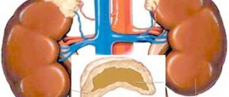
Hemolytic anemia, whose causes and symptoms will be described below, is a disease in which the life cycle of red blood cells is significantly shortened. In this case, blood cells are destroyed (this process is called “hemolysis”) in much greater quantities than they are produced. Decay occurs in just 2-3 weeks instead of the 3-4 months allotted by nature. This happens both directly in the vessels and in the following organs:
- bone marrow;
- spleen;
- liver.
In general, we are not talking about one pathology, but about a group. The disease is not detected too often - no more than 1 percent of the population. Among other types of anemia, hemolytic anemia accounts for 11%.
Causes of hemolytic anemia and risk factors
Hemolytic anemias develop either under the influence of extracellular (external) factors or as a result of red blood cell defects (intracellular factors). In most cases, extracellular factors are acquired, and intracellular factors are congenital.
Red blood cell defects are an intracellular factor in the development of hemolytic anemia
Intracellular factors include abnormalities of red blood cell membranes, enzymes, or hemoglobin. All these defects are heritable, with the exception of paroxysmal nocturnal hemoglobinuria. Currently, over 300 diseases associated with point mutations of genes encoding the synthesis of globins have been described. As a result of mutations, the shape and membrane of red blood cells change, and their susceptibility to hemolysis increases.
A larger group is represented by extracellular factors. Red blood cells are surrounded by endothelium (the inner lining) of blood vessels and plasma. The presence of infectious agents, toxic substances, and antibodies in the plasma can cause changes in the walls of red blood cells, leading to their destruction. By this mechanism, for example, autoimmune hemolytic anemia and hemolytic transfusion reactions develop.
Defects in the endothelium of blood vessels (microangiopathy) can also damage red blood cells, leading to the development of microangiopathic hemolytic anemia, which occurs acutely in children in the form of hemolytic-uremic syndrome.
Hemolytic anemia can also be caused by taking certain medications, in particular antimalarials, analgesics, nitrofurans and sulfonamides.
Provoking factors:
- vaccination;
- autoimmune diseases (ulcerative colitis, systemic lupus erythematosus);
- some infectious diseases (viral pneumonia, syphilis, toxoplasmosis, infectious mononucleosis);
- enzymopathies;
- hemoblastoses (myeloma, lymphogranulomatosis, chronic lymphocytic leukemia, acute leukemia);
- poisoning with arsenic and its compounds, alcohol, poisonous mushrooms, acetic acid, heavy metals;
- heavy physical activity (long skiing, running or walking long distances);
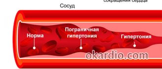
- malignant arterial hypertension;
- malaria;
- DIC syndrome;
- burn disease;
- sepsis;
- hyperbaric oxygen therapy;
- prosthetics of blood vessels and heart valves.
Classification
Based on the etiology or cause of the disease, the following nosological types are distinguished:
Hereditary or congenital, which include:
- Neuroferocytic. Insufficient enzyme activity obtained according to the chromosome set from one of the parents is dominant. The result is a significant decrease in the life of red blood cells. Normally, the life cycle of red cells is from 90 to 120 days. With this form, the decrease occurs for 12-40 days.
- Microspherocytic (Minkowski-Choffard anemia). A genetic defect manifested by damage to the membrane of red blood cells. The spherical shape of red blood cells lacks elasticity, which significantly reduces blood flow in bottlenecks in the circulatory system. Deformation of red blood cells causes small pieces called microspherocytes to break off. The spleen is forced to constantly utilize these microfragments. The increased load on the organ leads to a compensatory increase in the latter with the progression of hemolytic crises.
- Sickle cell. Hereditary destruction of cells due to mutation of the gene responsible for the formation of hemoglobin. The latter has an abnormal S-shape, giving the red blood cell a sickle shape. In small capillaries, this form is not viable and quickly collapses.
- Thalassemia. Destruction of the cell structure is observed due to a defect in hemoglobin chains inherited from one or both parents (homo or heterozygous pathology).
- Hemolytic anemia of the fetus or newborn occurs due to incompatibility of the Rh antigen of the mother and child.
Purchased:
- Autoimmune. It occurs due to the body’s own immune system attacking the cell membrane and destroying it.
- Traumatic. External causes (artificial heart valves, vascular prostheses, or abnormalities of large vessels) mechanically damage and disrupt the circulation of normal red blood cells.
- Acute paroxysmal nocturnal hemoglobinuria (according to the author, Marchiafava-Miceli disease). A rare type of disease caused by a mutation in bone marrow stem cells. In this form, leukocytes and platelets are also partially affected. Activates mainly at night. The causes of its occurrence are not fully understood; provoking factors may include surgical interventions, severe infectious diseases, blood transfusions, and heavy menstruation in women.
- Toxic hemolysis. Under the influence of toxic substances: chemical poisons, some long-term medications, biological poisons of insects and reptiles, the lipids of the erythrocyte membrane dissolve.
Depending on the severity of the disease, three forms are distinguished:
- Mild degree.
- Moderate weight.
- Heavy current.
According to the course of the disease, they are distinguished:
- Latent forms. Compensated conditions with a minimal clinical picture.
- Chronic. Unexpressed, sluggish symptoms with periods of exacerbation.
- Crisis. Characterized by an acute condition requiring emergency care.
Forms of the disease
All hemolytic anemias are divided into acquired and congenital. Congenital or hereditary forms include:
- erythrocyte membranopathies - the result of abnormalities in the structure of erythrocyte membranes (acanthocytosis, ovalocytosis, microspherocytosis);
- enzymopenia (enzymopenia) – associated with a lack of certain enzymes in the body (pyruvate kinase, glucose-6-phosphate dehydrogenase);
- hemoglobinopathies - caused by a violation of the structure of the hemoglobin molecule (sickle cell anemia, thalassemia).
The most common hereditary hemolytic anemia in clinical practice is Minkowski–Choffard disease (microspherocytosis).
Acquired hemolytic anemias, depending on the causes that caused them, are divided into the following types:
- acquired membranopathies (spur cell anemia, paroxysmal nocturnal hemoglobinuria);
- isoimmune and autoimmune hemolytic anemia - develops as a result of damage to red blood cells by own or externally obtained antibodies;
- toxic - accelerated destruction of red blood cells occurs due to exposure to bacterial toxins, biological poisons or chemicals;
- hemolytic anemia associated with mechanical damage to erythrocytes (marching hemoglobinuria, thrombocytopenic purpura).
Prevention
The use of personal protective equipment is mandatory!
The main preventive measures against the disease are the prevention of severe hypothermia and overheating, strengthening the immune system to reduce the risk of infectious pathologies and observing safety measures in areas where poisonous snake bites are possible. When working with chemicals that can cause intoxication, the use of personal protection is required. It also helps to promptly identify abnormalities in the composition of the blood and regular preventive medical examinations.
Symptoms of hemolytic anemia
All types of hemolytic anemia are characterized by:
- anemic syndrome;
- enlarged spleen;
- development of jaundice.
Moreover, each individual type of disease has its own characteristics.
Symptoms of hemolytic anemia
Hereditary hemolytic anemias
The most common hereditary hemolytic anemia in clinical practice is Minkowski–Choffard disease (microspherocytosis). It is traced in several generations of the family and is inherited in an autosomal dominant manner. A genetic mutation leads to insufficient levels of certain types of proteins and lipids in the red blood cell membrane. In turn, this causes changes in the size and shape of red blood cells, their premature massive destruction in the spleen. Microspherocytic hemolytic anemia can manifest in patients at any age, but most often the first symptoms of hemolytic anemia appear at 10–16 years of age.
Microspherocytosis is the most common hereditary hemolytic anemia
The disease can occur with varying severity. Some patients experience a subclinical course, while others develop severe forms, accompanied by frequent hemolytic crises, which have the following manifestations:
- increased body temperature;
- chills;
- general weakness;
- dizziness;
- pain in the lower back and abdomen;
- nausea, vomiting.
The main symptom of microspherocytosis is jaundice of varying severity. Due to the high content of stercobilin (the end product of heme metabolism), the feces are intensely dark brown. In all patients suffering from microspherocytic hemolytic anemia, the spleen is enlarged, and in every second patient the liver is enlarged.
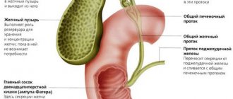
Microspherocytosis increases the risk of formation of stones in the gallbladder, i.e., the development of cholelithiasis. In this regard, biliary colic often occurs, and when the bile duct is blocked by a stone, obstructive (mechanical) jaundice occurs.
The clinical picture of microspherocytic hemolytic anemia in children also contains other signs of dysplasia:
- bradydactyly or polydactyly;
- gothic sky;
- malocclusion;
- saddle nose deformity;
- strabismus;
- tower skull.
In elderly patients, due to the destruction of red blood cells in the capillaries of the lower extremities, trophic ulcers of the feet and legs, resistant to traditional therapy, occur.
Hemolytic anemia, associated with a deficiency of certain enzymes, usually manifests itself after taking certain medications or suffering from an intercurrent illness. Their characteristic features are:
- pale jaundice (pale skin color with a lemon tint);
- heart murmurs;
- moderately severe hepatosplenomegaly;
- dark color of urine (due to intravascular breakdown of red blood cells and the release of hemosiderin in the urine).
In severe cases of the disease, pronounced hemolytic crises occur.
Congenital hemoglobinopathies include thalassemia and sickle cell anemia. The clinical picture of thalassemia is expressed by the following symptoms:
- hypochromic anemia;
- secondary hemochromatosis (associated with frequent blood transfusions and unreasonable prescription of iron-containing drugs);
- hemolytic jaundice;
- splenomegaly;
- cholelithiasis;
- joint damage (arthritis, synovitis).
Sickle cell anemia occurs with recurrent pain crises, moderate hemolytic anemia, and increased susceptibility of the patient to infectious diseases. The main symptoms are:
- retardation of children in physical development (especially boys);
- trophic ulcers of the lower extremities;
- moderate jaundice;
- pain crises;
- aplastic and hemolytic crises;
- priapism (spontaneous erection of the penis, not associated with sexual arousal, lasting several hours);
- cholelithiasis;
- splenomegaly;
- avascular necrosis;
- osteonecrosis with the development of osteomyelitis.
Sickle cell anemia – congenital hemoglobinopathy
Acquired hemolytic anemia
Of the acquired hemolytic anemias, the most common are autoimmune ones. Their development is caused by the production of antibodies by the patient’s immune system directed against their own red blood cells. That is, under the influence of certain factors, the activity of the immune system is disrupted, as a result of which it begins to perceive its own tissues as foreign and destroy them.
In autoimmune anemia, hemolytic crises develop suddenly and acutely. Their occurrence may be preceded by precursors in the form of arthralgia and/or low-grade body temperature. Symptoms of a hemolytic crisis are:
- increased body temperature;
- dizziness;
- severe weakness;
- dyspnea;
- heartbeat;
- pain in the lower back and epigastrium;
- rapid increase in jaundice, not accompanied by itching of the skin;
- enlargement of the spleen and liver.
There are forms of autoimmune hemolytic anemia in which patients do not tolerate cold well. When hypothermia occurs, they develop hemoglobinuria, cold urticaria, and Raynaud's syndrome (severe spasm of the arterioles of the fingers).
Features of the clinical picture of toxic forms of hemolytic anemia are:
- rapidly progressive general weakness;
- high body temperature;
- vomit;
- severe pain in the lower back and abdomen;
- hemoglobinuria.
On 2-3 days from the onset of the disease, the patient’s level of bilirubin in the blood begins to increase and jaundice develops, and after another 1-2 days hepatorenal insufficiency occurs, manifested by anuria, azotemia, fermentemia, and hepatomegaly.
Another form of acquired hemolytic anemia is hemoglobinuria. With this pathology, massive destruction of red blood cells occurs inside the blood vessels and hemoglobin enters the plasma and then begins to be excreted in the urine. The main symptom of hemoglobinuria is dark red (sometimes black) color of urine. Other manifestations of pathology may include:
- Strong headache;
- a sharp increase in body temperature;
- tremendous chills;
- joint pain.
Hemolysis of erythrocytes in hemolytic disease of the fetus and newborns is associated with the penetration of antibodies from the mother's blood into the fetal bloodstream through the placenta, i.e., according to the pathological mechanism, this form of hemolytic anemia is classified as an isoimmune disease.
Normally, the average lifespan of red blood cells is 110–120 days. With hemolytic anemia, the life cycle of red blood cells is shortened several times and amounts to 15–20 days.
Hemolytic disease of the fetus and newborn can occur in one of the following ways:
- intrauterine fetal death;
- edematous form (immune form of hydrops fetalis);
- icteric form;
- anemic form.
Common symptoms characteristic of all forms of this disease are:
- hepatomegaly;
- splenomegaly;
- an increase in erythroblasts in the blood;
- normochromic anemia.
Hemolysis what is it
Hemolysis is the mass death of blood cells. At its core, this is a pathological process that can occur in two spaces of the body.
- Extravascular, that is, outside the vessels. Most often, the foci are parenchymal organs - liver, kidneys, spleen, as well as red bone marrow. This type of hemolysis proceeds similar to physiological;
- Intravascular, when blood cells are destroyed in the lumen of blood vessels.
Massive destruction of red blood cells occurs with a typical symptom complex, while the manifestations of intravascular and extravascular hemolysis are different. They are determined during a general examination of the patient; a general blood test and other specific tests will help establish the diagnosis.
Why does hemolysis occur?
Non-physiological death of red blood cells occurs for various reasons, among which one of the most important places is iron deficiency in the body. However, this condition should be differentiated from disorders of the synthesis of red blood cells and hemoglobin, which is helped by laboratory tests and clinical symptoms.
- Yellowness of the skin, which is reflected by an increase in total bilirubin and its free fraction.
- A somewhat distant manifestation is increased viscosity and density of bile with an increased tendency to stone formation. It also changes color as the content of bile pigments increases. This process is due to the fact that liver cells try to neutralize excess bilirubin.
- The stool also changes its color because bile pigments “reach” it, provoking an increase in the levels of stercobilin and urobilinogen.
- With extravascular death of blood cells, urobilin levels increase, which is reflected by darkening of the urine.
- The general blood test reacts with a decrease in red blood cells and a drop in hemoglobin. Young forms of cells – reticulocytes – grow compensatory.
Types of red blood cell hemolysis
The destruction of red blood cells occurs either in the lumen of blood vessels or in parenchymal organs. Since extravascular hemolysis is similar in its pathophysiological mechanism to the normal death of red blood cells in parenchymal organs, the difference lies only in its speed, and it is partially described above.
When red blood cells are destroyed inside the lumen of blood vessels, the following develop:
- an increase in free hemoglobin, the blood acquires a so-called varnish tint;
- change in urine color due to free hemoglobin or hemosiderin;
- hemosiderosis is a condition when iron-containing pigment is deposited in parenchymal organs.
Diagnostics
Patients with hemolytic anemia are examined by a hematologist. When interviewing the patient, they find out the frequency of hemolytic crises, their severity, and also clarify the presence of similar diseases in the family history. During the examination of the patient, attention is paid to the color of the sclera, visible mucous membranes and skin, and the abdomen is palpated to identify possible enlargement of the liver and spleen. Ultrasound of the abdominal organs can confirm hepatosplenomegaly.
Changes in the general blood test with hemolytic anemia are characterized by hypo- or normochromic anemia, reticulocytosis, thrombocytopenia, leukopenia, and an increase in ESR.
Blood testing is a method for diagnosing hemolytic anemia
A positive Coombs test (the presence of antibodies in the blood plasma or attached to the surface of red blood cells) is of great diagnostic importance for autoimmune hemolytic anemia.
During a biochemical blood test, an increase in lactate dehydrogenase activity and the presence of hyperbilirubinemia (mainly due to an increase in indirect bilirubin) are determined.
A general urine test reveals hemoglobinuria, hemosiderinuria, urobilinuria, and proteinuria. There is an increased content of stercobilin in feces.
If necessary, a puncture biopsy of the bone marrow is performed, followed by histological analysis (hyperplasia of the erythroid lineage is detected).
Hemolytic anemia affects approximately 1% of the population. In the overall structure of anemia, hemolytic ones account for 11%.
Differential diagnosis of hemolytic anemia is carried out with the following diseases:
- hemoblastoses;
- hepatolienal syndrome;
- portal hypertension;
- cirrhosis of the liver;
- hepatitis.
Pathogenesis
https://www.youtube.com/watch?v=upload
At the heart of G. b. n. lies the incompatibility of the fetal blood with the mother’s blood, the consequence of which is hemolysis (see), which occurs in the fetus in utero and sharply increases immediately after birth.
Pathogenesis: Fetal red blood cells contain the Rh factor (Rh), inherited from the “Rh-positive” father, who can be homozygous (Rh Rh) or heterozygous (Rh rh); the mother is “Rh negative” (Rh), her red blood cells do not contain the Rh factor, and therefore it is an antigen in relation to the mother’s body.
Through the placenta, the Rh factor enters the mother's blood from the fetus and causes her to form anti-Rh agglutinins. Isoimmunization of the “Rh-negative mother” by the “Rh-positive fetus” occurs. The resulting anti-Rh agglutinins pass through the placenta from the mother to the fetus and cause hemolysis in it with the subsequent development of anemia.
In this case, they speak of a “Rh conflict” between mother and fetus. During childbirth, when the integrity of the placental barrier is disrupted, especially a lot of anti-Rh-agglutinins enter the blood of the newborn child, the hemolysis of his erythrocytes sharply increases and the wedge and the phenomena of G. b. n. Anti-Rh agglutinins, received by the child with mother's milk, further enhance his hemolysis.
Impaired bilirubin metabolism, caused by intense hemolysis, which begins during the period of intrauterine development, is aggravated by the imperfection of the conjugation processes of indirect bilirubin due to the immaturity of the enzyme glucuronyltransferase in the liver, as a result of which the detoxification of indirect bilirubin and its conversion into non-toxic direct bilirubin (diglucuronide bilirubin) is impaired.
AHTn-Rh agglutinins are formed in approximately 3-5% of women with Rh-negative blood during pregnancy with a fetus with Rh-positive blood. Repeated pregnancies of a “Rh-negative woman” with fetuses with Rh-positive blood, as well as repeated intravenous or intramuscular transfusions of Rh-positive blood made to such a woman earlier, sometimes even in childhood, contribute to a more intense formation of anti-Rh agglutinins in her during pregnancy “ Rh-positive fetus."
Also important are the state of reactivity of the organism of the “Rh-negative mother”, manifested in increased sensitivity to immunization with the Rh factor, the state of the endocrine system, existing chronic diseases, or acute diseases suffered during pregnancy that increase the permeability of the vascular wall, especially the vessels of the placenta, frequent artificial interruptions pregnancy, etc.
The so-called immunological tolerance to the Rh factor: “Rh-negative women” born from “Rh-positive mothers” rarely experience isoimmunization during pregnancy with a “Rh-positive fetus” (R. A. Avdeeva, 1965).
Apparently, these factors are associated with the fact that G. b. n. does not develop in every child born from a “Rh-negative mother” and “Rh-positive father”, but only in one out of 25-30.
Treatment of hemolytic anemias
Approaches to the treatment of hemolytic anemia are determined by the form of the disease. But in any case, the primary task is to eliminate the hemolyzing factor.
Treatment regimen for hemolytic crisis:
- intravenous infusion of electrolyte and glucose solutions;
- transfusion of fresh frozen blood plasma;
- vitamin therapy;
- prescription of antibiotics and/or corticosteroids (as indicated).
Transfusion of fresh frozen blood plasma is one of the methods of treating hemolytic anemia
For microspherocytosis, surgical treatment is indicated - removal of the spleen (splenectomy). After surgery, 100% of patients experience stable remission, as increased hemolysis of red blood cells stops.
Therapy for autoimmune hemolytic anemia is carried out with glucocorticoid hormones. If it is insufficiently effective, it may be necessary to prescribe immunosuppressants and antimalarial drugs. Resistance to drug therapy is an indication for splenectomy.
For hemoglobinuria, transfusion of washed red blood cells, infusion of plasma expander solutions are carried out, and antiplatelet agents and anticoagulants are prescribed.
Treatment of toxic forms of hemolytic anemia requires the introduction of antidotes (if available), as well as the use of extracorporeal detoxification methods (forced diuresis, peritoneal dialysis, hemodialysis, hemosorption).
Scarlet fever. Chicken pox.
- Choose one correct answer.
- The causative agent of chickenpox
a) Giardia
b) staphylococcus
c) virus
d) streptococcus
- A pinpoint rash on a hyperemic background is a symptom
a) chickenpox
b) measles
c) whooping cough
d) scarlet fever
- Incubation period for chickenpox days
a) 1 - 7
b) 10 - 21
c) 22 - 30
d) 30 - 40
- False polymorphism of the rash is characteristic of
a) hemophilus influenzae infection
b) MS infections
c) chickenpox
d) scarlet fever
- Used to treat vesicles in chickenpox
a) syntomycin emulsion
b) sodium tetraborate
c) aniline dyes
d) sodium sulfacyl
- Sore throat is a typical symptom
a) flu
b) chickenpox
c) scarlet fever
d) MS infections
- Causative agent of scarlet fever
a) virus
b) -hemolytic streptococcus
c) Staphylococcus aureus
d) hemophilus influenzae
- Quarantine is imposed on the entire children's institution when
a) scarlet fever
b) hemophilus influenzae infection
c) chicken pox
d) pneumonia
- Vesicular rash - symptom
a) mumps
b) whooping cough
c) diphtheria
d) chickenpox
- The main mechanism of transmission of scarlet fever
a) transmission
b) contact
c) airborne
d) fecal-oral
- Quarantine for scarlet fever days
a) 7
b) 14
at 21
d) 30
- Chickenpox rash
a) vesicular
b) small punctate on a hyperemic background
c) hemorrhagic stellate, with necrosis
d) maculopapular, confluent
- “Crimson tongue” - a symptom
a) scarlet fever
c) hemophilus influenzae infection
d) adenoviral infection
- Absence of rash in the area of the nasolabial triangle is a symptom
b) scarlet fever
c) MS infections
d) hemophilus influenzae infection
- Vaccination against chickenpox is carried out with a vaccine
a) BCG
b) pentaxim
c) influvac
d) okavax
- Isolation period for a patient with chickenpox
a) 21 days
b) 10 days from the onset of the disease
c) until the 5th day after the last rash
d) before the 5th day from the onset of the rash
- It is not advisable to prescribe antibiotics for
a) pneumonia
b) chicken pox
d) scarlet fever
- The pathogen is highly volatile
a) flu
c) adenoviral infection
d) hemophilus influenzae infection
- Trim the patient’s nails and ensure they are clean when
d) scarlet fever
- Incubation period for scarlet fever days
a) 1 - 7
b) 10 - 21
c) 22 - 30
d) 30 - 40
- A sign of scarlet fever that appears from the 2nd week of the disease is
a) white dermographism
b) lamellar peeling
c) purulent sore throat
d) pinpoint rash
- White dermographism - a symptom
b) measles
c) whooping cough
d) scarlet fever
Forecast
Children who have suffered G. b. n. and promptly treated with sufficient exchange blood transfusions, usually develop well in the future. With mild and moderate forms, the prognosis is favorable. Patients G. b. n. in the form of severe jaundice, not treated in a timely manner, usually die during the first days of life.
In most cases, those who survived subsequently showed damage to c. n. With. in the form of bilirubin encephalopathy, which is manifested by a lag in physical and mental development, athetosis, extrapyramidal rigidity, hearing damage and speech impairment. In the edematous form of G. b. n. babies are usually stillborn or die shortly after birth.