A non-infectious dermatological disease is eczema. It appears as a rash in the form of blisters filled with serous contents (liquid).
It has a wave-like course, periods of exacerbations are replaced by periods of remission. The nature of the disease is associated with disorders of the nervous system and autoimmune reactions, but the development of the pathological process begins for various reasons.
The body's sensitivity to allergens increases.
It can appear throughout the body, but most often affects the palms, head (including the face), and folds of the body.
Causes and risk factors
Microbial eczema on the hands or feet develops for obvious reasons.
The provoking factor is damage to the skin, which is colonized by pathological microorganisms. The following microbes contribute to the development of microbial eczema:
- staphylococci;
- group A beta-hemolytic streptococcus;
- Klebsiella;
- Proteus;
- diplococci of gonorrheal or meningeal type, etc.
Viral eczema is also encountered in clinical practice. Often the causative agent is a fungus of the genus Candida, and then the disease remains infectious, but not bacterial, but fungal.
Microbial eczema has a variety of clinical symptoms. Develops against the background of decreased immune defense. The pathology is characterized by a wide range of manifestations. To confirm the diagnosis, a culture of wound discharge is performed. The incidence rate of microbial eczema among other skin pathologies is quite high.
In case of prolonged contact with the patient, it is necessary to take a prophylactic dose of an antibiotic.
Often the causative agents are Staphylococcus aureus and β-hemolytic streptococcus. Each pathogen has its own specific manifestations. Chronic and acute eczema is also caused by other microbes:
- Staphylococcus epidermidis;
- Klebsiella;
- Proteus;
- gonorrheal or meningeal diplococcus;
- fungus of the genus Candida.
When the lesions are exposed to antibacterial drugs, erosions heal. Some pathogens are highly contagious (infectious).
The main provoking factor is a malfunction or weakening of the immune system, which is unable to withstand the onslaught of a bacterial infection from the outside. An exhausted immune system begins to react inadequately to any irritants. Such reactions on the skin can lead to:
- hormonal imbalance,
- metabolic disease,
- endocrine system failure,
- hereditary factor in case of gene mutation,
- stagnant processes in the blood vessels of the skin,
- phlebeurysm,
- lack of personal hygiene,
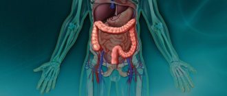
- presence of internal chronic diseases,
- bad ecology,
- use of low-quality cosmetics,
- cosmetic procedures leading to abrasions, cuts (tattoos, peeling),
- diseases of the neuroendocrine system,
- lymphostasis,
- mycosis.
The pathology is often detected in children with an unformed immune system, when an infectious agent enters the blood through scratches and cracks in the skin. Even a mosquito bite can cause bacteria to multiply. After scratching the affected area, an inflammatory reaction occurs, tissue swelling, itching, and irritation.
The most common pathogen identified in microbial eczema is β-hemolytic streptococcus. However, the development of microbial eczema can be associated with epidermal or Staphylococcus aureus, Proteus, Klebsiella, Neisseria gonorrhea or meningitis, Candida fungi and other pathogens. An underlying disease (varicose veins, lymphedema) significantly reduces the barrier function of the skin, and chronic exposure to microbial agents causes sensitization of the body and the occurrence of autoimmune reactions. Together, these processes lead to the development of microbial eczema.
Causes of seborrheic eczema
Seborrheic eczema is considered an infectious-allergic disease. This was confirmed by laboratory tests, which in some cases detected the following infectious agents: Pityrosporum ovale, Candida and even staphylococci. Predisposing factors for life activity and the development of infection are some conditions of the body:
- Increased work of the sebaceous glands
- Hormonal disorders, diseases associated with the production of hormones (diabetes, obesity, etc.)
- An organism weakened by long illnesses
- Diseases of the gastrointestinal tract (ulcers, gastritis)
- Depression, stress and other nervous disorders
- Weakened immunity (including AIDS) and many others
The exact cause of the appearance of seborrheic eczema in general is quite difficult to establish, but individually in each case considered, it is almost always possible to trace the source of its appearance. Since pathogens take root only in certain individuals, and some are not at all susceptible to skin diseases, even despite the presence of the above conditions. An exacerbation occurs in the winter season, with insufficient consumption of healthy foods by the body, which leads to a weakening of the immune system.
Back to contents
What does microbial eczema look like?
Infectious eczema has polymorphic clinical manifestations. Often develops against the background of bacterial-inflammatory or fungal skin lesions. Bacterial eczema ranks third among all infectious skin diseases. Typically, sites of chronic infection, atrophy, postoperative wounds, fistulas, and injuries are affected.
Manifestations depend on the state of local immunity and the protective properties of the skin. Large weeping erosions appear on the affected areas, which spread to healthy skin. Focal inflammatory changes with characteristic elements of a rash appear.
The rash is characterized by intense skin itching. The elements become covered with crusts, after which they are rejected leaving scars.
The epidermis is covered with papules, vesicles with transparent contents, as well as pustules filled with pus. The disease is characterized by abundant exudation (wetting). Inflammatory areas are surrounded by scalloped whitish edges. The epithelium around them becomes keratinized and rejected. The elements of the rash merge with each other.
Red, inflamed spots spread further and further from the entrance gates of the infection. The appearance of acute pain indicates damage to the nerve endings. Inflammation of the hair follicles is often observed, and furunculosis begins. Healthy skin becomes covered with “dropouts” (small papules with a tendency to suppurate).
Photos of eczema treatment
Treatment of thrombophlebitis - classification and symptoms of thrombophlebitis. Treatment options with drugs and traditional methods (110 photos)How to treat lichen - a review of the most effective drugs at home (115 photos and videos)
Wormwood medicinal properties - treatment and cleansing of the body with the help of wormwood. Tips for collecting, storing and preparing medicines (95 photos)
Read here! Hawthorn tinctures and properties: benefits, harm, instructions and contraindications for use (120 photos)
Please repost
0
Types of pathology
Eczema is classified according to location and symptoms:
- Varicose. The formation of weeping ulcers on the body, severe itching, swelling, inflammation due to venous insufficiency, varicose veins on the legs.
- Sycozyform type of eczema. Weeping, inflamed lesions appear under the armpits, on the chin, upper lip, and around the hair follicles.
- Post-traumatic. The pathogen penetrates through injuries, skin cuts, postoperative wounds, and the healing processes become protracted.
- Nummular. The appearance of round, inflamed plaques up to 3 cm in diameter, the formation of purulent crusts on the legs and arms with clear boundaries and a weeping surface.
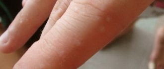
- Dyshidrotic. Papules and blistering rashes are localized on the fingers, palms, and soles of the feet. Provoking factors are impaired metabolism, weak immunity, predisposition to allergies, changes in the functions of the sweat glands.
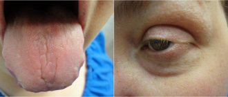
- Eczema of the nipples. Lesion of the breast in women, with thickening or cracking of the delicate skin. The reason may be insufficient hygiene during the lactation period.
The main symptom is the appearance on certain areas of the skin of weeping papules and vesicles with scalloped edges. They gradually increase in size, merge with each other, and turn into a large bleeding spot. Without treatment, the lesions will turn into weeping, erosive areas that quickly spread to healthy skin.
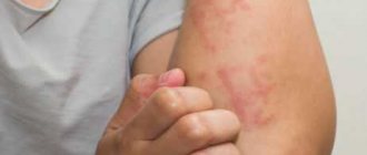
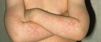
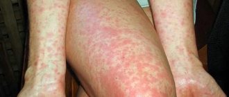
The main manifestations of eczema are severe itching, irritation, and peeling of the skin. Visual signs of the disease can be seen in the photo below.
Frequent localization
The most common location for an infectious rash is the legs. Microbial damage occurs in places of increased mechanical stress: on the feet, palms, knees and elbows. The location of the rash depends on the type of pathology.
The following types of microbial eczema are distinguished:
- Nummular. On the skin there are coin-shaped lesions (no more than 3 cm in diameter) with scalloped borders. The surface of the erosions is red with abundant exudate. Often covered with dry plugs. Microbial eczema appears on the arms, back and abdomen.
- Varicose. A prerequisite for development is varicose veins with vascular disorders. The entrance gate is a trophic ulcer. This type is characterized by a variety of rash elements, their clear edges, minor discomfort and dryness.
- Post-traumatic. Appears around areas of skin damage. Usually associated with immunodeficiency or decreased defenses.
- Sycosiform. The pathology is characterized by the appearance of exudative, single or connected itchy papules of a purple hue. Localized on the nasolabial triangle, armpits and groin.
- Nipple eczema occurs in women due to trauma to the areolas during breastfeeding. It can also occur in both sexes when scratching (scabies or skin mites, allergies) of the nipples. The areolas become covered with round lesions of a purple hue. The skin on the nipples is very delicate; secondary erosions and cracks quickly appear on it. This type of disease is characterized by severe itching.
Inadequate treatment of this type of eczema contributes to the appearance of new elements. Often, against the background of microbial inflammation, an allergic rash is observed, which can be of different types. Microbial widespread eczema is manifested by a spotty rash, blisters, ulcers and dense tubercles. Progression of the disease can lead to true eczema.
Consequences
The purulent-inflammatory process itself is not so terrible, but if the infection spreads into the deep layers of the epidermis and occupies large areas, the patient faces infection of internal organs. Lesions on the body can become multiple, difficult to treat, and have a severe course.
The disease is fraught with the following complications:
- shingles,
- eczema herpetiformis,
- cytomegalovirus infection,
- mononucleosis,
- chicken pox.
The advanced form and late treatment lead to the formation of unattractive scars and cicatrices in place of protracted ulcers.
With adequate treatment, the prognosis of the disease is favorable. However, the diagnosis is often made at an advanced stage. Foci of microbial infection cover a significant part of the body. Such lesions can lead to sepsis (blood poisoning). This condition is life threatening.
With incorrectly selected antibacterial therapy, the appearance of secondary elements is often noted. Purulent lesions can penetrate deep into the tissues, down to the infected periosteum and joints. Severe bone pain indicates osteomyelitis.
Eczematous microbial dermatitis in children is characterized by a high risk of complications. The child’s immunity is not able to withstand massive intoxication. Foci of microbial erosion increase in volume, sepsis is often inevitable.
Mechanism of development of eczema
Eczema is a disease that has several clearly defined stages of development. Each stage is characterized by its own symptoms and can occur at different times in different areas.
The beginning of the development of eczema is a symbiosis of that same “unfortunate coincidence of negative factors” and the presence of an allergen, which is a trigger for the development of the disease.
The initial stage (erythema) is characterized by redness of the skin at the location of the eczema. The temperature of the affected area of the skin rises and it begins to itch very much. The skin seems to swell. Not everyone can cope with severe itching and resist scratching the lesion. When scratching the lesion, the course of the disease worsens.
The next stage of eczema is papular . It is characterized by the appearance of small nodules (papules) with a pink-red color on the affected area of the skin. These nodules rise above the surface of the reddened skin. In appearance, the focus of the disease at the papular stage is an inflamed area of skin covered with a rash.
Following the papular stage, the vesicular stage of eczema . The resulting papules increase in size and after a few days they become small blisters filled with serous fluid (vesicles). Over time, these blisters enlarge, resulting in increased redness, swelling and itching. Some vesicles increase significantly in size, and the serous contents press on the epidermal cells with such force that the upper layers of the skin crack and the serous fluid pours out of the vesicle.
Next comes the pustular stage . Its peculiarity is the change in vesicles. The fact is that at the vesicular stage, not all vesicles can burst. Sometimes it happens that the vesicles do not burst at all. In this case, after some time the contents of the vesicles begin to darken. The serous fluid accumulated in them degenerates into purulent fluid. An abscess (pustule) forms in place of the vesicle. Sooner or later, the pustule must burst and then its purulent contents pour out.
Skin with burst pustules and vesicles is a truly unpleasant sight. The skin remains the same red and irritated. In place of the pustules, erosions form from which exudate constantly oozes. Hair growing on the damaged area of the skin either falls out or is glued together by the exudate produced. This stage of the disease is called the stage of weeping eczema . Damaged human skin at this stage is most susceptible to the risk of infection by all kinds of infections.
the crust stage begins . Redness goes away and itching is significantly reduced. The release of exudate is reduced. The affected skin begins to crust over. If purulent discharge is present, the crusts become yellowish in color and begin to peel off. The crust stage can be called the recovery stage. With normal access to oxygen, the skin is quickly restored, blood circulation is normalized, and the crusts quickly begin to be rejected. As a rule, the skin underneath is shiny, slightly different in color from normal skin and flaky.
The final stage of eczema is the scaly stage . At this stage, in place of the fallen off crusts, small scales form, which also fall off over time. If during the emergence and development of an eczema lesion part of the hairline was lost, then at the scaly stage the hair begins to grow again.
From the beginning of the development of the eczematous process to its end, 2-4 weeks pass. In rare cases, it may drag on for longer periods. If, after completing the scaly stage of eczema, eczema begins to develop again after some time, this is considered a relapse of the disease. Each new relapse causes significant damage to the health of the skin and can lead to significant irreversible changes in the structure of the skin.
Similar diseases and diagnosis
It is necessary to distinguish microbial eczema from the following diseases:
- streptoderma;
- candidiasis;
- trophic ulcers;
- psoriasis;
- atopic dermatitis;
- reticulosis;
- pemphigus;
- true eczema;
- allergic diathesis;
- basal cell skin cancer.
Microbial damage is characterized by secondary morbidity. To determine the pathogen and its sensitivity to antibiotics, culture of the fluid released from the plaques is carried out. They also take a scraping of the affected epithelium and examine it under a microscope. If cancer is suspected, a cell morphology study is performed.
When analyzing biopsy material, the doctor can detect swelling of the deep layers of the skin, excessive pigmentation of the epithelium, microscopic vesicles, and infiltration of lymphocytes with plasma cells.
Diagnosis of microbial eczema is not difficult in most cases. This is ensured due to the secondary nature of the disease. Damage to the skin due to trauma, varicose veins, streptoderma, candidiasis allows one to suspect the nature of the pathology.
However, it is not enough to determine the nature of the disease. It is necessary to understand which specific pathogen caused the development of symptoms. To establish the involvement of microorganisms, a technique is used to inoculate discharge from formations on the skin. The discharge is placed in a special medium, and the doctor monitors which colonies of microorganisms have sprouted. Antibiotic susceptibility testing can also be done at the time of culture.
If the case is considered complex, the doctor may recommend a histological examination. With its help, swelling of the dermis, the formation of blisters in the epidermis and a number of other characteristic changes in the skin.
An important stage of diagnosis is differentiation. It is necessary to distinguish eczema caused by different types of pathogens from each other, as well as from eczema caused by other factors, pemphigus, dermatitis and a number of other diseases.
To correctly classify a skin disease, in this case microbial eczema, you need to consult a dermatologist who can prescribe all the necessary examination options. Having studied the patient’s entire history with concomitant diseases, injuries and other changes, a medical specialist will be able to correctly make an accurate diagnosis.
To diagnose microbial eczema, skin cells are examined under a microscope for the presence of mycotic abnormalities. Bacteriological studies check a smear of discharge and a scraping from the affected skin for the sensitivity of pathogens to antibacterial drugs, as well as for the presence of pathogenic fungi.
If it is difficult to determine the true diagnosis, a histological examination of tissues/cells from a focal lesion of microbial eczema is prescribed. The material is also examined for:
- swelling of the dermis and intercellular space;
- skin hyperpigmentation;
- the process of formation of bubbles in the epidermal layer of the skin;
- the degree of infiltration into the lymphatic system in the presence of plasma cells.
An expanded diagnosis of microbial eczema is carried out in relation to other forms of eczema, other skin diseases, such as:
- psoriasis;
- dermatitis;
- skin reticulosis;
- benign pemphigus and others.
These symptoms develop at a particularly rapid rate and may appear in the first days:
- formation of red spots on the skin;
- the appearance of rash-like surfaces;
- bubble formation;
- death of the epidermis.
Symptoms characteristic throughout the course of bacterial eczema:
- skin lesions of various types begin;
- Mainly purulent areas, ulcers, and scratches are affected;
- the affected areas are sharply delineated;
- the top layer of skin cells dies;
- the center of the inflammatory process is covered with elevations, papules or vesicles filled with liquid;
- wet, purulent surfaces form;
- the outer layer of the epidermis is easily removed by touch;
- lesions are formed by small surfaces separated by small distances, which will decrease over time;
- Basically, the inflammation has a coin-shaped shape, but it also happens differently, the edges are uneven;
- the itching and burning are quite severe.
You should not self-medicate; you should consult a dermatologist for tests and therapy. First, the doctor will conduct an external examination of the skin, then refer you for instrumental and laboratory tests. Scrapings and washings from the affected areas will definitely be taken to study the biomaterial under a microscope.
It is important to identify the primary pathogen that led to hemorrhages and weeping ulcers. It could be mycosis, virus, streptococcus, staphylococcus. When the deep layers of the epidermis are affected, histology is performed: a biopsy is taken from the area of affected skin, the infiltrate and the antibodies produced are studied.
Particular attention is paid to differential diagnosis, since several types of bacterial eczema are known, with similar symptoms to psoriasis, dermatitis, and allergies.
To make an accurate diagnosis, patients are redirected for examination to specialized doctors - an endocrinologist, an allergist, an immunologist, and a nutritionist.
The secondary nature of eczema, its development against the background of varicose veins, streptoderma, candidiasis, areas of infection or trauma to the skin allow the dermatologist to suggest microbial eczema. To determine the pathogen and its sensitivity to antibacterial therapy, bacteriological culture of discharge or scraping from the area of skin lesion is carried out. If a fungal infection is suspected, a scraping is taken for pathogenic fungi.
In difficult diagnostic situations, a histological examination of a biopsy taken from a focus of microbial eczema can be performed. When examining the drug, swelling of the dermis, spongiosis, acanthosis, the formation of blisters in the epidermis, pronounced lymphoid infiltration with the presence of plasma cells are determined. Differential diagnosis of microbial eczema is carried out with other types of eczema, psoriasis, dermatitis, primary skin reticulosis, benign familial pemphigus, etc.
What is eczema and its pathogenesis
Eczema is a chronic disease caused by inflammatory processes in the epidermis and dermis and characterized by:
- a large number of different provoking factors;
- many variants of rash, the elements of which are simultaneously at different stages of their development;
- a frequent tendency to frequent relapses and an increase in the severity of the clinical course;
- highly resistant to many therapeutic methods.
The pathogenesis (mechanisms of occurrence and development) of the disease is based on:
- Polyvalent sensitization (reactive susceptibility) to allergenic factors. It is of predominant importance in the development of contact and microbial forms of eczema, which is confirmed by the positive results of skin allergy tests. In addition, such patients exhibit a relative deficiency of the immune system, resulting in a pathological chronic inflammatory response to microbial antigens.
- Increased sensitivity to central and/or peripheral neurogenic stimuli. The role of the central nervous system is confirmed by the symmetry of skin lesions, itching, the occurrence of relapses after psychological stress and emotional stress, and the positive effect after hypnotherapy sessions. The occurrence or recurrence of the disease after injury or other strong mechanical, temperature or chemical irritation, as a result of dysfunction of the digestive system, liver, kidneys, indicates the participation of the peripheral and autonomic nervous system in the mechanisms of disease development. This connection is realized according to the type of viscero-cutaneous reflexes, that is, along a reflex arc connecting the skin with the spinal cord and with internal organs (irritation of internal organs is accompanied by a skin reaction).
- Hereditary predisposition: the factors listed in the first and second paragraphs do not lead to a response from the body in the form of eczema in all people.
Is a person with infectious eczema contagious?
Microbial eczema is a disease caused by pathogenic microorganisms. Penetrating through damaged skin, they cause specific changes in it. It is not eczema itself that is contagious, but these microbes. But many of them constantly live on human skin, being its normal microflora.
However, contact with someone who is sick can be dangerous for young children, older people and pregnant women. People with weak immune status are also at risk. This category includes patients with primary or secondary immunodeficiency (AIDS). The risk group includes patients undergoing chemotherapy or immunosuppressive therapy.
With timely diagnosis, the disease responds well to treatment. It is important to verify the pathogen and conduct an antibiotic sensitivity test. Microbial eczema can be cured in 7-10 days, provided there are no complications.
Medicines
There are two approaches to treatment: systemic and local. Systemic therapy is used for severe disease. It must be prescribed to patients at risk.
Drugs for systemic therapy:
- antibiotics (cephalosporins, macrolides, fluoroquinolones);
- antiallergic drugs (Suprastin, Loratadine, Diazolin);
- sedatives (Glycine, valerian infusion, Adaptol).
Local medications must be prescribed. These include creams and ointments with an antibacterial component (Levomekol, Sintomycin, Metrogyl-gel). If a fungal infection is suspected, use antifungal ointments (Clotrimazole, Exoderil, Fucis-gel). To cure microbial eczema on the hands, after washing, wipe the skin with disposable wipes. After which ointments with a drying effect are applied. Ointments containing zinc (Baneocin) are used.
Traditional methods
Treatment with folk remedies is not an alternative. It is used only as an adjunct to drug treatment.
How to treat eczema on legs at home:
- Infusion of celandine. Pour 50 g of celandine herb into 1 liter of boiling water. Leave for 2-3 hours. Apply lotions with infusion to the affected areas. After half an hour, remove the gauze and apply a drying ointment.
- Pour 100 g of pine (spruce) needles into 1 liter of boiling water. Leave for 2-3 hours. Soak a sterile piece of gauze and apply it to the affected skin. After half an hour, apply antibiotic powder.
- Nettle decoction with the addition of propolis. Pour 50 g of nettle leaves into 1 liter of boiling water and cook over low heat for 30 minutes. Cool, add 1 tsp. propolis tinctures. Prepare baths. Keep the affected area of skin in the infusion for 40 minutes. Then get wet and apply antibiotic gel.
Other measures
Treatment of microbial eczema on the legs and arms requires an integrated approach. Magnetic therapy, ultraviolet irradiation, ozone and laser therapy are used.
Be sure to prescribe a gentle diet. All potential allergens are excluded from the diet. Patients should avoid trauma to the skin. Compliance with personal hygiene rules and timely treatment of diseases is a reliable prevention of microbial eczema.
Prognosis and prevention
Microbial eczema is an unpleasant pathology that is easier to prevent than to treat it later. To do this, it is recommended to promptly treat diseases that contribute to the development of skin infections (injuries, varicose veins, etc.), and observe the rules of personal hygiene. It is necessary to carefully care for skin wounds, especially if a person has a tendency to have a weakened immune system. Treatment of acute infectious diseases is another mandatory stage of prevention.
Infectious eczema requires careful monitoring to avoid becoming chronic. At the first changes, you should seek medical help.
The disease itself is not contagious, but elements of the rash can be a source of infection.
For prevention purposes, you need to:
- promptly disinfect and treat wounds, cuts, burns,
- prevent the development of chronic internal diseases,
- review your diet, eliminate harmful allergenic foods if you are prone to allergies,
- limit your intake of sweets,
- include dairy and plant foods in the diet,
- get rid of bad habits (alcohol, smoking),
- properly care for the affected areas, applying bandages to prevent bacterial infection.
If treatment for microbial eczema is started in a timely manner, the prognosis will be positive. The main thing is to achieve a reduction in the frequency of relapses in the future.
The prognosis of microbial eczema with adequate treatment is favorable. Prolonged and persistent course of eczema can be observed in weakened patients and elderly people. In the prevention of microbial eczema, the main importance is the identification and treatment of those diseases against which it can develop, the prevention of wound infection, and compliance with hygiene rules.
The predicted cure for microbial eczema is favorable with a properly selected set of therapeutic and dietary measures. Weakened and elderly patients have to undergo longer and more persistent treatment.
Preventive measures should consist of treating diseases that provoke microbial eczema, eliminating damaging factors, careful personal hygiene, and timely treatment of skin lesions with antiseptics.
Types of coin-shaped eczema
Coin-shaped eczema, the causes and treatment of which are described above, can occur in 3 main forms:
- Spicy. Various elements of the rash (nodules, blisters) are observed on the reddened skin.
- Subacute. Characteristic signs of this form are redness, thickening of the skin, crusting, and peeling.
- Chronic, in which there is weeping, increased skin pattern, change in skin color (pallor or, conversely, darkening), pain.
Symptoms
The area of skin lesions in microbial eczema is most often located in the lower extremities. It represents large foci of acute inflammatory changes in the skin with serous and purulent papules, blisters (vesicles), and weeping erosions located on them. The lesions are characterized by large scalloped edges. They merge with each other and have no areas of healthy skin separating them. The rash is usually accompanied by significant itching. Inflammatory foci of microbial eczema are covered with a large number of purulent crusts. They tend to grow peripherally and are surrounded by an area of sloughing stratum corneum. On apparently healthy skin around the affected area, individual pustules or areas of peeling are observed - screenings of microbial eczema.
Clinical dermatology distinguishes several types of microbial eczema: nummular, varicose, post-traumatic, sycosiform and nipple eczema.
- Coin-shaped eczema (nummular or plaque) is characterized by round lesions 1-3 cm in size with clear edges, a hyperemic and edematous weeping surface, covered with layers of serous-purulent crusts. The usual localization of coin-shaped eczema is the skin of the upper extremities.
- Varicose microbial eczema develops with varicose veins with symptoms of chronic venous insufficiency. Factors contributing to the occurrence of microbial eczema may be infection of a trophic ulcer, trauma to the skin in the area of varicose veins, or its maceration during dressings. This form of the disease is characterized by polymorphism of elements, clear boundaries of the focus of inflammation and moderate itching.
- Post-traumatic eczema develops around areas of trauma to the skin (wounds, abrasions, scratches). It may be associated with a decrease in the body's defense response and a slowdown in healing processes.
- Sycosiform microbial eczema in some cases can develop in patients with sycosis. This type of microbial eczema is characterized by weeping and itchy red lesions, which have a typical location for sycosis: beard, upper lip, armpits, pubic area. In this case, the inflammatory process often goes beyond the boundaries of hair growth.
- Nipple eczema occurs in women with frequent trauma to the nipples during breastfeeding or with constant scratching in patients with scabies. In the area of the nipples, bright, clearly demarcated red lesions with weeping and cracks form. Their surface is covered with crusts. There is severe itching. Nipple eczema, as a rule, is characterized by a persistent process.
Microbial eczema is manifested by symptoms such as a sharp onset against the background of an existing skin disease, manifested by bright redness, pinhead-sized blisters that burst, releasing clear liquid onto the eroded surface of the skin. This is the stage of weeping, which can last a long time with varying intensity.
Microbial eczema has sharply limited asymmetrical rounded shapes of large lesions with serous-purulent crusts that are susceptible to rejection, most often on the arms and legs. Under the layering of such crusts, weeping erosive skin lesions with severe suppuration are found. After cleansing the affected area from the cortical masses, the absence of the epidermal layer of the skin becomes visible, as if it were a varnished red wound, prone to bleeding.
Bacterial eczema has a fairly pronounced clinical picture. Large lesions can be noted on the skin, where there is active rejection of the stratum corneum of the epithelium.
The inflamed areas are affected by blisters containing an unpleasant serous fluid. Burst of the vesicle leads to the formation of a painful, itchy, weeping type erosion. The lesions are usually located asymmetrically and can merge with each other.
Doctors distinguish several subtypes of the disease:
- coin-shaped, characterized mainly by damage to the hands, easily spreading throughout the body without timely treatment, difficult to respond to therapy;
- varicose veins develop in the lower extremities and often act as a precursor to trophic ulcers with a constant lack of venous blood flow;
- paratraumatic – a consequence of infections affecting skin injuries, postoperative wounds, etc.;
- mycotic occurs on the feet and palms due to fungal contamination;
- sycosiform, characterized by damage to the hairy areas of the body if the skin is colonized by group A streptococcus;
- Nipple eczema develops when women breastfeed their children and do not properly care for the nipple area.
Eczema is as common in adults as in children. Moreover, adults with strong immunity should not be afraid of infection, but a small child can catch an infection from a sick person.
If a child develops eczema, it is recommended that you do not hesitate to seek medical help. The pathological process can greatly reduce the quality of life of young patients and requires thoughtful correction.
Which doctor treats microbial eczema
Infected eczema is a serious skin disease that causes not only physical but also mental discomfort.
Patients are advised to visit a doctor specializing in dermatology at the first sign of illness. The doctor will examine the skin, prescribe tests, and based on their results, make recommendations regarding therapy. Doctors specializing in infectious diseases, immunology, and allergology can also take part in the treatment of microbial eczema. In some cases (for example, with gonorrheal skin lesions), the help of a venereologist may be needed.