A child may successfully master one group of skills and lag behind in mastering another. Such unevenness in development may be the result of mother's upbringing. One mother believes that the main thing is to develop self-service skills in the baby, and there is no point in chatting; such a child may lag behind in speech development, but will learn to feed himself early. Another mother does everything for the child, but communicates and plays with him a lot, and accordingly, his speech develops early.
So, having a clear idea of the baby’s neuropsychic development, you can develop a training program. And we see the main task as making it easier for you and the specialist to choose the area of educational activity that you need to pay attention to first.
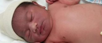
Already in the neonatal period, you can assess the state of the baby’s nervous system.
An important indicator of the maturation of a child’s nervous system is a group of unconditioned reflexes . From a fairly large number of reflexes, we have identified a main group that can give the most complete description of the state of the nervous system from the first hours of a child’s life. From the point of view of convenience of examination, we present the reflexes according to the positions (reflexes in the position on the stomach, back, in an upright position) in which they are examined. Unconditioned reflexes are labile and easily depleted, so it is better to carry out the examination 2 hours after feeding, when the child is in a calm state.
Moro reflex (grasp reflex)
More often it is caused in various ways: by hitting the changing table 20-30 cm from the child’s head or by quickly straightening bent legs, as well as raising the child’s pelvis and legs above the bed. In response, the following occurs: spreading the arms (the first phase of the reflex) and bringing them together with a tendency to clasp the body (phase 2). Sometimes during the first week of a child's life only the first phase is observed. The later appearance of the second phase does not indicate pathology.
Normally, the reflex disappears by 4-5 months. The absence of the Moro reflex or the persistent disappearance of the second phase is a suspicion of damage to the vestibular nerve. A delay in the reflex is observed in children born with asphyxia, and a decrease in newborns with birth trauma. The revival of the Moro reflex and its spontaneous appearance indicate increased neuro-reflex excitability and convulsive readiness. Asymmetry of the reflex indicates hemiparesis. Asymmetry and even disappearance of the reflex indicate Erb's palsy.
Content
- 1 Spinal motor automatisms 1.1 Protective reflex of the newborn
- 1.2 Support reflex and automatic gait of newborns
- 1.3 Crawling reflex (Bauer) and spontaneous crawling
- 1.4 Grasp reflex
- 1.5 Galant reflex
- 1.6 Perez reflex
- 1.7 Moro reflex
- 2.1 Sucking reflex
Support reflex
Consists of 2 phases:
phase - if you take a newborn baby under the arms, he reflexively bends his legs at the hip and knee joints;
phase - placed against a support, he straightens his legs and firmly rests his entire foot on the surface of the table.
Normally, the reflex lasts up to 1.5-2 months. It is absent in paresis and paralysis of the legs. Relying on your toes, especially with crossed toes, gives reason to suspect spastic diplegia, which is characterized by a sharp increase in the tone of the leg muscles (especially the adductor muscles of the thighs and shin flexors); muscle strength and range of motion are reduced.
Babkin's palmo-oral reflex
This funny reflex is triggered like this: you need to lightly press your thumb on the baby's palm, and in response the baby turns his head and opens his mouth. After two months this reflex decreases, and by three months it disappears completely.
The palmar-oral reflex is usually well expressed and constant. It decreases with some damage to the nervous system, especially with birth injury to the cervical spinal cord.
Grasp reflex
In response to touching the palm, the fingers bend and the object is grasped into a fist.
Before feeding and during eating, the grasping reflex is much more pronounced. Normally, this reflex is well evoked in all newborns.
A decrease in the grasping reflex is most often observed on the side of the lesion of the cervical spinal cord.
Robinson reflex
Sometimes, when this reflex is evoked, the child grabs the object or the doctor’s finger so tightly that such a clinging child can be lifted up by the finger. Thus, it turns out that a newborn, outwardly seeming to be a completely helpless creature, can develop such “muscular strength” in his hands that keeps his body suspended.
Normally, the Robinson reflex should be considered mandatory in all newborns. By 3-4 months of life, on the basis of this unconditioned reflex, a purposeful grasping of the toy is formed, and the good expression of this reflex subsequently contributes to the more rapid development of fine manual skill.
Inferior grasp reflex
This reflex is caused by light pressure from the tips of the fingers on the front of the newborn's sole, in response to which the child flexes his toes. In healthy children, this reflex persists until 12-14 months of life.
The inability to evoke this reflex occurs when the spinal cord is damaged at the lumbar level.
Moro reach reflex
This reflex is triggered like this: if you unexpectedly clap with both hands on both sides near a lying child, he will spread his arms half-bent at the elbows and spread his fingers, and then the arms will move in the opposite direction.
Normally, the Moro reflex lasts up to 3-4 months. In all healthy newborns, the Moro reflex is evoked quite well and always equally in both hands. With flaccid paresis of the arm, the reflex is reduced or completely absent on the affected side, which indicates that the spinal cord in the cervical region was injured during childbirth.
Perez reflex
In order to evoke this reflex, the doctor places the child face down on his palm. Then, pressing lightly, he runs his finger along the child’s spine from bottom to top from the tailbone to the neck. In response to this, the spine bends, the arms and legs extend, and the head rises. Testing this reflex gives the doctor information about the functioning of the spinal cord along its entire length.
In newborns with birth damage to the cervical spinal cord, there is no raising of the head, that is, the Perez reflex appears to be “decapitated.”
Support reflex
The support reflex is very important for assessing the state of the central nervous system of the newborn. Normally, the reflex looks like this: if you take a newborn under the arms, he reflexively bends his legs at the hip and knee joints. At the same time, if he is placed against a support, he straightens his legs and firmly rests his entire foot on the surface of the table and “stands” like this for up to 10 seconds.
Normally, the support reflex is constant, well expressed and gradually disappears by 4-5 weeks of age. When the nervous system is injured, the child may lean on his toes, sometimes even with his legs crossed, which indicates damage to the motor (pyramidal) pathway running from the cerebral cortex to the spinal cord.
Automatic walking reflex or stepping reflex
When resting on the feet and slightly tilting the body forward, the child makes stepping movements. This reflex is normally well evoked in all newborns and disappears by 2 months of life.
Assessing the automatic walking reflex is very important for the doctor, as it helps to identify the location of damage to the nervous system and its degree.
Alarming signs are the absence of an automatic walking reflex or walking on tiptoes with legs crossed.
Bauer crawling reflex
This reflex is caused as follows: a hand is placed on the feet of a newborn, laid on his stomach, in response to this the child begins to perform crawling movements. This reflex is normally evoked in all newborns and lasts up to 4 months, and then fades away. Assessment of the reflex is of great diagnostic importance for the doctor.
Defense reflex
The essence of the reflex is that a newborn laid on his stomach quickly turns his head to the side and tries to lift it, as if providing himself with the opportunity to breathe.
This reflex is expressed from the first day of life in all healthy newborns without exception. A decrease or disappearance of this reflex can occur either with particularly severe damage to the upper cervical segments of the spinal cord, or with pathology of the brain.
Assessing the protective reflex will help the doctor timely identify pathology of the nervous system in a newborn.
14 stages of speech formation. The role of the environment, care and education in the neuropsychic development of a child
From the moment of birth, the child develops vocal reactions: screaming and crying. True, they are still very far from the sounds of human speech. However, both screaming and crying contribute to the development of subtle and varied movements of the three sections of the speech apparatus: respiratory, vocal, articulatory. After two weeks, you can already notice that the child begins to respond to the speaker’s voice: he stops crying, listens when he is addressed. By the end of the first month, he can already be calmed down with a melodic song (lullaby). Next, he begins to turn his head towards the speaker or follow him with his eyes. Soon the baby already reacts to intonation: to a gentle one he becomes animated, to a harsh one he cries. About 2 months humming appears and by the beginning of the 3rd month - babbling (agu-huh, cha-cha, ba-ba, etc.). Babbling is a combination of sounds that are vaguely articulated. From 5 months the child hears sounds, sees articulatory movements of the lips of others and tries to imitate. Repeated repetition of a specific movement leads to consolidation of a motor skill. From 6 months The child pronounces individual syllables by imitation (ma-ma-ma, ba-ba-ba, cha-cha-cha, pa-pa-pa, etc.). Subsequently, through imitation, the child gradually adopts all the elements of spoken speech: not only phonemes, but also tone, tempo, rhythm, melody, intonation. In the second half of the year, the baby perceives certain sound combinations and associates them with objects or actions (tick-tock, give-give, bang). But at this time he still reacts to the entire complex of influences: the situation, intonation and words. All this helps the formation of temporary connections (memorizing words and reacting to them). At the age of 7-9 months. the child begins to repeat after the adult, more and more diverse combinations of sounds. From 10-11 months. reactions to the words themselves appear (regardless of the situation and intonation of the speaker). At this time, the conditions in which the child’s speech is formed (correct speech of others, imitation of adults, etc.) become especially important. By the end of the first year of life, the first words appear.
15 comprehensive assessment of the neuropsychological development of children
The main feature of assessing neurological status is a comprehensive assessment of behavioral and neurological signs. Assess the newborn's position, the baby's response on examination, and reflexes (Moro, grasping reflex, sucking reflex). Unconditioned reflexes are involuntary movements or actions that help us determine the state of the newborn’s nervous system. Some reflexes can only be evoked during a certain period of a child's development. The following reflexes are evoked only during the newborn period, and are also evoked symmetrically.
Search reflex.
Caused by touching the corner of a newborn's mouth.
He turns his head in the direction of the touch and opens his mouth - he begins to “search”. The search reflex helps the baby search for the breast. Sucking reflex.
The newborn tightly grasps the object placed between the lips. Grabbing the mother's nipple between her lips and holding it in the mouth, she simultaneously produces rhythmic sucking movements with her lips. The sucking reflex appears at 32 weeks of gestation and is fully developed after 36 weeks of gestational age.
Moro reflex.
Can be triggered from birth and is called the startle reflex. It disappears by 4-5 months after birth. Caused by a blow to the changing table or any sound or vibration stimulus. When hearing sharp sounds, the newborn closes his eyes, sometimes shudders and spreads his arms (the first phase of the Moro reflex) and straightens his legs, which were previously bent and pressed to the stomach. The second phase (clonic) is the abduction of the arms with a tendency to clasp the torso.
Robinson reflex (palmar grasp).
When touching the palm surface of a newborn, he closes his palm and squeezes his fingers - “grabs.” The reflex is more pronounced in premature infants.
Crawl on Bauer.
In the position on the stomach, the child reflexively pushes off with his legs from the palm placed on the soles. These reflexes disappear by the end of the first month of life.
Kernig reflex.
Inability to fully straighten the leg at the knee joint if it is bent at a right angle at the hip joint.
Babinski reflex.
When performing a stroke movement along the outer edge of the foot from the heel to the toes, plantar flexion of all fingers occurs - a negative symptom, or dorsal extension of the big toe while plantar flexion of the rest - a positive symptom.
These reflexes disappear in the first year of a child's life. The extinction of unconditioned reflexes in newborns indicates significant changes in the central nervous system. In addition, convulsions, severe muscle hypotonia, motor restlessness or adynamia, tension in the large fontanelle, and respiratory rhythm disorder also indicate changes in the central nervous system in children of this age.
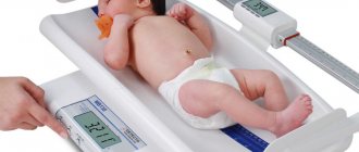
In infants, facial expression, body position, nature and range of movements, range of motor-static skills, and state of consciousness are assessed. A 1-month-old child fixes his gaze on an object and follows it. At 2 months, he holds his head, babbles, and laughs. At 3 months, he clumsily grabs objects, distinguishes the faces of close people, hums, and rolls over from his tummy to his back. At 4 months he picks up objects and plays with his hands. At 6 months, sits independently, grabs moving objects and throws them. At 8 months, he stands with support, crawls, examines a toy, moving it from hand to hand, and pronounces consonants. At 10 months he speaks individual words, stands up on his own, knows the meaning of individual words, and waves his hand. At 12 months he walks, has a vocabulary of 4-8 words, and knows the meaning of the word “impossible.”
The timing of development of motor-static skills and especially speech can fluctuate in normal healthy children depending on individual characteristics, characteristics of upbringing and regime. Therefore, when assessing neuropsychic development, it is necessary to focus on the entire complex: supporting signs and one cannot make a conclusion based on one or two signs
16 morphological and functional features of the skin in children
ANATOMICAL AND PHYSIOLOGICAL FEATURES OF THE SKIN IN CHILDREN.
The skin of a child, like that of an adult, consists of the epidermis, dermis (the skin itself) and hypodermis (subcutaneous tissue). However, in its morphological and functional characteristics it is distinguished by significant originality, especially in young children.
The epidermis has a very thin stratum corneum, consisting of 2-3 rows of loosely interconnected and exfoliating cells, and an actively growing main layer. The main membrane separating the epidermis and dermis in young children is underdeveloped and loose, as a result of which, in pathology, the epidermis can be separated from the dermis in layers (desquamative erythroderma). Children's skin is especially characterized by good blood supply associated with a dense network of wide capillaries, which gives the skin a bright pink, then soft pink color.
The dermis consists of papillary and reticular layers, in which elastic, connective tissue and muscle elements are poorly developed.
The baby's sebaceous glands function well already in utero, forming a cheesy lubricant that covers his body at birth. In newborns and children of the 1st year of life, yellowish-white dots are noticeable on the facial skin - excessive accumulation of secretions in the sebaceous glands of the skin. In children predisposed to exudative diathesis, a thin, so-called milky crust forms on the cheeks, and gneiss (oily seborrhea) forms on the scalp.
Sweat glands in newborns are formed, but during the first 3-4 months of life, some functional insufficiency is detected, which is associated with the imperfection of the thermoregulation center.
The hair on the head of newborn children is fully developed, but does not have a core, and is replaced several times in the 1st year of life. The skin on the back and shoulders is covered with fluff, which is more pronounced in premature babies. Eyebrows and eyelashes are weakly expressed, their growth increases in the 1st year, and by 3-5 years of life they become like those of adults. Nails are usually well developed and extend to the fingertips in full-term newborns.
Subcutaneous fatty tissue begins to form in the 5th month of intrauterine life, but is intensively deposited during the last 1.5-2 months of intrauterine life. In a full-term newborn, subcutaneous fatty tissue is well expressed on the cheeks, thighs, legs, forearms and weakly on the abdomen, and during the first 6 months of life it develops intensively on the face, limbs, and torso. Later, up to the age of 8, fluctuations occur in the formation of the fat layer, and then its growth begins again, more pronounced in girls. In young children, subcutaneous fat makes up about 12% of body weight, in adults this figure is more than 8%.
The composition of subcutaneous fatty tissue in children of different ages is different: in young children it contains more solid fatty acids (palmitic and stearic) and less liquid oleic acid, which causes denser tissue turgor in children 1 year of age, more a high melting point of fat and a tendency to form local compactions and swelling of the skin and subcutaneous tissue with the formation of sclerema and scleredema.
A peculiarity of young children is the presence of accumulations of brown adipose tissue in the posterior cervical region, supraileocecal zone, around the kidneys, in the interscapular space, around the great vessels. In a full-term newborn, its amount is about 1-3% of the total body weight. It provides a higher level of heat production due to the so-called non-contractile thermogenesis (not associated with muscle contraction).
17 thickness and distribution of the subcutaneous fat layer in children. Chemical composition of fat. The concept of normotrophy, paratrophy, hypotrophy
Normotrophy
State of normal nutrition - normotrophy
(eutrophy).
Indicators of weight and height correspond to age norms in normotrophy, development is proportional, the skin is clean, velvety, of normal color, mucous membranes are pink, muscle tone, skin elasticity, tissue turgor are preserved, the skeleton is correctly developed, appetite is preserved, physiological functions are normal, there are no pathological changes from the internal organs. Also, normotrophy
is accompanied by good resistance to infection, the child’s neuropsychic development corresponds to his age, and a positive emotional mood is observed.
Paratrophy is a chronic nutritional disorder in a child with a weight close to normal for his age and body length.
Hypotrophy (Greek υποτροφια, Lat. hypotrophia
) is an eating disorder characterized by varying degrees of underweight.
Development of malnutrition
§ With grade I malnutrition, the thickness of the subcutaneous tissue decreases in all parts of the body except the face. First of all, it thins on the stomach. Weight deficit is 11-20%
§ With II degree malnutrition, the loss of body weight is 25-30%. The child is lagging behind in growth and neuropsychic development. The subcutaneous layer is preserved only on the face; it is especially thin on the abdomen and limbs
§ With grade III malnutrition, body weight loss is more than 30%. Externally - extreme degree of exhaustion, the skin is pale gray in color, the subcutaneous fat layer is completely absent
18 morphological features of muscle structure and their development in children of different ages
There are about 600 skeletal muscles in the human body. The muscular system makes up a significant part of the total human body weight. If in newborns the mass of all muscles is 23% of body weight, and at 8 years old - 27%, then at 17-18 years old it reaches 43-44%, and in athletes with well-developed muscles - even 50%. Individual muscle groups grow unevenly. In infants, the abdominal muscles first develop, and later the chewing muscles. By the end of the first year of life, in connection with crawling and the beginning of walking, the muscles of the back and limbs noticeably grow. Over the entire period of child growth, muscle mass increases 35 times. During puberty (12-16 years), along with the lengthening of the tubular bones, the muscle tendons also intensively lengthen. The muscles at this time become long and thin, and adolescents look long-legged and long-armed. At 15-18 years of age, further growth in muscle diameter continues. Muscle development continues until the age of 25-30. A child's muscles are paler, softer and more elastic than the muscles of an adult.
19 features of embryonic bone tissue, epi- and endochondral ossification. Stages of Bone Formation
Bones develop in place of less specialized connective tissue in two ways. The first is due to the activity of osteoblasts capable of producing the protein substance ossein and mineral salts. Some bones are formed on the basis of membranous connective tissue and have a direct path of ossification, including two stages of development - membranous and osseous (primary bones). Other bones are formed in place of the cartilaginous models (secondary bones). This developmental specificity reflects the phylogenetic feature that is found in many animals that have a supporting skeleton made of connective tissue, others have a cartilaginous skeleton, and others have a bone skeleton. In embryogenesis, bone tissue appears at the 7th - 8th week of intrauterine development, when the embryo already has all other types of tissue. Formation of bone from membranous connective tissue
.
The formation of membranous (primary) bones (covering bones of the skull, all facial bones and clavicles) begins with the grouping of mesenchymal cells near small blood vessels. As a result of the activity of these cells, fibrous fibers are formed along the long axis of the bone, which are impregnated with osseomucoid (a substance resembling collagen). The fibrous substance serves as the basis for bone development. Around the fibers are bone cells (osteoblasts), which produce minerals to nourish the fibrous fibers. During development, osteoblasts gradually become embedded in bone tissue, turning into osteocytes. Bone plates are formed around the blood vessels, inserted into one another in the form of cylinders. The outer layer of primary bone develops into periosteum, which forms the outer common lamellae, allowing the bone to grow in thickness. Development of bone in place of cartilage
.
Bone development in the cartilaginous model (indirect ossification) occurs with the formation of a bone point inside (enchondral) and outside (perichondral) of the cartilage. Enchondral bone formation
. The development of bone in place of cartilage (secondary bones) is a more complex process than its development in place of connective tissue, and is associated with the restructuring of the cartilaginous anlage. Cartilage cells, which previously secreted the main substance of cartilage, destroy cartilage by secreting special enzymes in the 7th - 8th week of embryonic development. Blood vessels penetrate these cavities, accompanied by osteoblasts (Fig. 34). Osteoblasts surround blood vessels in dense rows. Initially, undestroyed strands of cartilage remain between them. Subsequently, the fibers are impregnated with mineral salts and the bone crossbars of the spongy substance are formed.
20 Features of phosphorus-calcium metabolism in children, its regulation. Normal levels of Ca and P in the blood.
The adult human body contains on average about 1-2 kg of calcium, of which more than 98% is found in skeletal bones. The mineral phase calcium on the surface of the crystals is in equilibrium with ions in the extracellular fluid, but only a small portion of the total calcium (approximately 0.5%) is exchanged. The processes of bone resorption and formation are closely related; skeletal bones absorb and release approximately 0.5 mg of calcium every day. Calcium intake is provided mainly through dairy products.
In children during periods of rapid growth, in women during pregnancy and lactation, calcium absorption increases, and with age it decreases by half. With a low calcium content in food (less than 500 mg per day), the absorption of more than 30–40% of calcium is required to maintain a positive calcium balance. Only in this case will absorption in the intestines be sufficient to compensate for losses through secretion by the gastrointestinal tract and excretion by the kidneys.
The calcium content in the body in children is about 200 mmol/l
Participates in physiological processes only in ionized form, an essential participant in the process of muscle contraction, an essential component of the blood coagulation system, is part of bones and cartilage in the form of apatites, is a stabilizer of cell membranes, and regulates the excitability of nerves and muscles. Calcium ionization depends on blood pH.
Phosphorus. About 70% of phosphorus is concentrated in bone tissue; it is part of the intercellular fluid and active biochemical compounds of every cell of the body. The phosphate requirement of an adult is about 1200 mg/day. Plays a key role in metabolic processes, being part of many coenzymes, nucleic acids and phosphoproteins.
21 structural features of the spine and chest in children. Pathological deformities
The spine is a structure of the body that is formed in the second week of intrauterine development of the fetus.
The neural tube (future spinal cord), developing from the ectoderm, provokes the formation of the notochord from the mesodermal layer. The notochord primordium is formed in the center of the embryo and then grows caudally (towards the tail, towards the legs) and cranially (towards the head). Subsequently, the chord acquires a segmental structure, and primary vertebrae are formed from it. By the fourth month of embryonic development, the cartilaginous model of the vertebra is completely consistent with that after the birth of the child.
In the second month of intrauterine development, primary islands of ossification and the structure of the spine arise in the thickness of the cartilaginous plates of the vertebrae. Ossification nuclei first arise in the cranial vertebrae, and only then does this process affect the underlying vertebrae. Initially, there are six ossification nuclei, after which they merge in pairs, forming three common nuclei. By the time of birth, any cartilaginous vertebra has two ossification nuclei in the vertebral arches and one ossification nucleus in the body.
There are three periods in the postnatal (postpartum) development of the child’s spine. The first period is characterized by a set of processes that contribute to an increase in the mass of the vertebral bodies and the complete replacement of vertebral cartilaginous tissue with bone. This period begins from the moment the child is born and lasts until 6–7 years. Already in the first year of life, the nuclei of the arches fuse with each other and the growth zones between the vertebral bodies and arches close. Fusion occurs first at the level of the thoracic vertebrae and gradually spreads up and down. The exceptions are the arches of the fifth lumbar vertebra and the sacrum. The arches of the fifth lumbar vertebra usually fuse only by the age of six years. The arches of the sacral vertebrae fuse by the age of three to eight years, and the vertebral bodies by the age of 14–16.
After the birth of a child, his spinal column is almost straight. Cervical lordosis develops in children when they begin to hold their head up. Thoracic kyphosis develops in a child by six months, when he begins to sit independently. Lumbar lordosis develops from the moment the child begins to gradually stand up. Sacral kyphosis appears after the child begins to walk and develops until the age of 6–8 years.
The next period of development of the spinal column occurs between the ages of six to eight and 12–14 years. During this period, in addition to the increase in size and formation of vertebral elements, ossification nuclei appear in the thickness of the epiphyseal zones (growth zones). They grow, covering the entire perimeter of the marginal zones of the vertebral bodies. Their complete fusion occurs by 14–15 years.
The third period of spinal development occurs between the ages of 14–16 and 21–25 years. This period is characterized by the formation of ossification nuclei in the apophyseal zones of the vertebral arches and processes. The final synostosis of the apophyses contributes to the formation of a fully formed vertebra, characteristic of an adult.
Kyphosis
(ancient Greek κύφος bent, humpbacked) - in general cases it represents a curvature of the upper spine. It can be either acquired or hereditary.
There are physiological kyphosis, which is observed normally in adults (thoracic and sacral), and pathological kyphosis, which develops as a result of diseases (for example, rickets, tuberculous lesions of one or more vertebrae), spinal trauma and postural disorders.
Lordosis (Greek λορδός - bent, stooped) is a curvature of the spine, convexity facing forward.
Scoliosis (Greek σκολιός - “crooked”, lat. scoliōsis
) - persistent lateral deviation of the spine from the normal straightened position.[
22 semiotics of lesions of the musculoskeletal system in rickets
Bone changes are especially pronounced: an increase in the size of the large fontanelle, pliability of its edges, sutures, costal “rosaries”, “bracelets” on the hands.
The head takes on a square shape, the bridge of the nose is sunken, the arch of the hard palate is gothic, the bite is incorrect, there is a deformation of the chest (“chicken breast” and “shoemaker’s chest”), “strings of pearls”, curvature of the humerus bones, flattening of the pelvis and narrowing of the entrance to the pelvis, curvature of the legs and spine (kyphosis, scoliosis, lordosis) as a result of bone tissue damage. Hypotonia of the muscles and ligaments increases, which leads to loose joints, the appearance of a “frog” abdomen, and flat feet.
23 order and timing of teething (deciduous and permanent). Timing of closure of fontanelles and cranial sutures. Their clinical significance. Definition of “bone age” as one of the indicators of biological age.
Defense reflex
A newborn laid on his stomach turns his head to the side and tries to lift it.
Normally, after 1-1.5 months, the child tries to hold his head up on his own. In children with damage to the nervous system, the protective reflex is delayed and sometimes does not appear, for example, in a child with a natal injury to the cervical spinal cord. In children at risk for developing cerebral palsy, there may be prolonged elevation of the head and even throwing it back (due to increased tone of the neck extensors).
Weakening and strengthening of reflexes
When conducting a study of reflexes, it is necessary to take into account that they can be used as a diagnosis only if other symptoms exist. That is, a change in reflexes in the absence of other neurological disorders in the baby is not a pathology. There are a number of problems that are indicated by the strengthening or weakening of certain reflexes.
- An increase in neuromuscular excitability can cause a slight increase in the main innate reflexes.
- Pathology of the nervous system, impaired muscle tone, infectious and inflammatory diseases can cause a complete absence of reflexes or their sudden suppression.
It is worth specially drawing the attention of parents to the fact that reflexes in a healthy baby should have a symmetrical appearance. In other words, they must be the same on both sides. This can be checked by studying the grasping reflex. If the baby grabs your finger tightly with one hand and weakly with the other, this is a reason to show the baby to a specialist. Since this situation may be an asymmetry of reflexes.
Therefore, if you notice any deviations in your child’s reflexes, do not put off visiting a specialist. Only he can adequately assess the situation and prescribe certain treatment or gymnastics.
Doctors often recommend inducing innate reflexes, which are a kind of gymnastics for the baby. These include the support reflex, crawling and others.
Perez reflex
It is caused by applying line irritation along the spinous processes from the coccyx to the neck. In response, the child’s legs are simultaneously bent and brought toward the stomach, the back is arched, the anal sphincter often opens, and urination may occur. If you induce the reflex with the child face down on the doctor’s palm, the response is a sharp cry, extension of the arms, legs, head, protrusion of the anus, and urination.
Normally, the reflex is evoked up to 3-4 months. It's better to check it last. Its suppression during the neonatal period or delayed reverse development is observed in children with damage to the central nervous system.
Based on materials from the book by O.V. Goncharova.
"Baby Care Benefit"
Oral segmental automatisms
Sucking reflex
When the index finger is inserted 3-4 cm into the mouth, the child makes rhythmic sucking movements. The reflex is unconditional and is absent in cases of paresis of the facial nerves, severe mental retardation, and severe somatic conditions. The sucking reflex in human children usually subsides between three and four years of age, which explains why in many cultures breastfeeding lasts until the age of three or four years, i.e. up to the age at which the child suckles on his own. US anthropologist Professor Katherine A. Dettwiler came to the conclusion that the need for sucking, i.e. The natural period of breastfeeding (expected by our children) can last from 2.5 to 7.0 years[1].
Search reflex (Kussmaul reflex)
When stroking the corner of the mouth, the lip lowers, the tongue deviates, and the head turns toward the stimulus. Pressing on the middle of the upper lip causes the mouth to open and the head to straighten. When you press on the middle of the lower lip, the lower jaw drops and the head bends. This reflex is especially pronounced 30 minutes before feeding. Pay attention to the symmetry of the reflex on both sides. The search reflex is observed up to 3-4 months, then fades away. Reflex asymmetry—unilateral paresis of the facial nerve. There is no reflex - bilateral paresis of the facial nerve, damage to the central nervous system.
Proboscis reflex
A quick tap on the lips with a finger causes the lips to stretch forward. This reflex lasts up to 2-3 months.
Palm-oral reflex (Babkin reflex)
Play media file
Video showing the Babkin reflex.
When pressing with the thumb on the area of the newborn's palm (both palms at the same time), closer to the thenar, the mouth opens and the head bends. The reflex is clearly pronounced in newborns. Sluggishness of the reflex, rapid exhaustion or absence indicate damage to the central nervous system. The reflex may be absent on the affected side with peripheral paresis of the arm. After 2 months it fades away by 3 months. disappears.