The process of giving birth to a child is a difficult physical test not only for the female body, but also for the child. At birth, the baby is capable of injury. The reasons may be either too strong mechanical influence of labor forces or obstetric manipulations - for example, the use of forceps if complications arise during childbirth. Trauma during childbirth is a fairly common occurrence among newborns. The soft tissues of the scalp with hair are most susceptible to damage. Such injuries can be abrasions or cephalohematomas - hemorrhages under the periosteum.
Causes of cephalohematomas
Injury to the head in newborns most often occurs during the passage of the fetus through the maternal birth canal. Usually everything happens spontaneously, in situations that are conducive to injury. As a result, when the skin on the head moves, the periosteum, the connective tissue that covers the flat bones of the skull, moves along with it. During this, the vessels are torn and damaged, and the space between the cranial bones and the periosteum is filled with blood, the amount of which is 5-150 ml.
There are a number of factors that increase the likelihood of injury. Cephalohematoma in newborns can occur when:
- fast, rapid labor;
- incorrect position of the fetus;
- large fruit;
- discrepancy between the size of the child’s head and the female pelvis;
- giving birth before the due date;
- protracted labor;
- breech presentation of the fetus;
- umbilical cord entanglement;
- incorrect insertion of the head;
- using forceps or a vacuum extractor.
There is a risk of the child receiving a birth injury even in a situation where childbirth proceeds normally. The causes of such injuries as cephalohematoma can also be the condition of the fetus and the course of pregnancy. For example, birth injuries are also likely in the case of:
- chronic or acute fetal hypoxia;
- prematurity;
- intrauterine infection;
- disorders of nutrition and fetal growth.
Cephalohematoma in newborns and its consequences
Ossification of cephalohematoma
7 days after the occurrence of a cephalohematoma, it may be replaced by dense connective tissue, resulting in bone deformation. The defect is only cosmetic and does not pose any harm to health. When the child grows up, the defect will not even be noticeable.
Anemia
If the cephalohematoma is large enough, the baby may experience posthemorrhagic anemia. Since the child's circulating blood volume is small, hemorrhage can cause changes in the peripheral blood.
Usually the anemia is mild and does not require additional treatment. Without continued bleeding, hemoglobin will recover on its own over time.
Jaundice
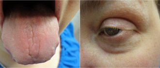
With a large size of the cephalohematoma and with its rapid resorption, the breakdown of red blood cells increases, which leads to an increase in bilirubin.
The baby’s body does not have time to completely remove it, and bilirubin is deposited in soft tissues, causing the skin and visible mucous membranes to turn yellow. This condition is temporary, similar to physiological jaundice and does not require additional treatment.
Symptoms
The region of the parietal bones is the most common site for the appearance of cephalohematoma. It can also be found on the forehead or back of the head, but not as often as on the right or left parietal bone. Hemorrhages of this kind can be large or small, unilateral or bilateral. In the latter case of hematoma, the shape of the fetal head becomes irregular, asymmetrical or ovoid. For comparison, there are many videos and photos on the Internet of children born with hematomas.
READ ALSO: why does a hematoma on a child’s head appear and how is it treated?
Due to the fact that the hematoma is located under the periosteum between the sutures of the cranial bones, its localization always remains within the bone over which it is located. Such an injury does not cause severe pain to the baby, pulsation is not typical for it, and the skin in its area does not change.
Cephalohematoma on the head often occurs within a few hours after the baby is born. Over the next few days, it continues to increase in size, but then its growth stops and the process of regression begins. At 6-8 weeks, the resorption process is completed, the baby’s head again becomes a normal round shape.
READ ALSO: A newborn has a lump on the head after childbirth - what could it be?
How to recognize
After the birth tumor subsides (on days 2–3), manifestations of cephalohematoma become noticeable. From the first day of birth, the size of the hemorrhage increases. A newborn does not have enough blood clotting factors, so it remains liquid - this makes it impossible for blood clots to thrombose damaged vessels. The tumor is elastic to the touch; when you press on it, you can feel fluid movement, pulsation and fluctuation.
Article on the topic: The effect of stress on the body: consequences for the body, brain and soul
If the hematoma is small, it shrinks within 7–8 days and goes away without leaving a trace. If the hemorrhage is large, the resorption process takes several months. Sometimes a crack or fracture of the bone is observed in the area where the problem is located. The boundaries of the formation are always clear and look like a compacted ridge around the circumference. The demarcation is associated with the tight fusion of the periosteum with the cranial bones in the area of the sutures, so the hematoma is located only in the area of one bone. The average tumor size is 3–7 cm. Large hematomas over 8 cm are subject to aspiration.
Establishing a diagnosis
Diagnosing this type of birth injury is quite easy. The absence of difficulties in establishing a diagnosis is explained by the fact that cephalohematoma has a very specific location, unique to it. Despite this, in the presence of a bilateral generic cephalohematoma, which has an impressive size, it would be useful to conduct additional studies. They will help rule out cracks in the skull and other possible brain damage. Auxiliary diagnostics include:
- radiography;
- neurosonography (ultrasound).
Having a specific clinical picture, a hematoma is sometimes mistaken for:
- aponeurosis;
- protrusion of the meninges and brain tissue through the fontanel;
- bone defects;
- birth tumor.
READ ALSO: what does a small fontanel mean in a newborn and does it need to be treated?
As for the birth tumor, it (just like cephalohematoma) appears when the child passes through the birth canal. It is a common swelling of the subcutaneous tissue, which resolves on its own in 2-3 days. One way or another, it is important to establish an accurate diagnosis in order to take the necessary measures and not harm the baby.
Cephalohematoma or birth tumor?
Sometimes young mothers feel that the doctor does not talk about the problem the baby has, although it is clearly visible. However, you should not think that medical workers are inattentive to the baby. Most likely, the vigilant mother confused the symptoms of two birth complications, mistaking the birth tumor for a cephalohematoma. This is the name for swelling that occurs on the baby’s head after childbirth. Subcutaneous swelling, which is not a cephalohematoma, is not limited to any bone of the skull, and usually disappears on its own, without any intervention, within 2-3 days.
Treatment of cephalohematoma
Birth cephalohematomas do not require any special therapy. If a child has a birth injury of this nature accompanied by complications, it is the responsibility of a neonatologist and, if necessary, a pediatric surgeon to treat it. Treatment of cephalohematoma in this situation comes down to:
READ ALSO: intracranial birth injury of a newborn: causes, symptoms and treatment
- feeding the baby with donor or expressed breast milk for 4-6 days;
- taking calcium supplements, for example, calcium gluconate, course 3-5 days;
- taking vitamin K for 3-5 days.
When a baby has an ossified cephalohematoma, resection of the periosteum down to the bone is required to correct the cranial deformity. Ossified means an ossified hematoma.
In some cases, it is necessary to resort to puncture of the cephalohematoma. It is carried out if:
- suppuration appeared;
- size exceeds 8 cm;
- the tumor does not begin to shrink or, on the contrary, may even increase in size after 10 days.
For the purpose of treating hematomas, it is not recommended to apply ice or suction the blood, since such procedures are quite dangerous for the baby. It is important that during this adaptation period mother and child are together. Staying together plus breastfeeding is the best way to speed up recovery.
After the birth hematoma has completely resolved, in order to promote the correct formation of the baby’s head, you can resort to the help of orthopedic pillows. However, this should be done only after consultation with a pediatrician or orthopedic doctor, neurologist and surgeon who observed the newborn.
About the consequences
In the vast majority of cases, there are no consequences or complications after the pathology. At an older age, it is impossible to even say whether the child had a hematoma or not. It is extremely rare that a slight change in the shape of the skull is possible; this happens when the blood not only comes out, but is replaced by connective tissue.
When the hematoma resolves, the only complications that can be identified are suppuration and jaundice, which is formed due to the release of a large amount of bilirubin into the blood. With adequate treatment, even such unpleasant symptoms quickly disappear.
Remember that it is extremely important not to refuse a caesarean section if your obstetrician recommends it. Also, a lot depends on your efforts; a quick birth means preventing many complications.
A case of suppuration of a birth injury
The purulent process begins as a result of the activity of bacteria that penetrate through abrasions, scratches or other damage to the skin in the area of the hematoma. For the sensitive body of a newborn, even a local infection is potentially dangerous. However, suppuration of birth injuries is a fairly rare occurrence. In this case, such a complication is accompanied by:
- redness of the skin over the hemorrhage;
- compaction of the above area;
- lethargy of the baby, which consists of either a complete cessation of crying, or the baby quickly gets tired when he starts screaming;
- breast refusal as a result of intoxication and loss of appetite;
- increase in body temperature to 38-39°C.
The appearance of any of these signs is a reason to consult a doctor. The treatment of a festering hematoma itself resembles the treatment of an abscess and boils down to:
- removing hair in the area of the hematoma and treating the area with an antiseptic;
- taking a puncture and suctioning the contents of the cavity;
- installing drainage, a rubber tube, with the help of which fluid is removed from the wound;
- doctor prescribing an adequate dose of antibiotics.
These procedures can be done as early as 10 days after birth. It is better not to delay therapy aimed at removing suppuration in the cephalohematoma. When the start of treatment is delayed, the risk of generalization of the process increases, which in turn can lead to tragic consequences.
Nuances in caring for a baby with cephalohematoma
Infants may feel uncomfortable due to the presence of a cephalohematoma. As a result, there are a number of nuances in caring for such children. Basically, these simple rules are aimed at reducing possible unpleasant sensations for the baby and creating a comfortable position for the child’s head. In addition, following them will prevent the development of complications. These rules are as follows:
- Compliance with medical instructions and refusal to use auxiliary means that disperse blood. This also applies to traditional methods. Due to the high sensitivity of the baby’s body to any chemically active substances, it is important to know when to stop when carrying out therapy. So, when a doctor prescribes Traumeel alone, it is worth using it exclusively. Self-medication is fraught with increased bleeding and other adverse consequences.
- Protecting the baby's head from additional external damage. As a result of a blunt impact caused by the fall of the baby, the periosteum can rupture, because it is already under tension due to the hematoma. The penetration of microbes and suppuration of the hematoma can be caused by a simple scratch, puncture, scraping or other damage at the site of hemorrhage;
- Minimum pressure in the area of the hematoma. A tightly tied hat or bonnet can increase pain in a baby (we recommend reading: what size bonnet should a newborn buy?);
- Control over the size of hemorrhage and their trends. If the size increases, you should immediately consult a doctor, as this is evidence that either the bleeding has not stopped, or the plasma has come out of the vessel wall;
- The use of special gel pillows designed to give a child's head a comfortable position. With their help, you can evenly distribute pressure between different parts of the head, thereby reducing discomfort.
READ ALSO: how is a hematoma on the head of a newborn treated?
Diagnostics
A cephalohematoma is a localized swelling that does not extend beyond the parietal, frontal, or occipital bones. This is due to the presence of limiting seams. With severe trauma, the formation of several subperiosteal hemorrhages with a volume of 5 to 150 ml is possible.
The presence of a hematoma can be suspected by visual examination and palpation (feeling) of the tissues of the cranial vault. The need for further examination is determined by the presence of the following criteria:
- swelling size 5 cm or more;
- continued increase in cephalohematoma over time;
- the swelling is not limited to one bone, which indicates pronounced changes.
Diagnostic tests are prescribed by a neonatologist or pediatrician. They include several basic techniques that have become widely used clinically.
For diagnostics the following is carried out:
- X-ray – despite the fact that X-ray examination carries a radiation load on the child’s body, it is prescribed for suspected bone fractures, including the skull bones.
- Ultrasound examination - a technique that allows you to visualize hemorrhage, assess its size and precise location.
- A coagulogram is a blood test that determines the state of the coagulation system based on several indicators. The study has diagnostic value, especially in cases of prolonged increase in size of the cephalohematoma, indicating ongoing hemorrhage under the periosteum.
Additional diagnostic tests are required before treating cephalohematoma, especially in cases where surgical intervention is necessary.
What consequences can occur after a birth head injury?
Typically, cephalohematoma in children does not lead to serious consequences. The most common complication that occurs if the hematoma is large or the blood contained inside it does not resolve for a long time is ossification, when such a hemorrhage becomes ossified. The skull may become deformed or become asymmetrical. Other possible complications include:
- Hemolytic anemia, caused by blood loss, as well as other types of anemia.
- Suppuration. It is caused by an infection that enters the hematoma through existing damage.
According to experts, including Dr. Komarovsky, cephalohematoma, as well as its purulent variant, with adequate and timely treatment, does not lead to damage to the brain or other parts of the nervous system. Being a simple accumulation of blood above the skull, such hematomas do not become causes of mental retardation, speech impairment, paralysis or other neurological problems in the future.
Loading…
Share with friends!
Types and degrees of subperiosteal hemorrhage
The classification of cephalohematomas lists their types depending on several factors. The most famous:
- By size: first degree (up to 4 cm), second (4.1–8 cm) and third (more than 8 cm), tumors are single and multiple, for the latter the total area is estimated.
- According to the combination of hemorrhage with other injuries: with a depressed fracture of the skull bones, with brain damage (edema, cerebral hemorrhage, epidural hematoma), with neurological manifestations (focal and general symptoms), with hemorrhagic stroke.
- By localization: parietal, frontal, occipital, temporal (very rare).