Complaints and symptoms of jaw fractures
It would seem that any fracture is equally unpleasant. Especially if its consequences are in plain sight and deform the face. But still, a broken jaw is more dangerous than other injuries, because in addition to problems in the field of aesthetics, it entails several more therapeutic and psychological ones.
Obvious signs will help you avoid confusing a fracture with a bruise. And this is not only and not so much a crooked jaw.
Fracture of the lower jaw
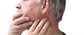
First, symptoms of a fracture include significant swelling that distorts the symmetry of the face. When a fracture occurs, it is much hotter to the touch than the surrounding skin.
Secondly, a blood clot visible in the soft tissues (with a closed fracture) or blood in the mouth (with an open one).
Thirdly, of course, pain that intensifies when talking, changes in the bite and increased sensitivity of the teeth.
Maxillary fracture
Signs of a rather rare fracture of the upper jaw are even more revealing: numbness and bruises under the eyes, nosebleeds and impaired sense of smell. In this type of fracture, part of the upper jaw is torn off from the base of the skull, and the fault line crosses the eye socket and the bridge of the nose.
The main thing in such a situation is to start treatment as quickly as possible, so as not to risk complications, which may include osteomyelitis or perimandibular phlegmon - in other words, inflammation of the bone or surrounding tissues.
Diagnostics and first aid
Before prescribing treatment, it is necessary to accurately determine the location of the fracture, as well as its severity. To do this, an X-ray scan, external examination and palpation of the affected area are performed.
Without detailed diagnostics, it is impossible to determine the extent of damage. But given the known symptoms, the victim can be provided with first aid.
The first aid provided at the scene of the incident should be as follows:
- Fixing the jaw with a tight bandage
Fixing (a kind of splinting) of the jaw with a tight bandage.
- Try to stop the bleeding: apply a sterile bandage, pack. In case of arterial bleeding, you need to apply pressure to the site of the rupture of the vessel.
- Carefully clean the oral cavity from blood clots, vomit, and tooth fragments.
- Fix your tongue, otherwise breathing may become difficult.
- Provide the victim with maximum immobility.
- Apply a cold compress to the injury site.
Getting help immediately after an injury can be invaluable!
Once precautions have been taken, the person should be taken to the nearest medical facility immediately. There he will be provided with professional medical care. When transporting to a medical facility, certain rules should also be followed. It is recommended to transport the victim sitting or in a lying lateral position, with the head kept tilted down. In an unconscious state, patients are transported lying down with their head on the side.
Already in the hospital emergency care continues:
- wound treatment;
- constriction of large blood vessels;
- installation of a tracheal breathing catheter;
- antishock treatment.
The first diagnosis is external examination and palpation. After this, radiography is performed to obtain a complete clinical picture of the injury and the method of treatment is determined.
Classification of jaw fractures
The most common fracture is a mandibular fracture. A fracture of the upper jaw is much less common.
In addition, there are complete and incomplete fractures, closed and open. Complete is considered to be the one in which the fragments have been displaced, open - when the bone has touched the soft tissues of the face or mucous membranes.
But even complete and open fractures are not comparable in danger to splintered fractures, which require immediate surgical intervention. Fortunately, this type of injury is rare and requires incredible force to occur—much more powerful than an opponent's fist, a volleyball, the weight of one's own body in a bad fall, or a collision with an airbag.
There are typical fracture sites. As a rule, they are located in those places where the jawbone experiences the greatest load, or where its strength is lower. The most common fractures include:
- angles of the lower jaw (in the area of wisdom teeth);
- in the projection of the mental foramen (in the area behind the fangs);
- middle (center) of the body of the lower jaw;
- articular process (in the area of the temporomandibular joint).
Ask us a question
Treatment
Treatment of a victim with mandibular injuries is determined based on the characteristics of the injuries received. Activities include the following therapeutic solutions:
- surgery to align bone fragments (local anesthesia);
- splinting for the period of rehabilitation and recovery after injury (full splinting is required if the fracture of the lower jaw is displaced);
- creating and maintaining favorable conditions in the area of damage contribute to faster and better tissue fusion;
- medication (antimicrobial, restorative drugs) to prevent the development of infectious and inflammatory processes in the affected area;
- physiotherapy (at the last stage of treatment) to restore jaw function.
Prompt surgical care may also include:
- installing staples at the site of a bone fracture, “stitching” with wire or nylon thread;
- fixation with metal knitting needles;
- fixation of fragments with bone metal plates;
- fixation of bones with special extraoral structures.
The most severe cases may, after urgent measures, require further measures to restore facial plastic surgery and install jaw prostheses.
All assistance offered to the patient (first aid, pre-medical, surgical, specialized) has several important goals:
- the fastest and most effective restoration of bone and soft tissues;
- restoration of normal bite;
- restoration of jaw functionality.
Treatment of jaw fractures, depending on the strength and complexity of the injury, lasts from 4 weeks. During the rehabilitation period, splinting is mandatory; after removing the bandage, therapeutic exercises and mechanotherapy are mandatory.
It is better not to neglect broken bones in the facial area and to seek qualified help in a timely manner, because in addition to aesthetic unattractiveness due to displacements, there may be more disastrous consequences, even death.
17.4. Treatment of patients with mandibular fractures
The goal of treating patients with fractures of the lower jaw is to create conditions for the fusion of fragments in the correct position in the shortest possible time. In this case, the treatment should ensure complete restoration of the function of the lower jaw. To carry out what was previously said, the doctor must: firstly, carry out reposition and fixation of jaw fragments for the period of consolidation of the fragments (includes removal of the tooth from the fracture line and primary surgical treatment of the wound); secondly, creating the most favorable conditions for the course of reparative regeneration in bone tissue; thirdly, prevention of the development of purulent-inflammatory complications in bone tissue and surrounding soft tissues.
Before considering methods of immobilizing fragments for fractures of the lower jaw, I want to express my opinion in relation to the tooth that is located in the fracture gap. There can be a wide variety of options for the location of teeth in relation to the fracture gap (Fig. 17.4.1).
To be deleted:
• broken roots and teeth or teeth completely dislocated from the socket;
• periodontitis teeth with periapical chronic inflammatory foci;
• teeth with symptoms of periodontitis or moderate to severe periodontal disease;
• if an exposed root is located in the fracture gap or an impacted tooth that interferes with the tight (correct) juxtaposition of the jaw fragments (a tooth wedged into the fracture gap);
• teeth that are not amenable to conservative treatment and support inflammatory phenomena.
Subsequently, primary surgical treatment of the wound is performed, i.e. delimit the bone wound from the oral cavity. In this way, an open fracture is converted into a closed one.
Sutures made of chrome-plated catgut are placed on the mucous membrane. They try to close the hole tightly so that there is less chance of infection of the blood clot and the development of purulent-inflammatory complications.
Rice. 17.4.1.
Possible options for the location of teeth in relation to the fracture gap
lower jaw (submitted by radiographs): a, b) in the area of the body of the jaw; c) in the mental department; d, e, f, g, h) in the area of the angle of the jaw.
Rice. 17.4.1.
(continuation).
♦ Temporary immobilization of fragments
It is carried out at the scene of an incident, in an ambulance, in any non-specialized medical institution by paramedics or doctors, and can also be performed as mutual aid. Temporary immobilization of fragments of the lower jaw is carried out for a minimum period (preferably no more than a few hours, sometimes up to a day) before the victim is admitted to a specialized medical institution.
Rice. 17.4.2.
Intermaxillary ligature binding of teeth with wire: a) application of a ligature; b, c) intermaxillary binding.
The main purpose of temporary immobilization is to press the lower jaw against the upper jaw using various bandages or devices. To temporary (transport)
• circular parietomental bandage;
• standard transport bandage (consists of a rigid splint - Entin's sling);
• soft chin sling Pomerantseva-Urbanskaya;
• intermaxillary ligature binding of teeth with wire (Fig. 17.4.2).
♦ Permanent immobilization of fragments
Conservative methods are used to immobilize fragments of the lower jaw.
(orthopedic) and
surgical
(operative) methods.
Most often, dental wire splints
(a conservative method of immobilization).
During the First World War, for the treatment of wounded with maxillofacial injuries, S.S. Tigerstedt
(dentist of the Russian army) in 1915,
aluminum dental splints were proposed,
which
a smooth
spacer bend) and double-jaw
Fig. 17.4.3) .
Rice. 17.4.3.
Variants of aluminum dental splints proposed by S.S. Tigerstedt (1915):
a) smooth tire - bracket; b) tire with a spacer (spacer bend);
c) double-jaw splints with hooking loops and intermaxillary rubber traction.
Rice. 17.4.4.
Appearance of a smooth splint applied to the lower jaw for a fracture between the lateral incisor and the canine.
Rice. 17.4.5.
Appearance in the oral cavity of a two-maxillary aluminum splint with hooking loops and an intermaxillary rubber rod.
Smooth tire - bracket
used for linear fractures of the lower jaw located within the dentition (from central incisors to premolars), for fractures of the alveolar process of the upper and lower jaws (the intact area of the jaw must have at least 3 stable teeth on each side), for fractures and dislocation of teeth.
The technique for making a splint is as follows: from annealed aluminum wire 1.8-2 mm thick, using crampon forceps, bend a splint along the dental arch (Fig. 17.4.4) and insert ligatures (made of bronze-aluminum wire) into the interdental spaces, covering each tooth from the lingual or palatal side and bend the medial end of the wire up, and the distal one down (to differentiate the medial ends of the ligatures located between the central incisors, which are both bent upward, you must always bend one end of the wire on the left or right side down); after the splint is placed on the dentition, the ends of the wire ligatures are twisted together (the medial end with the distal), the twisted ligatures are cut off, leaving a free end up to 5 mm long, and they are folded into the interdental space in the medial direction (towards the midline) .
Tire with spacer
(Fig. 17.4.3-6) are made in the same cases as a smooth tire.
The indication for its manufacture is the absence of one or more teeth at the site of a fracture or an existing defect in the bone tissue. The spacer bend is always located only in the area of the jaw fracture.
The edges of the spacer bend rest against adjacent teeth (to avoid displacement of fragments), and its depth should correspond to the width of the lateral surface of the tooth located along the edge of the defect.
Tire with hook loops
(Fig. 17.4.5) is applied to both jaws. Indications for its manufacture are fractures of the lower jaw within the dentition or beyond, both without displacement of fragments and with their displacement, as well as for fractures of the upper jaw (in the latter case, a parietal-mental bandage or a standard chin sling and headband must be additionally applied cap).
On each aluminum splint, 5-6 hooks (loops) are made, which are placed in the area of the even teeth (second, fourth and sixth). The length of the loops is about 3-4 mm and they are at an angle of 35-40° to the axis of the tooth. The splints are attached to the teeth in the previously described manner (see splint making technique). On a splint attached to the upper jaw, the loops (hooks) are directed upward, and on the lower jaw - downward. Rubber rings are put on the hook loops (they are cut from a rubber tube with a diameter of about 8 mm). The ligature wires need to be tightened every 2-3 days, and the rubber rod needs to be changed every 5-6 days (or as needed).
Wire aluminum bars are currently the most widely used because of their availability and ease of manufacture. It is necessary to strive to ensure that the contours of the dental splint correspond as much as possible to the bends of the dental arch. But along with the advantages of bent wire splints, there are a number of their disadvantages: injury to the mucous membrane of the lips and cheeks due to hooks (loops); due to the oxidation of tires and their clogging with food debris, difficulties arise with the hygienic maintenance of the oral cavity; the need for individual production; with a deep bite, they interfere with the correct closure of the dentition; the presence of additional retention points; cutting through soft tissues with ligatures; the appearance of galvanic currents, etc.
Standard dental band splints
made of stainless steel with ready-made hooking loops were proposed by BC Vasiliev in 1967 (Fig. 17.4.6-17.4.7). Tire thickness 0.38-0.5 mm. The splints are fixed to the teeth using a ligature wire in the previously described manner. Standard dental band splints are free from some of the previously listed disadvantages and are widely used. Indications for the use of tape splints are the same as for wire splints.
Rice. 17.4.6.
Appearance of Vasiliev's tires.
Rice. 17.4.7 .
Fixation of the Vasiliev splint to the teeth in case of a fracture of the lower jaw.
Gorobets E.V. and Kovalenko V.V. (1997) proved that dental aluminum splints cause the occurrence of galvanic currents in the oral cavity of patients, the severity of which is 8-10 times higher than that in victims who were treated with dental tape splints made of stainless steel (Vasiliev splints). This indicates the advantages of the latter over Tigerstedt tires.
Less commonly used are dental splints made of quick-hardening plastic.
VC. Pelipas proposed such a tire in 1969. The application technique is as follows: steel hooks are tied to individual teeth with nylon thread, and then quick-hardening plastic is placed on the dentition on the vestibular side, covering the base of the hooks attached to the teeth. To date, other tires made of quick-hardening plastic have been proposed (M.B. Shvyrkov and others).
Compression-approximating dental splinting using double polyamide thread and steel wire according to V.G. is used. Centilo et al. (1997) and splinting with quick-hardening plastic according to M.B. Shvyrkov.
Rice. 17.4.8.
Dental and supragingival splints: a) Weber splint; b) Port bus; c) Vankevich tire.
Rice. 17.4.9.
Using the patient's removable dentures as a splint. Both prostheses are connected into a monoblock: a) with a ligature wire; b) fast-hardening plastic.
, dentogingival and supragingival splints are made (in the laboratory) .
The most commonly used
is the Weber splint.
A plastic splint covers the teeth, fits tightly to the gingival edge of the alveolar process and rests on the latter. The chewing surfaces and cutting edges of the teeth are not covered by the splint. Indications for its use are fractures that occur within the dentition and each fragment of the jaw has several stable teeth (Fig. 17.4.8-a).
Tire Porta
(supragingival splint) is used for fractures of the lower jaw in patients with toothless jaws.
It consists of base plates on the alveolar process of the upper and lower jaws, which are fastened into a single block, and
in the anterior section of this splint there is a hole for eating (Fig. 17.4.8-6). Using a Porta splint to firmly fix the fragments of the lower jaw, it is necessary to apply a parietal-mental bandage or a standard chin sling and a head cap.
Shinu Vankiewicz
should be used in patients with a fracture of the lower jaw with a bone tissue defect and the absence of teeth on the fragments. The splint is fixed on the teeth of the upper jaw, and the side wings - pilots (processes) are lowered down and rest against the inner surfaces of the broken fragments, which keeps them in the correct position (Fig. 17.4.8-c). The basis for the Vankevich splint is a plastic palatal base, and for the Vankevich-Stepanova splint it is a steel arch.
The patient's removable dentures can be used as a splint (Fig. 17.4.9). The dentures of the upper and lower jaws are connected to each other using ligature wire or quick-hardening plastic.
♦Osteosynthesis
Osteosynthesis
- a surgical method of connecting bone fragments and eliminating their mobility using fixing devices.
Indications for osteosynthesis:
• insufficient number of teeth for splinting or absence of teeth in the lower and upper jaws;
• the presence of mobile teeth in patients with periodontal diseases that prevent the use of a conservative method of treatment;
• fractures of the lower jaw in the area of the neck of the condylar process with an irreducible fragment, with dislocation or subluxation (incomplete dislocation) of the head of the jaw;
• interposition - the introduction of tissue (muscles, tendons, bone fragments) between fragments of a broken jaw, preventing reposition and consolidation of fragments;
• comminuted fractures of the lower jaw, if the bone fragment cannot be placed in the correct position;
• bone fragments of the lower jaw that are not comparable as a result of displacement.
Classification
modern methods of osteosynthesis of fragments of the lower jaw, taking into account devices for its implementation Yu.D. Gershuni (1986) presented in the following form
Diagnostic measures
An orthopedic doctor or surgeon makes a preliminary diagnosis based on a survey and examination of the patient.
To clarify the diagnosis, instrumental studies are carried out:
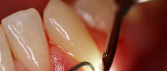
- Extraoral examination. An open fracture of the jaw is characterized by trauma to the skin, swelling and bleeding.
- Intraoral examination. The lower dental arch is examined. The movement of the lower jaw is assessed. The oral cavity is inspected for damage to the mucous membrane and teeth.
- X-ray examination. An X-ray of both jaws is taken in several projections. Radiography allows you to determine the location of the fracture, the number of fractures, the presence of fragments, and the degree of displacement.
- MRI. This method allows you to assess the condition of blood vessels and nerves, the degree of damage to muscles and ligaments, and identify hemorrhage in soft tissues.
Based on clinical and instrumental studies, a diagnosis is made and treatment is prescribed.
Instrumental examination allows a more complete assessment of the severity of injury