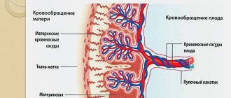
The placenta performs many important functions during pregnancy. First of all, it carries out gas exchange, transporting oxygen to the fetus and removing carbon dioxide. Through the placenta, the fetus receives the nutrients it needs for development and gets rid of waste products. The placental barrier protects the baby from viruses and bacteria. Finally, the placenta serves as the endocrine system for the fetus.
What features of this organ will doctors pay attention to during an ultrasound examination? We have already talked about the degrees of maturity of the placenta and problems with its location. Let us recall that usually the baby's place is attached to the back wall of the uterus, and develops according to a certain pattern, providing at each stage all the needs of the fetus. By week 36, the placenta reaches its full maturity and aging begins. By the end of pregnancy, the placenta normally has a diameter of 15-18 cm and its thickness is 2-4 cm.
Another issue that will be of interest to doctors is the thickness and structure of the placenta. These indicators will be discussed in today’s article.
What is placental hyperplasia?
Hyperplasia is an ancient Greek word meaning “excessive development”, “enlargement”. This term applies to any organ of the human body. In the case of the placenta, the term hyperplasia usually refers to an increase in its thickness, mass and circumference. However, with an ultrasound scan it is possible to accurately measure only the thickness of the placenta, so they start from this indicator. I would like to note that a single ultrasound examination is not competent to diagnose placental hyperplasia. Dynamic monitoring of the pregnant woman, the opinion of several doctors, and extensive studies are necessary. Moreover, independent assessment of ultrasound results is unacceptable.
Hyperplasia or diffuse thickening of the placenta implies its swelling, as well as a compensatory increase in the number of structural units. There are several reasons for this:
- Infections. This is perhaps the most common cause of thickening of the placenta. Bacteria, viruses and other foreign agents can penetrate the uterine cavity, amniotic membranes and waters both ascending from the vagina and through the bloodstream from other foci. The placenta is rarely infected in isolation. Inflammation of the baby's place is called placentitis and very often it is combined with inflammation of the membranes and intrauterine infection of the fetus. With the development of the inflammatory process, swelling of the placenta occurs and an apparent increase in its thickness.
- Immunological conflict between mother and fetus, for example due to the Rh factor. In this case, the picture will be similar to an infectious process, only the placental tissue is affected not by viruses and bacteria, but by the mother’s antibodies. In simple terms, in this case the mother’s body tries to kill the fetus and all temporary organs of pregnancy, mistaking them for foreign.
- Severe or long-term gestosis. With gestosis, blood pressure rises, protein appears in the urine and swelling increases, including hidden ones. Such hidden edema also includes placental edema. In addition, gestosis affects the vasculature of the placenta, also leading to edema.
- Severe anemia in the mother. When the hemoglobin level drops below 80 g/l, the fetus begins to experience oxygen starvation. In this case, the placenta begins to grow compensatoryly in order to increase the area of gas exchange and thus help the fetus.
- Natural feature. There is no need to exclude the possibility of placenta enlargement simply as a development option. Often large children have massive placentas, or this feature is passed on through generations.
Very often, thickening of the placenta is combined with polyhydramnios or oligohydramnios, as well as expansion of the intervillous spaces (IVS) of the placenta. In the first two cases, these are additional manifestations of infection or immune conflict. The expansion of the urinary tract indicates that the placenta is trying to compensate for its function. Isolated expansion of the urinary tract is not a diagnosis, but may only imply an infectious process, anemia, fetoplacental insufficiency, or simply a developmental feature.
Symptoms and diagnosis of placental hyperplasia
The placenta is deprived of pain innervation, therefore, with the initial signs of its damage, nothing bothers the pregnant woman. Typically, the main symptoms appear weeks or even months later.
- The main symptoms of dysfunction of the placenta are signs of oxygen starvation of the fetus: growth retardation, disruption of fetal-uterine blood flow, acute hypoxia. The woman notes a decrease in fetal movements and a decrease in its activity. During the examination, the doctor pays attention to a decrease in the growth rate of the pregnant woman’s abdomen, poor heart rate or cardiotocography.
- Manifestations of the infectious process can also act as the main complaints. A pregnant woman complains of fever, weakness, headaches and muscle pain, chills, and discharge from the genital tract. Often, when questioned, a woman recalls a recent exacerbation of a chronic inflammatory process (otitis, sinusitis, pyelonephritis) or an acute illness (ARVI, influenza, tonsillitis, thrombophlebitis).
If thickening of the placenta is detected at the next ultrasound, it is necessary to perform a so-called diagnostic search - a series of examinations to identify possible causes:
- Complete blood count, urine test and blood biochemistry to look for inflammatory changes, as well as determine hemoglobin and ferritin levels.
- Vaginal culture and smear, testing for sexually transmitted infections and the TORCH group.
- Determination of the level of antibodies to the Rh factor and blood groups to exclude Rh sensitization.
- Blood for glucose.
- Consultations with a therapist and cardiologist to exclude gestosis, an infectious disease specialist in complex cases of intrauterine infection.
Continuous CTG monitoring and Doppler ultrasound of the fetus are included in the monitoring protocols for such pregnant women.
Placental hyperplasia: consequences for mother and baby
Since the placenta is a temporary organ for the existence of the fetus, the consequences of disruption of its functions affect mainly the child:
- chronic;
- fetal growth restriction;
- acute fetal hypoxia;
- intrauterine fetal death;
- premature birth.
The danger to the mother is not so much the placental hyperplasia itself as the cause that caused it. Preeclampsia and eclampsia, infectious process, severe anemia certainly threaten the health and life of a woman.
Treatment of placental thickening
Therapy for placental hyperplasia consists of treating the immediate cause that caused this complication:
- Antibacterial and antiviral therapy in case of infection.
- Treatment of gestosis, as well as early delivery.
- Treatment of Rh conflict, consisting of periodic intrauterine blood transfusions to the fetus and plasmapheresis of the mother. In this case, the fastest possible delivery is also indicated with mandatory prevention of Rh conflict with anti-Rhesus immunoglobulins in the next pregnancy.
- Therapy with iron supplements for anemia in a pregnant woman, as well as red blood cell transfusion if hemoglobin decreases below 75 g/l.
Considering the impaired function of the placenta, the use of various drugs that improve placental blood flow is indicated: Curantil, Actovegin, Piracetam, Pentoxifylline.
Alexandra Pechkovskaya, obstetrician-gynecologist, especially for the site
Useful video:
The placenta performs many important functions during pregnancy. First of all, it carries out gas exchange, transporting oxygen to the fetus and removing carbon dioxide. Through the placenta, the fetus receives the nutrients it needs for development and gets rid of waste products. The placental barrier protects the baby from viruses and bacteria. Finally, the placenta serves as the endocrine system for the fetus.
What features of this organ will doctors pay attention to during an ultrasound examination? We have already talked about the degrees of maturity of the placenta and problems with its location. Let us recall that usually the baby's place is attached to the back wall of the uterus, and develops according to a certain pattern, providing at each stage all the needs of the fetus. By week 36, the placenta reaches its full maturity and aging begins. By the end of pregnancy, the placenta normally has a diameter of 15-18 cm and its thickness is 2-4 cm.
Another issue that will be of interest to doctors is the thickness and structure of the placenta. These indicators will be discussed in today’s article.
Causes
Thickening of the placenta indicates that some negative changes are occurring in the mother’s body. They may be of a different nature, but in any case, this fact cannot be ignored, since it is dangerous, first of all, for the child.
Placental hyperplasia can be caused by:
- infectious diseases;
- anemia (hemoglobin level less than 80 g/l);
- diabetes;
- Rh conflict, provoked by different Rh factors of the mother and fetus;
- late gestosis.
Sometimes doctors are unable to establish an objective reason for the development of such a complication.
Structure of the placenta
Until 16 weeks, the baby's place is just forming, so the heterogeneous structure of the placenta is not a particular cause for concern. After this period, up to 30 weeks, normally the placenta should not change until the process of its maturation begins. However, in some cases, doctors talk about the heterogeneous structure of the placenta - its increased echogenicity and the detection of various inclusions. This may indicate a disruption in its normal functioning.
One of the common features of structural changes is the expansion of the intervillous spaces in the embryonic organ. This place is intended for the exchange of substances between mother and fetus. When there is a need to increase the area of this exchange, the profit centers expand accordingly. This phenomenon does not lead to fetoplacental insufficiency, and usually does not require additional research.
Another option for heterogeneity of the structure is the inclusion of calcifications. They can prevent the embryonic organ from functioning normally. However, shortly before birth, starting from the 37th week, the presence of calcifications is normal and indicates aging of the placenta.
Functions of the placenta
The placenta is also called the baby's place. Translated from Latin, placenta means “cake”. It actually looks like a flatbread.
With the help of the placenta, gas exchange occurs between the organisms of the child and his mother. Oxygen is sent to the fetus, and from it carbon dioxide enters the maternal bloodstream. It’s the same with other substances the baby needs: he receives them from the mother’s body, and waste products come back through the placenta.
The placenta also has a barrier function, preventing, for example, toxins, drugs or antibodies from the mother from passing through. Unfortunately, it cannot trap drugs and alcohol, nicotine and viruses. This organ produces hormones that are necessary for the normal course of pregnancy. Based on the above, the placenta has the function of immune protection for the child.
Placenta thickness
This parameter must be examined by a doctor after 20 weeks of pregnancy. Normally, the placenta has a thickness corresponding to the duration of pregnancy - for example, 20 mm at 20 weeks, 25 mm at 25 weeks, and so on. These values are approximate and may vary. But if the thickness of the placenta does not greatly correspond to the gestational age - for example, 30 mm at 20 weeks, this indicates certain problems with its functioning.
When the thickness of the baby's place is too small, we are talking about its hypoplasia, and when it is too large, we are talking about placental hyperplasia. A small thickness is not at all uncommon, and it does not cause any special problems. Only a significant decrease in the thickness and size of the embryonic organ can seriously affect the fetus. A woman’s weight and build also matter—petite women may have a smaller placenta.
Thickening of the placenta can occur for a number of reasons:
- Preeclampsia in a pregnant woman;
- Viral infections;
- Sexual infections (chlamydia, ureaplasma, toxoplasma, etc.);
- Diabetes mellitus in the mother;
- Rhesus conflict between mother and fetus;
- Low hemoglobin and anemia in a pregnant woman.
Thickening of the placenta is a serious problem that can even lead to termination of pregnancy. Therefore, monitoring the condition of the fetus and timely treatment is necessary.
Why is placental hyperplasia dangerous? She grows too quickly and matures and ages prematurely. Calcifications form on the surface, and the structure becomes heterogeneous. The fetus does not receive enough oxygen and nutrients and experiences hypoxia. The hormonal function of the placenta is also disrupted, which can lead to premature birth.
If a woman has a thickened placenta, doctors will monitor the baby’s condition using cardiotocography and Doppler sonography. Treatment may be required to restore normal blood supply to the fetus.
The placenta is an important organ of the female body that facilitates the transport of nutrients to the fetus and provides sufficient oxygen. The final formation of the organ occurs at 6 weeks of pregnancy, so the heterogeneous structure of the placenta should not bother the mother.
What does the heterogeneous structure of the placenta mean?
During the process of bearing a child, the placenta changes structure and is characterized by the degree of maturity at a certain period. It also happens that the second stage of maturity occurs earlier than 34 weeks. This does not indicate an uneasy development of pregnancy.
Structure. The condition of the organ can be clearly seen at 12 weeks. The homogeneous structure of the placenta is characterized by a smooth chorale covering. The indicator refers to zero degree and indicates homogeneity.
Thickness. Using an ultrasound examination, the thickness of the organ is determined. Up to 32 weeks it increases and normally is no more than 30 mm. Rapid thickening or, conversely, a decrease indicates the process of premature aging of the organ. The reasons are: mismatch of the Rh factor, development of an infectious disease, diabetes of the woman.
Indicators of heterogeneity can be inclusions of various natures and areas of echo-negative state. They indicate a disruption in the functioning of the placental organ. A heterogeneous placenta with hyperechoic inclusions indicates serious disorders that affect the development of the child.
During the examination, great attention is paid to the thickness of the placenta at 20 weeks of pregnancy. The normal value is from 1.5 cm to 5 cm. If the placental organ is thin and less than 1.5 cm, then there is a risk of fetal underdevelopment. But this phenomenon is quite rare.
Development and maturation
To determine the parameters of development and maturation of the organ, you will need to perform an ultrasound procedure, which measures the following indicators:
- transverse and longitudinal segments, which are drawn through the farthest points, these segments must form a perpendicular to each other;
- thickness of the organ.
Most often, the child's organ is fully formed by 16 weeks. If the process of bearing a baby goes without pathological problems, then it will grow until 37 weeks, gradually reaching its maximum size.
The first thickness measurement by week is carried out at 20 weeks, and then according to the doctor’s indications.
It is worth knowing that by the end of the 3rd trimester its thickness decreases, but these indicators should change within normal limits, which correspond to the period of gestation.
An equally important indicator of a full and healthy pregnancy, along with the thickness of the baby’s place, is the degree of its maturity.
If thickening and early aging occur, this indicates the presence of pathology.
There are 4 degrees of maturity , each of which is observed at a certain time in fetal development:
- 0 degree – the formation of the organ occurs (if the pregnancy proceeds normally, then this stage lasts until the end of the 2nd trimester);
- 1st degree – growth and active development are observed, in which echogenic inclusions appear;
- 2nd degree - a mature organ changes its own structure;
- 3rd degree – aging, the structure becomes lobulated.
For example, the thickness of the placenta at 20 weeks is approximately 20 mm. During an ultrasound scan at 20 weeks, the thickening can normally range from 16.5 to 28.5 mm. Moreover, this will continue almost until the end of pregnancy, after which it stops.
The standard thickness of a child seat is indicated in the table:
If the thickening has larger or smaller parameters, this indicates a pathology that requires treatment.
Why is a thick or thin placenta dangerous during pregnancy, and how does it affect the condition of the mother and the unborn baby. If both thickening and thinning of the baby's place are diagnosed, an additional ultrasound will be required. After examining its results, the doctor will prescribe the necessary treatment.
Diagnostics
The ultrasound procedure is an important stage in monitoring the condition of the body during pregnancy. Diagnostics allows us to detect the formation and accumulation of calcifications, tumors and the threat of detachment.
Degrees of heterogeneity:
- 1st degree. Homogeneity is noticeably lost. Characteristic changes in the structure appear;
- 2nd degree. Formation of areas similar to a comma;
- 3rd degree. The process of salt deposition on dead parts of tissue is significantly enhanced. Calcification occurs.
During the ultrasound, the location of the placenta in the uterus is determined with millimeter precision. Starting from the 10th week, the doctor records the location of the fetus. The third examination procedure shows the stage of development of the child and identifies initial pathologies.
The ultrasound examination method allows us to identify not only changes in the embryonic organ, but also determine the thickness, exact location, and stage of maturity. Since the final development takes place at 16 weeks of gestation, the structure should not change until the 8th month.
The practice of conducting ultrasound examinations includes cases of expansion of the intervillous space. MVP is the space in the embryonic organ where metabolism between mother and fetus occurs. As the child grows, the amount of nutrients consumed increases.
Timely diagnosis makes it possible to detect calcifications on dead tissues. This is a common phenomenon associated with the natural aging of the embryonic organ. The doctor keeps the volume of salt formation under control. After 33 weeks of gestation, the number of calcifications may increase sharply. The rate of growth and development of the child must correspond to the degree of deterioration of the placenta. Only in this case there is no need to worry.
Causes of hyperplasia
To the main reasons
, causing an increase in the size of the placenta include:
- Severe anemia
; - Diabetes
; - Development of Rh conflict and conflict according to the ABO system
with incompatibility of blood groups; - Late gestosis
; - Infectious diseases of the genital organs
; - Any infectious diseases of the mother suffered during pregnancy
.
Causes and symptoms
The main reasons for heterogeneity of the placenta may be infectious diseases, even those that existed before, or the negative consequences of drinking large quantities of alcohol and smoking. This leads to disruption of blood flow from mother to baby. Oxygen starvation of the fetus or the fading of the pregnancy process occurs. A striking symptom is a change in the movement of the child inside. Quitting smoking and alcohol reduces the risk of placental defects
If changes occurring in the structure are detected by a doctor at week 30, this fact is normal in development. The main condition is the absence of serious abnormalities in the fetus. The causes of heterogeneity are most often a combination of incorrect actions of the mother, including nervous experiences. The more comfortable and calm the mother is, the better the unborn baby will feel. The psycho-emotional state determines the state of the female body.
You should refrain from contact with sick people, as you can become infected with an infectious disease. And this is another negative factor leading to improper development of the structure.
20 - 27 weeks. A heterogeneous structure of the placenta at 20 weeks is a common phenomenon among mothers. In case of a negative change in the condition of the organ, the ultrasound procedure will immediately report the initial stage of the pathology. The placenta may remain heterogeneous until the 27th week; after this period it returns to normal. Do not panic, each individual organism has its own developmental characteristics.
30-32 weeks. Upon reaching 30 weeks, the structure of the organ should become homogeneous. If at 30-32 weeks the structure remains heterogeneous, then this indicates the presence of pathology in the female body. A heterogeneous placenta at 32 weeks indicates a serious pathology occurring in the body.
Norms and deviations
What does the heterogeneous structure of the placenta mean?
During the process of bearing a child, the placenta changes structure and is characterized by the degree of maturity at a certain period. It also happens that the second stage of maturity occurs earlier than 34 weeks. This does not indicate an uneasy development of pregnancy.
Structure. The condition of the organ can be clearly seen at 12 weeks. The homogeneous structure of the placenta is characterized by a smooth chorale covering. The indicator refers to zero degree and indicates homogeneity.
Thickness. Using an ultrasound examination, the thickness of the organ is determined. Up to 32 weeks it increases and normally is no more than 30 mm. Rapid thickening or, conversely, a decrease indicates the process of premature aging of the organ. The reasons are: mismatch of the Rh factor, development of an infectious disease, diabetes of the woman.
Indicators of heterogeneity can be inclusions of various natures and areas of echo-negative state. They indicate a disruption in the functioning of the placental organ. A heterogeneous placenta with hyperechoic inclusions indicates serious disorders that affect the development of the child.
During the examination, great attention is paid to the thickness of the placenta at 20 weeks of pregnancy. The normal value is from 1.5 cm to 5 cm. If the placental organ is thin and less than 1.5 cm, then there is a risk of fetal underdevelopment. But this phenomenon is quite rare.
Treatment and childbirth
Complete restoration of homogeneity with the help of drugs is impossible. The absence of stress and anxiety has a beneficial effect throughout pregnancy.
A heterogeneous placenta during pregnancy is directly related to hypoplasia and hyperplasia. The doctor prescribes medications to improve uterine tone, blood circulation and proper oxygen delivery to the fetus. As an additional method - a healthy lifestyle and giving up bad habits. An ultrasound scan before the due date is also a necessary aspect of health monitoring.
Treatment of heterogeneity of the placenta:
- eliminating stress and depression;
- decreased tone, hypoxia;
- therapy of diseases: hypoplasia and hyperplasia;
- delivery.
A woman should strictly follow the doctor’s recommendations and take medications if they have been prescribed. Monitoring of fetal development should take place at all stages of gestation. If you follow the rules and lead a healthy lifestyle, then there is no need to fear for the unborn child and childbirth.
The tactics for managing childbirth with placental insufficiency are prescribed by the attending physician based on the condition of the woman’s body. Premature aging of the placental organ is an abnormal indicator that even causes concern to the doctor. With proper treatment, the problem can be corrected.
Prognosis and prevention
Prognosis will depend solely on the reasons that could cause hyperplasia. If the expectant mother took adequate treatment in combination, then the prognosis will be favorable. To prevent any complications, women who are planning a pregnancy need to check their health with specialists in advance. Pregnant women are also advised to avoid crowds of people during an unfavorable epidemiological situation. Proper nutrition and walks in the fresh air will also have a beneficial effect on the health of the expectant mother.
Calcifications
Calcifications are salt formations that are deposited on areas of dead organ tissue. The placental organ contains multiple blood vessels. They transport vital substances to the unborn child. In case of vessel spasm or failure, it leads to the death of the area. Calcium salt deposits form on dead areas of tissue.
The deposition of calcifications in the placenta can result from:
- pathological process of the uterus;
- severe anemia;
- gestosis and its manifestations;
- infection and chronic diseases;
- sexually transmitted diseases;
- bad habits.
In order for the pregnancy to proceed positively, periodic examinations by a doctor are required. A modern way of observing the process is the ultrasonic method, that is, ultrasound. With its help, pathology is identified in the early stages and calcifications are detected in areas.
Symptoms. In the case of a single deposition of calcifications, the woman will not feel discomfort. This will not harm the unborn child. If multiple foci of salt deposits form on dead tissue, this seriously harms the fetus. With a strong phenomenon, the woman notices a change in the nature of the child’s behavior inside. He becomes too active or, on the contrary, calms down. Impaired performance results in failure to perform adequate life support functions. The child does not receive additional oxygen. In severe cases, the death of the fetus inside the female body occurs.
Treatment. It is impossible to completely remove salt deposits inside the body. To begin with, the doctor establishes the specific cause of the appearance of salts and tries to eliminate the possibility of relapse. If salts were noticed late in pregnancy, and their quantity is small, then periodic examinations of the woman in labor are prescribed. The doctor monitors the situation using ultrasound and monitors the heterogeneous placenta with calcifications. The examination reveals a large number of foci of salts, but the functioning of the placental organ can proceed without disturbance. If the fetus continues to receive the necessary vital substances, then taking therapeutic drugs is not required.
Complications. If dysfunction is detected, if the fetus suffers from lack of oxygen and nutrition, the doctor prescribes appropriate treatment. It includes taking medications that normalize blood circulation, as well as vitamins. Additional drugs in treatment are stimulants of the metabolic process.
Close monitoring of the structure of the placenta during pregnancy is required. Also, do not forget about the normal, comfortable development of the fetus inside the mother’s body. Based on his behavior and well-being, conclusions are drawn about the normal process of gestation.
The placenta is the most valuable organ of the female body during pregnancy. Its main functions are to deliver the required amount of nutrients to the fetus and provide it with oxygen. The normal course of pregnancy, as well as childbirth, directly depends on the state of development of the child's place. In addition, the embryonic organ performs protective functions, preventing various infections from reaching the child, and ensures that it is supplied with a sufficient amount of hormones that allow the fetus to grow and develop.
Diagnosis of the condition of the placenta is extremely important and should be carried out throughout pregnancy using ultrasound (ultrasound).
This method makes it possible to timely detect any abnormalities in the development of an embryonic organ. And the faster pathologies are detected, the greater the chances of successful treatment. Using ultrasound, doctors determine its structure, location,... During such a study, the expectant mother may be diagnosed with a heterogeneous structure of the placenta.
Symptoms
In the early stages of pregnancy, hyperplasia practically does not manifest itself in any way and does not particularly affect the condition of the child. In the last trimester, a woman may independently suspect the occurrence of complications due to excessive or, conversely, insufficient activity of the fetus. As for clinical studies, at all stages hyperplasia is visible on ultrasound. Also, thickening of the placenta negatively affects the baby’s heartbeat. During the test, the doctor may detect tachycardia, bradycardia, or muffled tones.
Reasons for heterogeneity
In most cases, the heterogeneous structure of the embryonic organ should not be a cause for concern, because is the norm. The final formation of the placenta occurs at , after which its structure should remain unchanged until the thirtieth week. And if at this time any changes occur in the structure of the baby’s place (the doctor may detect echo-negative areas or other inclusions), then these changes can cause serious disturbances in the functioning of the placenta.
The main reasons for such disorders may be the presence of any infection in the mother’s body or the consequences of drinking alcohol, anemia, smoking, etc. The heterogeneous structure of the placenta can provoke a disruption of blood flow between the pregnant woman and the fetus, which will result in a slowdown in the intrauterine development of the child and a difficult birth. If, during an ultrasound, changes in the structure of the baby's place are detected, then there is nothing to worry about, and the pregnancy proceeds normally. Sometimes changes in the placenta that are detected on are considered normal, provided that the child continues to develop fully and according to his due date. Ultrasound examination can detect the following phenomena associated with a violation of the structure of the child's place:
- Structure of the placenta with expansion of the intervillous spaces (intervillous spaces). The urinary tract is the place in the embryonic organ where metabolism occurs between the mother and the fetus. As pregnancy progresses, the baby requires more and more nutrients to grow and support vital functions. To meet its needs, profit center increases. The expansion of this space does not lead to, but does not exclude it either. Additional research is not carried out when expanding the MVP.
- Heterogeneous structure of the child's place with calcifications. These accumulations of calcium salts prevent the embryonic organ from performing its functions effectively enough. As a rule, calcifications are detected in the placenta after (in 50% of cases). At later stages (after 37 weeks), the presence of calcifications does not pose any threat to pregnancy, because this is due to the natural aging of the placenta.
Prevention
The best prevention of placental thickening is a responsible attitude towards pregnancy planning. You should think about possible complications even before the moment of conception and try to protect yourself and your unborn child from troubles as much as possible.
A comprehensive examination will help determine the presence of infections and other diseases in the body and provide timely treatment. Those with serious illnesses such as diabetes and anemia should additionally consult with specialists in these areas.
To minimize all risks, you should be careful about your body. A healthy lifestyle and following medical recommendations will help not only prevent hyperplasia, but also, if necessary, cope with it as soon as possible.
Treatment
The heterogeneous structure of the child's place may be associated with or, as well as it. In this case, additional studies may be prescribed, such as Doppler, cardiotocography.
Complete restoration of the heterogeneous structure of the placenta is impossible. However, there are medications that normalize the tone of the uterus, oxygen delivery, and improve blood circulation. The absence of stressful situations and all kinds of worries also have a positive effect. And the key to a normal pregnancy is the timely implementation of the necessary diagnostics, proper nutrition and a healthy lifestyle.
Ultrasound diagnostics
To determine the thickness of the baby's place, ultrasound is performed starting from 18–20 weeks. There is no point in delaying this study, since placental abnormalities detected early are easier to compensate for. The most important indicators:
- Correspondence of the degree of maturity of the placenta to the gestational age.
- Uniformity of structure.
- Physiological tissue density.
- The presence or absence of additional segments.
If the structure is preserved and the placenta is slightly thickened, no measures are required; dynamic monitoring is carried out.
The most important indicator is the condition of the fetus and what effect thickening of the placenta has on its growth and development, and whether it affects the normal course of pregnancy.
There is no more informative method for identifying placental pathology than ultrasound. Therefore, the timing of research cannot be neglected.
Complications
Information An increase in the thickness of the placenta leads to disruption of the uteroplacental blood flow, as a result of which the placenta cannot cope with its basic functions.
Complications of placental hyperplasia
:
- Fetoplacental insufficiency;
- Intrauterine growth retardation;
- Chronic fetal hypoxia;
- Intrauterine fetal death;
- Change in the volume of amniotic fluid (polyhydramnios or oligohydramnios);
- Premature termination of pregnancy.