What it is?
The position of the baby in the mother's womb is very important. It largely determines the course of pregnancy as a whole. So, if the baby is located physiologically, then the period of bearing the baby is less complicated by the development of any dangerous pathologies.
The presentation of the fetus in the uterus is also an important condition for choosing the method of delivery. Head presentation, according to statistics, occurs in obstetric practice in the overwhelming majority of cases. The occipital option is the most favorable.
With an occipital presentation, the baby in the mother's womb is in a slightly bent position. Moreover, his head, or rather the back of his head, is closest to the birth canal. During birth, the occipital part of the head will be the first to be born, and then other parts of the body.
Doctors distinguish several options for occipital presentation:
- anterior, which most often develops in the first position;
- posterior, which develops in the second position.
Which way to give birth?
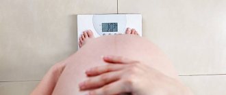
This issue is usually resolved at 35-36 weeks of pregnancy. It is by this time, according to doctors, that any unstable position of the fetus in the mother’s womb becomes stable and permanent. Of course, there are isolated cases when an already large fetus literally a few hours before birth changes the incorrect position of the body to the correct one, but counting on such an outcome is at least naive. Although it is recommended that both the pregnant woman and her doctors believe in the best.
The choice of delivery tactics is influenced by many factors. The doctor takes into account the size of the expectant mother’s pelvis - if the fetal head, according to ultrasound, is larger than the size of the pelvis, then with a high degree of probability the woman will be offered a planned caesarean section for any fetal presentation. If the fetus is large, then this is the reason for prescribing a planned cesarean section for breech and transverse presentation, and sometimes for cephalic presentation, it all depends on what weight ultrasound specialists “predict” for the baby.
An immature cervix may also be a reason for prescribing a cesarean section, regardless of presentation. In addition, doctors try not to take risks and perform surgery on women who become pregnant as a result of IVF - their birth can present a lot of unpleasant surprises.
With a breech presentation, natural childbirth is possible if the fetus is not large, the birth canal is wide enough, and the size of the pelvis allows the baby’s bottom and then his head to pass through unhindered. Natural childbirth is allowed for women with complete breech presentation, and also sometimes with mixed presentation. If the child is low weight, has signs of hypoxia, or is entangled, they will not be allowed to give birth.
We invite you to familiarize yourself with the features of the liver structure: fine-grained and medium-grained
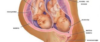
With frontal cephalic presentation, doctors also try to prescribe a cesarean section so as not to risk the life and health of the baby. If one of the two babies is in the wrong position during a multiple pregnancy, a caesarean section is also recommended, especially if the baby who will be born first is sitting or lying across the uterus. For transverse and oblique presentations, they most often try to prescribe a planned caesarean section. Natural childbirth is very dangerous.
A planned caesarean section is usually performed at 38-39 weeks of pregnancy, without waiting for the onset of spontaneous labor. The central importance in choosing a method rests on the individual characteristics of the female body and the anatomical characteristics of her baby. There is no universal risk assessment system. There can be so many nuances that only an experienced doctor can take them into account.
To see what fetal presentation is like, see the following video.
Biomechanics of childbirth
During its passage through the birth canal during birth, the child performs a number of active and passive actions. This entire complex biological process is called the biomechanism of childbirth. During its movement, the fetus performs extension, flexion, and rotation around its own axis.
The anterior and posterior types of occipital presentation have several features in the biomechanism of labor. Conventionally, the entire process of bringing a child into the world can be divided into several successive events.
Anterior type of occipital presentation
The onset of labor is accompanied by flexion of the fetal head. The baby brings his chin closer to his chest, and the position of his body begins to change little by little. Thus, the first moment of labor is associated with the fact that the fetal head begins to descend to the entrance to the pelvis.
The next point is the internal rotation of the head. As the head continues to move along the birth canal, it is forced to pass through a series of obstacles and constrictions. So that the child’s head continues to move, and its internal (correct) rotation occurs around its own axis. It occurs in the place where the wide part of the female pelvis turns into a narrow one.
Then gradually the head begins to unbend. This happens already at the exit from the small pelvis. At the same time, the fetus begins to tilt its head slightly towards the sacrum. Gradual advancement and extension of the head leads to its birth. First, the back of the head is born, then the parietal part of the head, then the forehead, the main parts of the face, and then the chin.
After the baby's head is born, active birth of the rest of the body begins. To do this, first there is an internal rotation of the baby's shoulder joints and an external rotation of the head. Next, the upper end of the fetal body begins its movement along the birth canal. When a shoulder is born, the baby's head turns toward his mother's left or right leg.
Further, under the influence of active contractions of the uterus, a strong flexion of the fetal body occurs in the thoracic spine. This contributes to the fact that the front shoulder appears first, and then the back one. After the appearance of the arms, the birth of the other half of the body begins. This happens much easier now.
Posterior type of occipital presentation
Obstetricians-gynecologists consider this option to be the position of the fetus when the back of its head faces the sacrum. It is believed that various conditions can lead to the development of this variant of the location of the fetus in the uterus. These include:
- anatomical features of the structure of the female pelvis;
- decreased functionality of the muscular apparatus of the uterus;
- individual shape of the fetal skull.
An obstetrician-gynecologist can determine the posterior type of occipital presentation in a fetus even during a routine vaginal examination. At the same time, he determines that the small fontanel on the child’s head is located in the sacral area, and the large one is located closer to the womb.
The onset of labor causes the fetal head to flex. In this case, its movement occurs in such a way that it moves through the wide part of the pelvis with its oblique size. On average it is about 10.5 cm.
The next important stage in the biomechanism of childbirth is the internal rotation of the head. In contrast to the anterior type, with the posterior type of occipital presentation, the head is rotated incorrectly. However, it only rotates 45 or 90 degrees.
The next stage of labor is gradual maximum flexion of the head. In this case, the point of fixation is the forehead. The consequence of this movement is that the back of the head comes to light up to the area of the suboccipital fossa.
After this, another stage of birth begins. It consists in the fact that the fetal head begins to gradually unbend. It is important to note that there are two points in this process - support and fixation. The fulcrum is the anterior surface of the coccyx, and the fixation point is the suboccipital fossa. Active contractions of the uterus contribute to the appearance of the forehead, and then the rest of the face. In this case, they are located towards the womb. Further stages of the birth of the remaining parts of the baby’s body occur almost the same as with the anterior type of occipital presentation.
Gymnastics for turning the fetus over
A few simple exercises will help the fetus change its position. But you can do this gymnastics only after consulting a doctor and making sure there are no contraindications: complete or partial placenta previa, threat of miscarriage.
It is better to do gymnastic exercises on an empty stomach or a few hours after eating. Don't tense up, relax your body and calm your nerves.
- Lie on your side for 7-10 minutes, take a deep and calm breath, turn over to the other side. Perform 3-4 passes during the day. It is better to lie on the elastic surface of a sofa or couch, rather than on a soft bed.
- Place one pillow under your lower back and several under your legs so that they are 20-30 cm above your head. Lie in this position for 10-15 minutes 2-3 times a day.
- A stand in the knee-elbow position is useful, which also needs to be done 2-3 times a day for 15-20 minutes.
The sleeping position is on the side towards which the baby's head is facing. Swimming is very effective. During water procedures, muscles that do not work “on land” are activated. The general tone of the body rises, blood flow to the internal organs improves and the fetus is stimulated to assume the correct vertical position.
- Lie on the side opposite the fetal head, bend your legs at the hip and knee joints. Spend about 5 minutes in this position, turn on the other side.
- Lying on your side, straighten your legs one by one. Lying on the right side - left, on the left - right.
- In a sitting position, grab the bent knee opposite the side to which the baby's head is adjacent. Gently lean forward, making a semicircle with your knee and touching the front wall of your abdomen. Take a deep and calm breath, straighten your leg and relax.
When the baby takes the desired position, it is advisable to put on and wear a special prenatal bandage throughout the day.
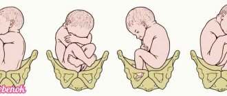
Two types of occipital presentation
During occipital presentation, the fetal head is in a bent position, and the lowest area is the back of the head. There are two types of such fetal arrangement:
- anterior view of occipital presentation; - posterior view of occipital presentation.
It is the occipital presentation that characterizes 96% of all births occurring in the last decade. The entire biomechanism of future birth will depend on the type of presentation.
Posterior occipital presentation - biomechanism of childbirth
If the occipital position of the fetus , then regardless of how it was turned at the beginning of labor, by the period of expulsion it is usually located under the articulation of the womb; accordingly, the child is born in an anterior view.
But in 1-2%, the fetus appears in the posterior view, that is, the back of the child’s head faces the sacrum during birth. The reasons for the formation of such an arrangement may be in the functional inferiority of the uterine muscles or in the physiological characteristics of the fetus.
What is childbirth with an occipital presentation?
To answer this question, it is important to first understand what an occipital presentation is. This option assumes that the baby's back is turned to the mother's back.
In this position, the baby's chin often rises. Which is why it appears larger in size.
This type of labor is considered physiologically normal. With it, the head passes through the birth canal with the back of the head forward.
In the uterus, the baby is head down, which is slightly bent. The back of the head initially descends into the woman's birth canal. Therefore, this position is called occipital. It refers to a type of birth in the cephalic presentation.
Many mothers begin to worry when they see such a conclusion in their chart. But this is not necessary, because the occipital presentation of the child does not pose any danger.
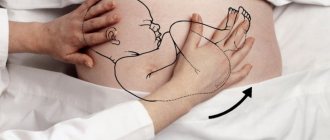
How to correct presentation
If your baby is positioned incorrectly, you can try adjusting his positioning. This is possible until the birth process begins. Unstable or incomplete cephalic presentation of the fetus in the uterus is subject to correction.
To do this, the mother must constantly change her position, more often taking exactly the position that provokes the baby’s movements. If the baby’s head is not located directly towards the exit of the uterus, but is slightly displaced, you should often lie on the side where the fetus is located.
Immediately after the position of the fetus is restored, a longitudinal presentation will take place - it must be secured. You can use a bandage for this. You can remove it only in extreme cases, when you need to change clothes or wash.
But the fetus can change its position at the very last moment. This occurs after the rupture of amniotic fluid. Then there is more space in the uterus, and the baby has room to carry out a revolution.
Classification of births in cephalic presentation
Head presentation is a common variant of the position of the baby in the uterine cavity.
Depending on which part of the head will first enter the birth canal, it is divided into:
In the facial position, the head is significantly extended. The child lowers his face into the basin.
This generic subtype is dangerous due to the development of asphyxia, and sometimes stillbirth. Therefore, most often delivery occurs by caesarean section.
Frontal presentation is one of the rarest head positions in obstetric practice. The head is lowered into the pelvis with the frontal part.
The anterior cephalic position of the child can be diagnosed during a vaginal examination. When lowered into the pelvis, the head slightly extends.
The occipital presentation of the fetus is the most correct for the birth process. In this position, the child lowers the back of his head into the pelvis and the risk of injury is minimal.
Danger and risks
This situation has little effect on the course of pregnancy itself.
However, it should be remembered that any incorrect position of the baby in the uterus is a significant risk factor for premature birth. In the case of the transverse position, this happens in 40% of cases. A child who is born much earlier than the scheduled obstetric date cannot always adapt to a new environment. Thus, if the lung tissue is immature, problems with independent breathing may arise, acute respiratory failure may develop, and with a low weight child born prematurely, it will be difficult for the baby to retain heat. If spontaneous labor begins, small parts of the baby's body and umbilical cord loops may fall out along with the rupture of water. This is fraught with the death of the child, injuries, deformities, disability, and the development of severe complications from acute hypoxia. For a woman, such childbirth is dangerous due to injuries to the pelvic bones, ruptures of the perineum, cervix and body of the uterus, vagina, and heavy bleeding. In severe cases, everything can end in the death of both the child and the mother in labor.
During rapid spontaneous labor, the baby’s shoulder is often “hammered” into the small pelvis, and this is how an advanced transverse position of the fetus develops, in which independent birth of a child is impossible. It is when the transverse position is neglected that traumatic uterine rupture most often occurs.
If the child’s position is oblique, then it is considered transitional. Theoretically, even during childbirth it can change to either longitudinal or transverse. Naturally, no one will wait to see how the baby turns out; the risks are too high.
Breech presentation during childbirth is dangerous due to the development of severe complications. The water may pour out prematurely, and along with it, it is possible that the umbilical cord, its parts, and even parts of the fetal body may fall out. Often women develop weakness of labor forces when contractions do not lead to dilatation of the cervix. Often, the birth of a child with the pelvis and legs forward leads to acute hypoxia, the death of the baby, and irreversible changes in his central nervous system.
During the birth process, the baby may throw back his arms and chin. The latter is most dangerous due to the development of disabling birth trauma associated with fractures, displacement of the cervical vertebrae, brain and spinal cord. For the mother, such childbirth is dangerous due to ruptures of the cervix, vagina, and severe bleeding.
For a child, the consequences of breech presentation can be quite unpleasant - congenital hip dislocation, pathologies of the gastrointestinal tract, kidneys and urinary system, injuries, and the development of cerebral palsy.
However, dangers lurk not only during childbirth, but also during pregnancy. In the first half of gestation, breech presentation of the fetus increases the likelihood of miscarriage and hypoxia; the risks of developing early gestosis are also considered increased. In the second half of pregnancy, a woman whose baby is positioned head up is at risk of premature birth, preeclampsia, including severe preeclampsia, and premature placental abruption.
Women with a breech presentation of the fetus have a 60% increased risk of developing fetoplacental insufficiency and subsequent fetal malnutrition. In a state of lack of nutrients, vitamins and oxygen, the baby’s nervous and digestive systems do not develop well and quickly, there are problems with the endocrine system and the functioning of the heart and blood vessels.
From 34-35 weeks of pregnancy, if the baby does not turn over to the head position, the rate of development of the structures of the medulla oblongata slows down, which leads to disruptions in the functioning of the pituitary gland and adrenal cortex. Negative changes in a child who occupies an incorrect position in space also occur in the genital area - swelling and hemorrhages occur, subsequently a girl may develop exhausted ovarian syndrome, and a boy may experience oligozoospermia or azoospermia. Among children with congenital heart defects, there are many who spent the entire nine months with their head up and bottom down.
Fetal position in occipital presentation
There are 2 varieties:
- anterior view of occipital presentation;
- posterior view of occipital presentation.
In the first option, the baby's head is in a bent position. The lowest part will be the back of the head. The child’s back is most often turned forward and slightly to the left.
Childbirth in the anterior form of occipital presentation is the most correct from a physiological point of view.
In the posterior version, the child is also positioned head down, with the chin pressed to the chest. In this case, the back of the head moves forward. The child's face is turned to the mother, and his back is to her spine.
Childbirth in the posterior form of the occipital presentation of the child is a little more difficult.
Possible complications during childbirth
Childbirth for a woman is a very difficult process, even though it is inherent in nature.
Complication of childbirth is understood as a process that occurs during a woman’s labor. It negatively affects its course and outcome.
Unfortunately, about 70% of all births are accompanied by complications, and 30% of them end with surgical intervention.
Main causes of complications
- Features of the structure of the mother's birth canal. This includes a narrow pelvis, a saddle-shaped uterus, decreased tissue elasticity, etc.
- The presence of inflammation in the mother's genital organs.
- Difficult pregnancy.
- Previous previous terminations of pregnancy, operations and surgical interventions that left scars and connective tissue formations.
- Injuries. This includes a variety of ruptures that occurred during the woman’s past births.
- Maternal hypertension.
- Diabetes.
- Myopia.
- Malposition.
- Pathological features of the child's structure. Large size and the presence of congenital defects can interfere with the normal course of labor.
- Multiple pregnancy.
- Incorrect structure and pathological functioning of the membranes around the fetus. Umbilical cord entanglement, polyhydramnios, premature rupture of the amniotic sac, low placentation.
- Incorrect monitoring of pregnancy.
All complications are divided into the following groups:
- Child infection;
- Severe fetal hypoxia;
- Bleeding;
- Injuries and ruptures of the mother during childbirth;
- Hypertensive crisis;
- Retinal detachment in the mother during a prolonged period of pushing;
- Embolism. With this complication, amniotic fluid enters the bloodstream of the woman in labor. 80% of such births end in death for both mother and child;
- Leaving parts of the placenta, blood clots and tissue in the uterine cavity.
Symptoms that signal possible complications
- Changes in contractions and pushing period;
- Strong pain;
- Uncharacteristic discharge of water from the vagina;
- Stopping the baby's movements;
- Changes in the state of health of a woman in labor.
If signs of complications appear, it is necessary to be examined by a doctor, who should conduct the following studies:
When determining the type of complication, the necessary therapy is prescribed or the process of labor is changed.
Prevention of complications
Each woman can independently reduce the risk of complications during childbirth. To do this, it is enough to visit a doctor in a timely manner and undergo the necessary examinations.
With correct diagnosis of any pathology, the risk of an unfavorable outcome is significantly reduced.
In the early stages of development of anomalies during gestation, it is possible to correct them.
Occipital presentation of the fetus is a variant of the normal physiological birth process.
If all recommendations from obstetricians are followed, childbirth in most cases occurs without complications.
The posterior view of this presentation is not a pathology and does not require special observation. However, if signs of abnormal progress of labor appear, timely informing the doctor makes it possible to correct them as much as possible.
Complications of childbirth are a common occurrence in obstetrics, and experienced doctors almost always know how to deal with these situations.
Author: Anita Igorevna, pediatrician
Especially for the site kakrodit.ru