Trichinosis is a disease caused by the parasite Trichinella spiralis, which is present as an adult in the intestines, and as larvae in the striated muscles of humans and various domestic and wild animals.
This parasite is the smallest representative of nematodes (roundworms). Cases of trichinosis are inextricably linked with the consumption of poorly cooked meat, which contains the larvae of spiral trichinella. Thus, the route of human infection is oral.
The causative agent of trichinosis
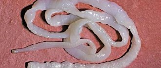
The causative agent of this helminthiasis is Trichinella, the genus of which includes more than 10 species (class of nematodes). But only one of them causes this disease - the parasite Trichinella spiralis. Sexually mature individuals enter the digestive tract, where (the small intestine) they begin to actively reproduce. Trichinella are viviparous parasites; one female can produce up to 2000 larvae over the entire period of its existence. The length of females does not exceed 1.8 mm, and after fertilization they reach 4.4 mm. In males, the body length is slightly less - 1.2 - 2 mm. It is typical that after fertilization, male parasites die, and females begin to secrete larvae. For Trichinella, humans are both the definitive (adults are parasitic in the intestines) and intermediate (the larvae are found in the muscles) host.
How can you get infected?
The mechanism of infection with trichinosis is nutritional, and the route of transmission is oral, through meat contaminated with trichinosis. The disease is a natural focal disease, although foci of infection can be not only natural, but also synanthropic.
In natural foci, helminthiasis is common among wild animals (source of trichinosis):
- wild boars;
- bears (both brown and white);
- foxes;
- seals;
- seals;
- nutria;
- badgers;
- whales.
Synanthropic foci are formed in human habitats after feeding game waste to domestic animals. Helminthiasis is common among pigs, dogs and cats. In this case, human infection with trichinosis occurs by eating infected pigs (in some areas, dogs).
Human susceptibility to this disease is quite high. When eating 10–15 grams of contaminated meat, parasite infestation occurs in 100% of cases. The development of helminthiasis in entire groups of people or families after a joint feast and eating game is typical.
Development cycle
1 - 1.5 hours after eating meat infected with parasite larvae, they are released from the capsule in the digestive tract with the help of enzymes and gastric juice and penetrate into the small intestine. In 1.5 days, the larvae reach the stage of sexually mature individuals, and on days 3–4, the female parasites begin to produce new larvae. The female can survive in the small intestine for 42–56 days. New larvae pierce the mucous membrane of the small intestine, penetrate the lymphatic system, and from there into the circulatory system. With the blood flow, the newly formed larvae are spread throughout the body and settle in the striated muscles (5 - 8 days after infection). As a rule, the muscles of all organs are affected, with the exception of the heart (muscles of the tongue and forearm, diaphragm and intercostals, gastrocnemius and deltoid). On days 17–18 after penetration into the muscles, the larvae mature and become infective. While the larvae are in the muscles, they actively begin to produce hyaluronidase, with the help of which they penetrate the muscle fiber where they become covered with a capsule (3 – 4 weeks). The capsule covering the larva provides it with nutrition and protection. After six months to a year, the capsule becomes calcified (calcium salts are deposited), which indicates the end of the development of the parasite. In this encapsulated form, the larvae retain their viability for 25–40 years.
Life cycle of a parasite
- Helminth larva. This intermediate form of the parasitic organism is formed after worm eggs enter the human gastrointestinal tract along with raw or undercooked meat or water.
- A healthy individual in the human body. The formation of a healthy individual occurs immediately after the appearance of the larva (active growth). In just a day, the protective shell disappears and a full-fledged parasitic individual appears.
The life cycle of Trichinella and the speed of its reproduction and the fact that a healthy individual lays up to two thousand eggs per day determines the rapid development of a dangerous disease. The parasite easily moves throughout the human body and infects its internal organs. After a week or a week and a half, there are a hundred helminth larvae inside a person.
Prevalence of trichinosis
Natural foci of helminthiasis have been recorded in North America, Germany and Poland, Ukraine and Belarus, as well as in the Baltic states. In the Russian Federation, trichinosis is most common in the Khabarovsk and Krasnoyarsk Territories, in the Magadan Region and in the Krasnodar Territory. In total, the disease is recorded everywhere, with the exception of the Australian continent.
Contribute to the spread of helminthiasis:
- the ability of the pathogen to tolerate high and low temperatures, which ensures its survival in any climatic conditions;
- high human susceptibility to trichinosis;
- group outbreaks - collective consumption of contaminated meat;
- unstable immunity, which provokes repeated cases of infection after the initial infection.
Phases of development of helminthiasis
The development of helminthiasis occurs in several stages:
- Enzymatic-toxic
The initial phase of the disease takes 7–14 days after infection. Infective larvae enter the intestinal mucosa, where they develop into adult Trichinella, which during their life processes form enzymes and metabolites, which causes intestinal inflammation.
- Allergic
The allergic phase develops 3–4 weeks after infection. The occurrence of allergic reactions is, firstly, associated with the effect of toxins and dead parasites on blood vessels and blood clotting, and, secondly, with the introduction of newly formed larvae into the striated muscles. A protective granulation ridge is formed around the larvae in the muscles, and protective antibodies appear and grow in the blood. As a result, massive death of larvae occurs, which leads to the development of allergic myositis and the formation of nodular infiltrates in the internal organs, which include eosinophils, plasma cells and lymphoid elements.
- Immunopathological
Develops by 5–6 weeks after invasion. Systemic vasculitis and associated severe organ damage occur. Complications of trichinosis such as meningoencephalitis, myocarditis, focal pneumonia and others appear.
Classification
Depending on the severity, the disease occurs in mild, moderate and severe forms. Typical and atypical forms of helminthiasis are also distinguished. Atypical include erased and asymptomatic trichinosis. There are acute and chronic trichinosis.
Clinical picture
There is no visible physical harm when infected with this helminthiasis. There are no changes in the muscles. The proteins of parasites, which are very strong allergens, have a negative impact, which provokes the development of severe allergic reactions, leading to damage to blood vessels and joints. Symptoms of trichinosis depend on the degree of parasite infestation. The incubation period is 10–25 days, but it can be extended to 45 days. It is typical that the more severe the helminthiasis, the longer the latent period.
Manifestations of mild and moderate forms
The disease includes 2 periods:
- acute (allergic reactions and damage to internal organs);
- recovery period.
The tetrad of symptoms of trichinosis in humans includes:
- fever;
- edema syndrome;
- pain syndrome (in muscles);
- high level of eosinophils in the blood.
Clinical manifestations:
- Fever
The temperature rises to 40 degrees for several days, then sharply decreases to 37, which lasts 7 - 10 days. In some cases, low-grade fever persists for several months. A mild course of the disease may not be accompanied by hyperthermia. Against the background of high temperature, signs of general intoxication appear: weakness, chills, sweating and nausea, headache.
- Swelling of the face
A characteristic sign of helminthiasis is the appearance of swelling of the eyelids and face as a whole, which is often combined with conjunctivitis. As a result, trichinosis is also called “puffiness.” Swelling occurs on the 1st 05th day of the disease and persists for up to two weeks. In cases of severe disease, swelling appears more slowly, but lasts longer. The sequence of appearance of edema: eyelids, brow ridges, entire face. In severe cases, swelling reaches the neck, torso, arms and legs, which is considered an unfavorable prognostic sign.
- Myalgia
Starting from the third day of illness, sometimes later, muscle pain occurs in various muscle groups. First of all, myalgia appears in the muscles of the legs (calf muscles), then spreads to the gluteal muscles, muscles of the back and abdomen, covers the arms and shoulder girdle, rises to the cervical and pharyngeal muscles, muscles of the tongue, chewing and oculomotor muscles. Such pain is very pronounced, intensifies with movement, and palpation of the muscles causes pain. In severe cases, myalgia with contractures develops, which leads to immobilization of the patient. Both myalgia and edema persist for up to 1–3 weeks and periodically recur, which is accompanied by an increase in temperature.
The acute phase of the disease is accompanied by abdominal syndrome (pain, nausea, vomiting, diarrhea) and allergic manifestations - rash (rosacea - pink blisters that turn pale when pressed, urticaria - spots merging with each other, urticarial - blisters raised above the skin).
- Eosinophilia
The level of eosinophils increases to 80%, which appears with the first symptoms of helminthiasis. The maximum increase in eosinophils is observed at 2–4 weeks of illness (mild up to 30%, moderate up to 60%). An unfavorable prognostic sign is an increase in eosinophils to 95%.
Severe course
A severe form of the disease is accompanied by the development of organ lesions that appear 3 to 4 weeks after infection. It is organ damage that often becomes the cause of death of the patient.
The following complications arise:
- allergic myocarditis is the leading cause of death of the patient (increased heart rate, decreased blood pressure, signs of acute vascular insufficiency, ECG symptoms of diffuse myocarditis);
- lung damage - the development of pneumonia in combination with pleurisy, which is caused by systemic vasculitis;
- brain damage (encephalomyelitis, encephalitis, meningitis);
- liver damage - hepatitis;
- thrombohemorrhagic syndrome (blood clotting disorder);
- kidney damage - nephritis.
The duration of the disease in the case of a mild course ranges from 7–14 days to 35–42. If treatment is started on time, the duration of each period is reduced by 3–5 days. The recovery period in case of severe helminthiasis is delayed to half a day or more. Prolonged muscle pain occurs in patients taking immunosuppressive drugs (glucocorticoids).
Symptoms depending on the stage of the disease
Infestation stage
This continues for a week from the date of consumption of contaminated meat. The parasite larvae enter the stomach, and from there into the small intestine, where they attach themselves to the mucous membrane and cause inflammation. At this stage, the disease is accompanied by diarrhea, alternating constipation, epigastric pain, nausea and vomiting, and lack of appetite.
Dissemination stage
Develops 14–28 days after infection. Newly formed larvae produced by mature parasites penetrate into the muscles with blood and lymph. There they take hold, grow and begin to release allergens into the blood. Intoxication of the body and allergic reactions develop. At this stage, the following signs of the disease appear:
- periorbital edema - the eye muscles affected by the larvae become inflamed, interstitial fluid accumulates in them - the eyelids, bridge of the nose swell, and eye movement is painful;
- hemorrhages in the retina, conjunctiva - allergic vasculitis, accompanied by itching and lacrimation;
- hyperthermia – caused by the action of parasite toxins;
- puffiness of the face - penetration of larvae into the lingual and masticatory muscles - rashes on the skin of the face;
- headache is a consequence of toxic brain damage;
- malfunction of the central nervous system - damage to brain cells by parasite toxins leads to insomnia and depression;
- dysphagia – difficulties with swallowing are caused by the penetration of larvae into the swallowing and chewing muscles;
- cough, breathing problems - as a result of an allergic reaction, a large amount of sputum is released;
- rash is an allergic response to the action of helminth toxins.
Encapsulation stage
Lasts 6 weeks - 6 months after invasion. At this stage, tissue regeneration begins. The larvae grow up to 0.8 mm and curl into a spiral. Each spiral-shaped larva is delimited by a capsule in the muscles and freezes in development. Parasite toxins cannot enter the body, and gradually all manifestations of the disease disappear. The deposition of calcium salts in the capsule can destroy the larva, but in general they retain their viability for up to 25–40 years. Encapsulated larvae do not affect the patient’s well-being.
At this stage, the functions of the affected organs are restored (15–20 days), muscle pain persists for up to 2 months, and eosinophilia for up to 3 months.
Routes of infection
Trichinosis - what is it and how does this pathology spread? Trichinosis is a disease that has a natural focal source of infection. This is due to the fact that the main carriers of these parasites are wild animals. Trichinae are also spread by insects, whose infected individuals come into contact with plants that are food for animals.
Infection of pigs with parasites can occur due to the consumption of slop or raw waste, as well as after contact with rats.
The ways of infecting humans with trichinosis are very simple; it is enough to eat raw or undercooked meat, and the parasites enter the body. The meat that is especially dangerous is not from livestock, but from animals that grew up in the wild. Interestingly, parasites are not able to survive where there is no meat. For example, they die in pure lard, but if there is even a small proportion of meat in it, there may be worms in it. The human body cannot acquire this disease from infected people.
Diagnostics
Diagnosis of trichinosis is difficult, since in the early stages of the disease it is often confused with influenza, colds and allergies of unknown origin. A thorough medical history plays an important role in diagnosis; the doctor should be alert to the patient’s consumption of game. At the same time, all participants in the feast are required to be examined.
Also, suspicion of trichinosis should be caused by a characteristic set of symptoms: periorbital edema, high fever and muscle pain.
Laboratory research methods:
UAC
A high content of eosinophils is found in the peripheral blood (50–80% of the total number of leukocytes), which indicates an allergic reaction of the body to the waste products of parasites. The level of leukocytes also increases, signaling an inflammatory process.
Serological diagnosis
It consists of identifying antibodies that are produced by the patient’s body after the introduction of parasites. The test for trichinosis involves the reaction of blood with administered antigens, which are isolated from Trichinella larvae. The following tests are performed as serological diagnostics:
- ELISA;
- RSK;
- RNGA;
- RIF and others.
Skin allergy test
The essence of the method is to provoke an allergic reaction as a response to the introduction of trichinosis antigen. For this purpose, a solution of antigens is injected subcutaneously. If hyperemia appears and a blister forms at the injection site, the test is said to be positive, which confirms the presence of parasites in the body. The test becomes positive from the 2nd week of illness and persists for 10 years.
Muscle biopsy
To carry out trichinoscopy - identifying larvae in the muscles, the deltoid and gastrocnemius muscles are examined. The material is obtained by puncture of the muscle, and then studied under a microscope.
Meat check
Mandatory diagnosis of suspected helminthiasis includes examination of the meat consumed by the patient and his family. Testing for trichinosis should be carried out for preventive purposes on all killed game and meat (pork) going on sale. Infected meat can contain up to 200 larvae per gram. Externally, such meat is no different from normal meat; the color and smell are identical to the meat of healthy animals. The larvae of the parasite can only be identified under a microscope in a laboratory setting. Meat that has been tested and is safe against trichinosis is marked accordingly.
Tests for this helminthiasis are carried out by veterinary laboratories available in markets and the laboratory of the sanitary and epidemiological station. Sampling is done from the entire carcass. Small pieces of meat (about 5 grams) are collected from the intercostal muscles, diaphragm, tongue and masticatory muscles. If at least one larva is detected, the meat is considered contaminated and destroyed. To destroy, they dig a hole at least one meter deep, douse the meat with kerosene and bury it. Or clean meat is covered with quicklime and buried.
Prevention measures
To prevent people from becoming infected with trichinosis, many recommendations have been developed around the world, some of them are approved in regulations. This primarily concerns the living conditions of farm animals and meat quality control. On the consumer side, we can only hope that manufacturers adhere to the standards
. It is also important to adhere to the rules of prevention at home.
Basic recommendations:
- Do not eat meat from adult carnivores. When a predator reaches the age of 6-7 years, the probability of infection is 80-90%.
- Do not allow pigs to eat raw carcasses of other animals, including rats, which may be infected with Trichinella.
- Analysis of the harvested animal carcass is carried out through a microscope (trichinelloscope), by cutting strips of tissue from different areas (tongue, larynx, abdominal muscles) 24 times longitudinally along the fibers with a size of 5x5x2 mm, the accuracy of this “field” method is 70-80%, operations are performed with disposable gloves, cutting is done with a disposable blade (stationery knife).
- If there is a suspicion that the carcass is contaminated, bury it to a depth of 40-60 cm with obligatory filling with bleach; boil the tools used for 1 hour.
It is necessary to cook meat from wild animals more carefully. Freezing bushmeat, as opposed to freezing pork products, even for long periods of time, may not be effective
.
This is because these Trichinella species are more resistant to freezing than the species that infect pigs
.
At the usual temperature of frozen meat (−10 °C), T. spiralis larvae survive for a long time, at −12 °C for up to 57 days, at −18 °C for up to 21 hours
.
T. nativa larvae can withstand freezing down to −23 °C for 3 days, and at −16 °C they remain invasive for 20 months
.
At temperatures above +50 °C, the larvae survive for several minutes, but you must remember that when preparing meat dishes, this temperature is not always reached in the depths of the piece
. When salted, the causative agents of trichinosis can persist in the depths of the piece for up to 1 year.
Treatment
Treatment of trichinosis in humans must be carried out inpatiently, with the exception of erased forms of the disease. Therapy of the disease includes 2 directions: etiotropic and pathogenetic. Etiotropic treatment of trichinosis includes the prescription of drugs that have a detrimental effect on parasites localized in the intestines, suppress their ability to produce new larvae, disrupt the encapsulation process and increase the death of helminths located in the muscles. For this purpose, use:
- Mebendazole (Vermox)
It prevents parasites from absorbing glucose, which disrupts the production of ATP in their bodies, leading to metabolic disorders and further death of Trichinella. The drug is not prescribed to pregnant and lactating women. Dosage: 1 – 2 tablets three times a day for 10 – 14 days.
- Albendazole
The effect of the drug is similar to Mebendazole, high activity was noted against parasite larvae. Dosage: 10 mg per kg of patient weight for 10 – 14 days.
- Tibendazole
The effectiveness of the drug is lower due to severe adverse reactions. Dosage: 25 mg per kg of patient weight. The course lasts 3–5 days, the medicine is taken twice a day. If necessary, repeated treatment is carried out (after 7 days).
Pathogenetic treatment includes the prescription of non-steroidal anti-inflammatory drugs that help reduce muscle pain, fight inflammation and reduce temperature (nurofen, paracetamol, diclofenac and others). In addition, antihistamines are necessarily prescribed, especially during treatment with antiparasitic drugs. Antihistamines reduce allergic manifestations and prevent the severity of allergic reactions in response to etiotropic treatment.
In severe cases, with the development of organ pathology, glucocorticoids are included in the treatment regimen. Glucocorticoids suppress the immune system, thereby reducing allergic manifestations.
The effectiveness of treatment is assessed after six months to a year.
In case of immobilization of the patient and the development of muscle contractures, massage, physiotherapy and special gymnastics are prescribed.
In parallel with the main treatment, the patient is prescribed hepatoprotectors and multivitamins, as well as drugs to improve microcirculation.
Consequences of the disease
It is already clear what the disease trichinosis is. It is clear how insidious it is, but we still need to know what consequences it has for our body, especially in cases of refusal to treat the disease. Because of this disease, serious problems develop with the respiratory system, disruptions in the central nervous system, disorders of the heart muscle, and problems also arise in the blood vessels.
Undergoing treatment for trichinosis is a huge blow to the immune system; the patient can lose almost all his strength. There are cases of signs of pneumonia, myocarditis or meningoencephalitis, but they do not appear as an independent disease, but as consequences after suffering trichinosis. Deaths are possible.
