4D ultrasound during pregnancy - negative reviews. Neutral. Positive. + Leave a review Negative reviews Nevskaya https://deti.mail.ru/forum/v_ozhidanii_chuda/beremennost/uzi_3dili_4d_stoit_li_delat/ made three-dimensional, they did not record it on disk. it was not useful, I did it for myself okssana1988 https://nashemnenie.com/detskoe/11255-4d-uzi-vo-vremya-beremennosti.html Almost all clinics now do 3D and 4D ultrasound in addition to regular ultrasound. They promise a super clear picture that you will be able to see the baby in almost the smallest detail and even understand what he will look like when he is born. 4D is also a super clear picture + also recording in four dimensions, including time. In St. Petersburg, the difference in prices is about 1000, respectively, 4D is the most expensive)) In fact, I didn’t notice much of a difference between a regular ultrasound and 4D, although I went for 4D already at 32 weeks, when, as they wrote on the Internet, you can consider almost the expression on the baby's face. In fact, everything looks almost exactly the same, there are no super-duper clear images, and the fact that the picture is three-dimensional is not particularly clear, well, unless they point at a face, and then you have to use your imagination))) In the end, I overpaid an extra 2000 and I received a disc with a recording of everything that happened on the screen. I have no idea where to put the disk, because... Without the doctor’s explanation “this is a pen, and these are fingers, and now I’m measuring the circumference of the head,” I simply don’t understand half of what is written on this disc. So my advice is to do a regular ultrasound and not bother with these fancy things, but rather spend the money on something more useful. ElenaRoll https://otzovik.com/review_4094049.html Advantages: None Disadvantages: Expensive, not informative Being pregnant for the first time at 35 weeks, I did as I was then told the necessary 4D ultrasound. Registration for it was two weeks in advance, it was done in only one place and for a lot of money. Being inexperienced, I agreed to such an ultrasound. Either the ultrasound specialist I came across was not qualified, or the 4D ultrasound was not as informative as they say, but in the end, when this ultrasound was done, they didn’t tell me anything about my daughter’s double heart defect. How it was possible not to see two holes in the heart of an already formed child still remains a mystery to me. If I had known about the defects, the birth process would have been completely different for my child. My opinion is that it is not worth paying so much money for an ultrasound that can be done with a regular ultrasound machine. The only plus from this ultrasound was good photos of my daughter when she was still in the tummy, but that also cost extra money. Neutral reviews [email protected] https://irecommend.ru/content/stoit-li-pereplachivat-za-4d-uzi-namnogo-li-ono-informativnee-obychnogo-uzi Good day everyone. During pregnancy, my husband and I were looking forward to the time when we could look at the baby with a 4D ultrasound. In my city they don’t do this kind of ultrasound for free, so I chose among private paid clinics. I chose a clinic that works on Voluson E8 Expert from General Electric. Expert device "Voluson" E8 - new possibilities for volumetric color ultrasound in 3D and 4D modes. What is 4D ultrasound? Doctors also call this type of examination “live” ultrasound. This newest method allows the specialists who perform it to see not a static picture on the monitor of an ultrasound machine, but a full-fledged three-dimensional image. In addition to the ultrasound image, you see the baby’s movements in real time. The images are different from what you are used to seeing on a regular ultrasound. But it’s not a fact that you will be able to understand something... Cost. In the clinic where I had an ultrasound, the pricing was as follows: 1700 cost of ultrasound + 500 rubles for 4D ultrasound + 500 rubles for recording on disk. First, the cost of a regular ultrasound was paid, then a 4D ultrasound was done, and if the patient was satisfied with everything, then after the ultrasound there was further payment. We decided that we wouldn’t record it on disk, so the ultrasound cost us 2,200 rubles. Process. Honestly, in essence this study is no different from a regular ultrasound. It just takes a little longer. But first of all, the service is aimed at excluding or identifying developmental pathologies. Only then for you to take a closer look at your baby. Images. The doctor can give you advice on when it is best to do this ultrasound. It all depends on what you want to see. If it’s a full baby, then it’s better to do an ultrasound at 22-24 weeks; if you do it closer to 30 weeks or later, you’ll only see the face. So, the choice is yours... We decided to see the baby in full and did an ultrasound at 22 weeks. The baby was spinning very actively, so in most of the photographs we saw only his sexual characteristics. There were also some photographs that were not very clear. Unfortunately, we were not able to see the facial features as such, but we really wanted to see what our baby looked like before birth. Later, at 32 weeks, I did a scheduled ultrasound as expected and was given a photo. In my opinion, it conveys the child’s facial features even better than the result that I got from a 4D ultrasound. Is it worth doing a 4D ultrasound? It’s up to you to decide whether to do such an ultrasound or not. I decided for myself that a regular ultrasound was enough for me. It is no less informative and will show how the baby is developing. I received absolutely the same information regarding development at the scheduled free ultrasound and at the paid 4D. Thank you for your attention. Positive feedback Anonim https://zoon.ru/msk/medical/detskij_meditsinskij_tsentr_preambula_v_novokosino/?zutm_source=zbd&zutm_medium=none Children's Medical Center "PreAmbula" on Novokosinskaya Street is a specialized institution specializing in the diagnosis and treatment of the youngest patients. In order to guarantee first-class results, the rooms are equipped with the latest technology, and a carefully formed staff of doctors practices an individual approach, uses advanced techniques and proven safe drugs. The consultation is conducted by experts in the field of pediatrics, cardiology, neurology, ophthalmology, allergology-immunology, orthopedics, dermatology, endocrinology, psychology, surgery, traumatology, urology and other areas. The center also offers to take the necessary tests, do an ultrasound, undergo comprehensive examination programs, issue a certificate for a swimming pool, for admission to a school or university, a sanatorium-resort card, and call a doctor at home. Tuesday, Thursday and Sunday are dispensary days when prompt access to the necessary specialists is available. You will receive answers to all your questions by calling the specified phone number. Contact PreAmbula, here your and your child’s health will be in safe and caring hands. Type honey institutions children's clinic, family medicine center Diagnostics vision diagnostics, laboratory diagnostics, comprehensive examination programs, ultrasound, pregnancy ultrasound, functional diagnostics, endoscopy General medicine allergology, andrology, gastroenterology, gynecology, immunology, cardiology, speech therapist, neurology, neonatology, nephrology, orthopedics, otorhinolaryngology, ophthalmology, pediatrics, psychology, urology, endocrinology Regenerative medicine vaccination, adult vaccination, therapeutic massage, manual therapy, osteopathy, treatment room, psychological assistance Medicine for children pediatric allergology, pediatric andrology, pediatric gastroenterology, pediatric gynecology, pediatric immunology , pediatric cardiology, pediatric neurology, pediatric orthopedics, pediatric otolaryngology, pediatric ophthalmology, pediatric psychology, pediatric urology, pediatric surgery, pediatric endocrinology, pediatric massage, pediatric medical examination, massage for infants, patronage of newborns, ultrasound for a child, ECG for a child Home medicine tests at home, calling a doctor at home, calling a pediatrician at home, calling a neurologist at home, calling a urologist at home, calling a surgeon at home, children's massage at home Medical examinations and certificates, medical certificates, sick leave, health resort card, certificate in swimming pool, certificate for kindergarten, certificate for camp, certificate for sports, certificate for admission to a university, certificate for admission to school Daria https://www.babyblog.ru/user/id2632991/12817 Just recently I started a diary here, so that later I can remember all the joyful moments of my pregnancy (and how else, if not joyful?). It’s only 25 weeks, and I’ve already had an ultrasound 9 times, well, I can’t sleep peacefully when I don’t know how our son is doing in his tummy and whether everything is fine with him. Of all the procedures, I remember two most of all: the first, when they said “Your baby is developing exactly on time, listen to his heartbeat” - at 7 weeks and 3 days and completely... Maria https://www.baby.ru/blogs/post/ 20648850-15095055/ I'm delighted! today... my daughter really looks like me... Dudls http://kupi-slona.com/catalog/4d-uzi-vo-vremya-beremennosti/889 During scheduled ultrasounds (and there were 8 of them throughout the entire pregnancy), they periodically offered me take a photo of the monitor screen. But I have a phone to make calls, not to take selfies. Therefore, every time I surprised the uzists with the answer that... Natalya Eremchenk https://galya.ru/clubs/show.php?id=964146 I have already done 3D-4D ultrasounds during pregnancy 2 times, and it seems to me that the main thing here is to choose a center with normal equipment and a professional doctor, so that there are no questions later “about the baby’s eyelids” - this is simply terrible! Both times I did a 3D ultrasound at Ultrasound 21 CENTURY in St. Petersburg, the first time at 12 weeks the baby was very small, the second time the screen fit somewhere up to the waist, and the facial features are already clear, we are waiting for the third trimester to make a good portrait! KatiaKaterinka https://ekodeti.ru/blog/analizy_beremennost/post158 My brother’s wife went to see me twice, but she had indications, low placenta previa + antibody titers. She was very upset, because she expected more, in the end she was given a photo that was practically no different from a two-dimensional image, the research itself was so fast that she did not have time to see anything on the monitor. Perhaps everything was like this, due to the fact that they were referred for free; paid patients most likely spent more time on the patient. Our homeopath is against doing an ultrasound again without indications, since it is still a small amount of radiation, and the baby is still 99% water, and as you know, ultrasound waves change the structure of water. Of course you want to look at him, but soon your meeting will be real) etolmacheva https://otzovik.com/review_3203877.html Advantages: allows you to get a good look at the child Disadvantages: expensive Good day to all readers of reviews! Continuing the topic of various studies that I underwent during pregnancy, I want to talk about 4D ultrasound. Of course, this is an expensive pleasure, I want to say right away, but it’s worth it. I did this research at the Interregional Medical Genetics Center GENOM (Ukraine, Donetsk). When I first came to this center, I had to undergo an ultrasound and biochemical screening of the first trimester, I sat in the corridor and waited for my turn, and in one room there was a 4D ultrasound sign. I thought this must be very interesting. But I didn’t intend to do such a study on purpose, because it said it was very expensive. But then, in the late stages of pregnancy, my doctor prescribed me an ultrasound with Doppler in the same center, and there such ultrasounds are performed using equipment with 4D ultrasound. So I was lucky, so to speak, for going through it. 4D ultrasound is performed almost the same as a regular one, only on the monitor we see not a flat image of the fetus, but a three-dimensional one. I underwent this examination twice. The first time I was lying on the couch and there was a monitor hanging above me that allows me to see everything. My husband and I were there, we really liked it - we saw our daughter’s face, it was so funny. They asked us to take several photos, they printed out 4 photos on a color printer, and I also asked to put them on a flash drive, for this pleasure we paid about 100 hryvnia, just for the photo, and the study itself, together with Doppler sonography, cost about 400 hryvnia, this was all in 2012. The second time I was already in another office and the monitor was not hanging above me, but the doctor then showed me the child, but the back of her head with ears, because she turned away. In general, it’s very cool to see the baby still in the belly, that’s how much progress has come. I recommend to everyone! See you soon! dzhonik35 https://otzovik.com/review_5420391.html Advantages: facial features Disadvantages: price More than a year has passed and now I decided to share my opinion about the well-known ultrasound procedure during pregnancy - 4D ultrasound... How great it is that progress does not stand still, but moves at low speed, but still. Eleven years ago, I couldn’t even imagine that it was possible to do such a realistic, let’s say, ultrasound... And a little over a year ago I had to do such an ultrasound of my own free will... Of course, the procedure is not cheap, not everyone can afford it, and We don’t do it in all places... In general, I did it and didn’t regret it - yes, indeed, how great it is when you can see your baby still in the stomach, and thanks to 4D, you can also see his face and facial features... I was lucky with a specialist , and the baby was lying well, so it was wonderful to look at him... Unfortunately, the photo has not survived to this day, but with the move many things would have to be left behind... I think that if you have the money, then it’s definitely worth doing a 4D ultrasound at the second screening -especially since the procedure is safe if you do it wisely, instead of the usual second screening, for example, do 4D, I recommend it! I am Mommy https://otzovik.com/review_5252243.html Advantages: informative, painless, visual, free procedure Disadvantages: the child is not pleased by the strong light rays. He constantly turns away. I had two pregnancies done with 4D ultrasound many times at the DIAGNOSTIC CENTER OF THE ALTAI REGION. We usually schedule this 4D ultrasound at 12-13 weeks, then at 20 weeks and then around 32 weeks. Referrals from the antenatal clinic are given free of charge, which is very pleasing. I usually arrive on the appointed day at the appointed time. I register and wait for an invitation to the office. A doctor and a nurse are sitting in the office at the computer. I lie down on the couch. The doctor begins to move the sensor along the lower abdomen. The doctor looks at the second and third 4D ultrasound first, passing along the abdomen, then intervaginally. In front of me, above the stream, hangs a large screen, where I see my baby, how he moves, moves his arms and legs. How it turns over. I see everything in volume, almost as if in reality. The only unpleasant thing is that this procedure is not very useful for the child. My children kept turning away from the strong light during the study. The procedure lasts about 5 minutes if everything goes well. Then the nurse and doctor fill out a protocol sheet and hand it over to me. If there are deviations, they are immediately referred to a geneticist. If not, go home. In the antenatal clinic I show the 4D ultrasound protocol. Informative, painless, visual. They also give you a black and white photo. I recommend! tapcherello https://otzovik.com/review_2532199.html Advantages: Positive emotions, excellent quality, good memories) Disadvantages: Didn’t notice) My husband and I went for a 4D ultrasound at 30 weeks of pregnancy, when the fetus is fully formed and you can see everything, and so before the appointment I ate a chocolate bar so that the baby would wake up and show himself to us, I went into the office, turned off the lights and started, the baby still turned his butt towards us, but we managed to see him), we looked at his nose, legs, arms, he showed us never mind) it was such a joy to watch how the baby in my stomach rubs his eyes, shows something on his fingers, stomps his feet) we also managed to see the floor, and it was the long-awaited boy), everything was recorded for us on video, about 10 photographs were printed, and I ran happy to show my parents the little one) by the way, my husband was also delighted, he couldn’t believe that such a baby could already do something in his tummy)) I advise everyone to go and get a lot of memories and joy) dj-tata https://otzovik. com/review_1709337.html Advantages: informative Disadvantages: not a beautiful color During my first pregnancy, I had routine ultrasounds at my antenatal clinic completely free of charge, at the first ultrasound at 12 weeks I still understood what I was seeing, at the second and third even though the ultrasound doctor showed me different parts the bodies of my baby, I looked at them with difficulty. My second pregnancy was long-awaited, after all, five years had passed. I also did the first ultrasound screening at 12 weeks for free in my consultation. I decided to go to the second ultrasound with my husband and show him our little miracle. I started looking for paid centers (husbands are not allowed into ultrasound consultations) I saw that they do 4D ultrasounds, the price is not significantly higher, and you can see the baby better, in addition, at the second ultrasound they tell you the gender of the child. And even if there are all kinds of pathologies, the doctor will see them better in Figure 4D. So the ultrasound, it goes exactly the same way as a regular ultrasound, but it is three-dimensional and even the baby’s movements are visible. Both my husband and I liked it, we saw the face and gender of our baby))) Of the minuses - it’s still not a camera, but the ultrasound and the body features are not even, but tubercles and the color is yellowish-brown - everything is scary. We liked this ultrasound so much that for the 3rd ultrasound I also did it in 4D format. Regarding danger and radiation: life in general is a dangerous thing, with food and chemicals there is more harm than a couple of ultrasound scans. And they also gave us photos and videos of our baby, I still keep them Dudls https://irecommend.ru/content/4d-uzi -pri-beremennosti-muzha-prishlos-podbirat-s-pola During routine ultrasounds (and there were 8 of them throughout the entire pregnancy), I was periodically asked to take a photo of the monitor screen. But I have a phone to make calls, not to take selfies. Therefore, every time I surprised the uzists with the answer that I did not have a camera. And I don’t carry a video camera with me because I’m not Dzhigurda. They blamed everything on a soggy pregnant brain and asked again. I really wanted to rejoice not alone, but together with my husband, but in a city antenatal clinic, an ultrasound with my husband is something utopian. I definitely wanted a 4D ultrasound during pregnancy (5D, 6! Let them also splash water, gas and wind in the face, like in the 4DX movie), so that it could be seen from all sides. In my husband's family, only boys were born. I was warned about this almost from the doorstep when I came to meet his parents. And I knew that I would have a girl. But the gender has not yet been determined in the residential complex. At what week of pregnancy can the gender of the baby be determined? I was told that 100% determination of the sex of the child is possible at 20 weeks. Just to be sure, I even added another week. Having barely made it to 21 weeks, we chose a center closer to home and went. Where can I get a 4D ultrasound inexpensively? Price of 4D ultrasound. A 4D ultrasound cost us 3,000 rubles. This comes with 3 photos and a video on the disc. Take a closer look at paid clinics and centers. It doesn't cost that much, it leaves wonderful memories, indelible for some. How it's going. During a routine routine ultrasound, my tone kept rising. I could barely crawl home. And this is no wonder! At first I had to travel a long distance by subway. No one gave up their seat for me because my stomach was so strange-looking that everyone thought I was just fat. Then the obstacle course. I had to get to the residential complex bypassing the fragrant and attractive smells of McDuck and alluring donut shops. Then it's the turn. And also pain! I always felt pain during the ultrasound! The ultrasound specialist pressed the sensor into my stomach to such a depth as if I had the Mariana Trench inside, and it was simply unbearably painful. In the paid clinic, everything is done for the comfort of the pregnant woman and the accompanying person. no queue, friendly staff, comfortable couch, dim lighting, relaxing music, huge TV in front of your eyes. You don't need to turn your head and bend like a snake to look into the monitor. The first photos and videos of the baby Affordable price How to make sure that the child moves and does not sleep during the ultrasound. It happened that the baby slept during the ultrasound. Then the procedure took too long. There is a little life hack - eat a chocolate bar before the ultrasound. It always helped me. I wonder why my husband almost fainted then? The ultrasound specialist shows where the baby’s head is, where the arms are, where the legs are, lets you listen to the heartbeat, and comments on all this. And of course, he answers the burning question: “Well, who do we have?!” In my pink dreams I pictured how we would hold hands and be touched and examine the fruit of our love to the accompaniment of romantic melodies. Instead, my outstretched hand soon became empty, and my husband, turning green, began to crawl sideways into the corner, where the screen was almost invisible and occasionally glanced from behind the curtain. During the usual 2 D ultrasound, and even in the pictures, I never saw anything. It reminded me of stereo - pictures that you have to look at for a long, long time to see the convex image underneath. Therefore, trying to see my child on these black and white cards, I even brought my eyes to my nose, as if watching stereo pictures, in order to at least see something. During a 4D ultrasound, everything is more than clear. The child is convex, although it seems as if he is sculpted from plasticine and it actually looks somewhat frightening... You can see his facial expressions! When we first saw our daughter, she was chewing her fist angrily. She stuffed it into her mouth almost entirely. And no bows, dresses, hair, no flair of cuteness. Just a naked baby gloomily chewing his hand. Gender determination using Ultrasound is 100% result. I was sure that 3D-4D ultrasound would give us 100% result. I have already mentioned that my husband’s family consisted of only boys! So my acquaintances, even casual ones, began to guess by looking at my stomach. Oh, you have such a neat round belly! There's definitely a boy there! Craving sprats? Yes, this is a boy! Boy, boy, boy, son! This made me terribly angry. As soon as I learned about pregnancy, I already began to mentally grab all the girls’ dresses from the store shelves. And then some neighbor, looking at my belly, began to say that I was definitely having a boy. After careful examination, during the ultrasound we were told that with 50% confidence we had a girl. Ahahaha. My daughter was spinning so hard as if she was trying to do a triple sheepskin coat in my womb. The ultrasound technician tried for a long time to see what was there below the belt, but it was all in vain. After the procedure, I did not have to dry myself with my own clothes; I was given napkins. Memories and material evidence. After a 3D/4D ultrasound, you will have not only memories (some pleasant, some terrible), but also photos and videos. 3 photos and a recording of the entire ultrasound appointment from start to finish. You can insert the photo into an album or hide it (like me), and show the video to relatives. There are people for whom 3D is like stereo images. For example, when our grandfather saw a beating heart in close-up on the video, he thought it was an eye. He's been through a lot of fear! Regular ultrasound pictures look more romantic. Yes, a 4D ultrasound can show your baby's features. You can look at them and say: “Oh, dear, I think that hooked nose is yours!” or “Oooh, honey! Look, she has your hamster cheeks that you can see even from the back!” And we played it. But I completely lack spatial imagination; for me, all this was like fortune-telling with semolina porridge with lumps in an attempt to discern the face of a future lover. It's useless, in short. Therefore, I can say that 4D ultrasound is a fun attraction, a photo for memory. And the gender can be determined using a regular ultrasound. Despite this, I liked it. IRecommend! Evgeniya23 https://irecommend.ru/content/raznitsa-mezhdu-3d-i-4d-uzi-kak-poluchit-chto-ozhidaesh-ot-uzi-foto-4d-uzi-moi-opyt-budet-in Very long I decided to do a 4D ultrasound because... I was afraid to see my baby, and all because throughout my pregnancy I was in danger of losing my baby! I was afraid to see him, because I couldn’t survive knowing what he was like, and if he were gone! But on the other hand, I understood that I NEEDED to see him in order to strengthen the connection. Anyone who has been in a similar situation will understand me! But first, I want to tell you what the difference is between 3D ultrasound and 4D ultrasound: 3D ultrasound is a VOLUMETRIC, color image that allows you to see the child’s facial features very well. But during such an ultrasound, you cannot see the child in action, you cannot see his movements, facial expressions, and all because such an ultrasound will look like viewing photographs. As the uzologist explained to me. Therefore, I decided to refuse this type of ultrasound. I want to watch a movie with my child and I chose 4D research! 4D ultrasound is a three-dimensional, color image of a child, in film mode. During such an ultrasound, you can see facial expressions, you can see how he moves, how he makes faces, and waves his arms! In general, you can watch ENDLESSLY! That's why I recommend 4D ultrasound! My impressions: The procedure occurs as during a regular ultrasound. All the same! It took us almost 1.5 hours. We did such a study at 28 weeks, so the features were already clearly visible, and the uzists themselves say that there is already something to look at, because the later the period, the more interesting. BUT there is a pitfall: the baby can lie with his back, and you won’t be able to see anything except the back. Yes, this happens, we were warned about this. Therefore, if anything happens, be prepared that you will have to come another day. The quality of the picture depends on the ultrasound specialist, and here is my big mistake! I chose a clinic where I could do a 4D ultrasound. but it was necessary to choose an ultrasound specialist who could do it efficiently and professionally! Bottom line, I chose an excellent clinic, but the ultrasound technician performed on me was not a very good one. And as a result, out of 30 minutes of research that is recorded on the disk, I can find a good and high-quality image in 4-5 minutes. And the photos he took of us were absolutely terrible! Complete horror!!!!! Not a single normal photo at all. Therefore, be careful when choosing an ultrasound specialist. Choose it based on reviews! And yet, we were satisfied with everything, because... I still saw my child! This is a very strong impression! unforgettable and unforgettable! Be sure to take your husband with you! I don’t regret one bit that we did this kind of ultrasound, because it’s a memory that will last a lifetime, not only for you but also for your child! This is what the image will look like, in motion. And I’ll remind you of the 3D ultrasound, it will also only be a photo! Without video! Therefore, definitely 4D ultrasound and nothing else. I paid 1,500 rubles for the ultrasound. But believe me, this will be your most interesting and favorite movie! And this brings mom and dad very close, and also helps mom psychologically during pregnancy, especially if the pregnancy is difficult! This ultrasound gave me a lot of strength! After all, I saw that all my efforts to maintain the Pregnancy were not in vain. I saw my child, my snub nose! I believed in my strength, that I CAN carry my child! After all, he became so real. And I did it! My son was born completely healthy at 39 weeks and 4 days! I wish it to all future mothers and already mothers. Good health! To you and your kids! You might be interested: My Pregnancy Story danielle_sun https://irecommend.ru/content/s-odnoi-storony-balovstvo-s-drugoi-balzam-na-serdtse When I was pregnant with my first child 10 years ago, there were already such ultrasounds in Russia, but they were very rare and mostly paid private clinics provided this service at exorbitant prices. Oh, how I dreamed of going to such a 3D ultrasound then! Moms will understand me, because everyone remembers that ultrasound examinations are incredibly interesting and trembling moments when you can see how your baby’s heart beats and waves its arms, and even the sex of the child is found out precisely during this procedure. So when I became pregnant for the second time, I clearly knew that I would definitely go for this procedure in 3D mode. The doctors told me at what time it was necessary to go for the procedure - now I don’t even remember how many weeks I had then, but I remember that for better visualization it is necessary to guess that the period was not very long, that is, definitely not the last weeks. I paid 1,300 rubles for this ultrasound (in 2011). My husband and I were there, and he was able to see the picture on the screen very well. The baby was sleeping and covering herself with her arms, even though the doctor tried to rouse her with the apparatus, gently pushing her on the side. During this time, the doctor wrote down the readings, after all, this is not only pampering for the parents, the doctor also had a picture of a regular ultrasound on the screen, and there was also some kind of Dopplerometry. As far as I understand, this is a check of the blood circulation of the fetus and placenta. So, the good doctor handed over papers with the ultrasound results so that I could give them to my doctor, and sent us for a walk so that the baby would wake up and we could finally look at her. And half an hour later we finally found our girl awake. The baby is so clearly visible. The doctor took about 20 photos for us and put them all on a flash drive. Wonderful memory! And at the same time I did an ultrasound, only 500 rubles more expensive than usual. I recommend it to everyone, it is an interesting procedure and wonderful memories. My review of the first birth. Happy upcoming year to everyone! )) Thank you for reading! bulechka12 https://irecommend.ru/content/vozmozhnost-poznakomitsya-so-svoim-rebenkom-do-rozhdeniya-ili-ocherednoe-vytyagivanie-deneg I got a 4D ultrasound by accident. I was 28 weeks pregnant and was in conservancy. My doctor let me go home for the weekend, but told me to return to the hospital with the results of the ultrasound. She didn’t give me directions, she told me to do it anywhere. Well, I went to my consultation. Without a referral, of course, there is a fee, so I took 200 UAH just in case. The doctor turned out to be very friendly. He took me into a room with some super-new equipment. With a regular ultrasound, the monitor is only in front of the doctor, and it does not always allow you to look at the baby. And here, in addition to the monitor in front of the doctor, there is another one - you lie on the couch, and there is a small screen next to you - admire the child as much as you want))) Of course, the impressions are wonderful, you look at the child, see the face, nose, hand, count the fingers. Lots of feelings! And then the doctor turns on the 4D mode. The child appears on the screen in full volume, you can see the facial features))) But even here there are many factors on which the quality of the picture depends: 1) the doctor is a good specialist, he must be able to catch the moment, how best to show the baby 2) the position of the baby - suddenly he is with his back will turn around and not want to show his face. 3) calmness of the mother - the woman should relax and try not to move at all, then it will be easier to see the child. When I turned on 4D, something happened to me and when I saw my baby I started laughing nervously with happiness, which made the whole image swim... So I couldn’t see much, but still I noticed that my little boy’s nose was dad’s))) Well, the price of everything This entertainment turned out to be 300 UAH. I had to run home to pay extra for the ultrasound. They gave me a disc with a video recording. Now I watch it sometimes. And what can I say: The baby was really born the way they showed me on the screen. The price for this pleasure is quite high. But the emotions that the mother and husband experience when watching this video are indescribable!!! Even before the baby is born, you already feel an even greater connection with him! So I didn’t regret going to such an attraction for pregnant women, despite the steep prices.


Of course, any expectant mother wants to see how the child develops in the womb, whether he has any defects or malformations. Lately many people have been talking about 3D ultrasound during pregnancy.

Some people are hesitant to do such an ultrasound, fearing that it will harm the baby. However, there is no need to dismiss the idea of ​​3D ultrasound outright. Let's figure out what it is and what the advantages and disadvantages of this procedure are.

Is 3D ultrasound harmful during pregnancy?

Most obstetricians claim that ultrasound cannot cause harm and is absolutely safe. During the study, the level of ultrasound exposure is about 1% of the duration of the procedure, and the remainder is spent on processing and developing the received signals.

This procedure must be carried out as directed and under the supervision of a specialist. Any ultrasound, no matter what it is performed for, to identify possible defects or just to get to know the baby, should not be performed independently. Sometimes it is not clear to mothers why the doctor prescribes the procedure within a strictly defined time frame and cannot carry it out right away. This is due to the fact that there are periods that are determined by the Ministry of Health and they are of the greatest importance during pregnancy.

Despite doctors’ assurances about the absolute safety of the procedure, let’s look at it from a slightly different angle. To do this, you need to understand what ultrasound is. It is an elastic sound wave that is not perceived by the human ear. However, ultrasound itself is not considered something foreign to the body. Any ultrasound has an irradiating effect on the body, but ultrasound devices operate impulsively, which reduces the total exposure time. In addition, the area under study reflects about half of the ultrasound energy directed at the object under study.

In the 3D ultrasound procedure itself, it is difficult to find any serious threat to the health of the mother and child . How does all this go? The ultrasound emitter is collected into a narrow beam and directed to a certain place, and at this moment the doctor moves the device over the skin of the pregnant woman. The device also functions as a kind of scanner, which receives a signal and then transmits it to the screen.

Benefits of 3D Ultrasound

The main advantage of 3D ultrasound is its information content and accuracy. With such a study, it is possible to examine the fetus in three-dimensional format, to more accurately and in detail assess the structure of its internal organs, their structure, location, size, and so on.

As an additional advantage, we can mention that after a 3D ultrasound, parents can be given a three-dimensional photo of the baby and a short video about his life inside the mother’s belly on an electronic medium.

What pathologies does 3D ultrasound detect?

When deciphering the results of a three-dimensional scan, the following pathologies can be identified:

  • Chromosomal disorders. Down syndrome, Edwards syndrome (trisomy on the 18th chromosome leading to a complex of developmental disorders of the cardiovascular system, central nervous system, gastrointestinal tract, etc.), Patau (trisomy on the 13th chromosome, also leading to a complex developmental disorder of most internal systems of the fetus) .
  • Various heart defects.
  • Malformations of facial bones and tissues – cleft lip, cleft palate.
  • Genetic abnormalities, such as Smith syndrome.
  • Defects in the development of the neural tube of the unborn child.
  • Pathological increase in the volume of the fetal head (hypertensive-hydrocephalic syndrome).
  • Complete absence or deformation, underdevelopment of internal organs.

What is the difference between ultrasound and 3D ultrasound?

The difference between 3D ultrasound during pregnancy and conventional two-dimensional ultrasound is that a special sonar is used for three-dimensional scanning. It combines three emitters and three receivers of reflected waves. A special algorithm is used for processing the signals received by the sensors, which makes it possible to form an accurate and stable high-resolution three-dimensional image of the fetus.

The fetus is examined in three traditional dimensions - height, depth, width.

High radiation intensity is also what distinguishes 3D ultrasound from conventional two-dimensional ultrasound. There is an opinion that, due to such intensity, 3D ultrasound can harm the unborn child, although in practice no reliable evidence of this fact has been found.

#!UZIseredina!#

3D ultrasound procedure and its advantages

3D ultrasound has its advantages:

  • makes it possible to identify various developmental defects that are not visible during a conventional ultrasound, for example, the number of fingers, facial defects, non-fusion of the spinal cord, etc.;
  • the device displays a clearer picture, which allows you to quickly take measures to eliminate diseases;
  • With a 3D ultrasound you can find out the sex of the child with 100% accuracy , since with a regular ultrasound you can confuse the genitals. Determining gender to a greater extent is not just a whim of parents, but to prevent the development of possible pathologies.

Is 3D ultrasound harmful during pregnancy?

By the way, during the procedure you can take photos and even videos of the baby in the womb. This is very unusual and interesting, and it will remain a memory for many years.

What the 3D study shows

In general, the objectives of 3D imaging are not very different from those of ultrasound diagnostics. So, ultrasound in obstetrics is used to detect pregnancy:

  • single and multiple;
  • establishing the gestational age and expected date of birth (DAD);
  • pathological conditions of the pregnant woman and the fetus (from congenital malformations of the child to inflammatory processes of the uterine appendages);
  • uterine;
  • determining the position of the baby in the uterus in the later stages.

3D scanning in early pregnancy (less than 12 weeks) is unlikely to be informative.

But when the twentieth or more weeks pass after conception, three-dimensional ultrasound will prove useful in diagnosing many anomalies. So, a 3D examination during pregnancy is indicated if external malformations of the fetus are suspected: dysplasia, facial and body deformities, altered number of fingers, etc.

These assumptions can arise both from data from other studies (perinatal screening) and from pathology in close relatives. In addition, three-dimensional scanning should be done for women with a burdened obstetric and gynecological history, patients after IVF, ICSI, MESA, and with multiple pregnancies. In addition, the procedure is also prescribed for those with a family history of sex-linked diseases. In this case, finding out who the future embryo is - a boy or a girl - will not be difficult already at 10-14 weeks. In the above conditions, pregnant women undergo 3D ultrasound of the fetus free of charge.

Price for 3D ultrasound

The procedure costs from 40 to 100 dollars , the amount depends on many factors - the region, the qualifications of the specialist, the reputation of the medical center, etc. There is still no unanimous decision among doctors, some believe that the procedure is extremely positive, and some believe that such an examination should be carried out only in case of special indications, but not during a standard examination.

Another important question that future parents face is where to get a 3D ultrasound ? Now most medical centers provide this service. The main thing is to find a good master whom you can trust and be confident in his professionalism. If in your city none of the clinics provides this service, then you can do a 3D ultrasound in Moscow or in another regional center.

3D and 4D ultrasound - what is the difference?

Ultrasound examination of the fetus is a mandatory examination included in the pregnancy management program.
At the AltraVita Human Reproduction Clinic, patients are recommended to undergo a comprehensive examination using expert-class devices. Thanks to the high resolution of the technique, the sonologist can clearly see congenital anomalies, multiple or ectopic pregnancies. According to current recommendations, every expectant mother should undergo this examination two to three times during the entire period of pregnancy. If necessary, the number of inspections is increased. In particular, the doctor may recommend additional ultrasound before childbirth to choose the method of delivery.

However, today, in addition to conventional ultrasound examination, 3D and 4D ultrasound are used. What is the difference between these procedures, and how necessary are they?

Reviews about 3D ultrasound

Mothers who have already done a 3D ultrasound are in a hurry to share their impressions with others. Here's what they say:

  • Maria: “I had this ultrasound at 19 weeks. The doctor who treated me was simply amazing. Everything was explained and explained in detail. I was lying down, and there was a monitor above my head, where the baby’s tiny arms and legs were visible. He even moved right in my tummy. The doctor took all the necessary measurements, and at the end they issued a conclusion about the ultrasound performed. The only thing I didn’t realize was that we could also make a video, but we still had some good photos. I paid 1,750 rubles for the entire service, which was quite acceptable. By the way, it is better to go for the procedure before 26 weeks, since then the baby simply may not fit on the screen. I advise everyone to try it, it’s great!”
  • Oksana: “We did a 3D ultrasound at week 20, but we didn’t get good photos. Our baby turned away from us and covered her face with her tiny hands, feeling shy. The attitude at the hospital was very good. True, I didn’t like that the procedure lasted about half an hour, which was quite a long time for a baby, she even frowned and covered her ears with her hands. So if you do an ultrasound, it’s better to do it once; after all, it’s stressful for the baby.”

3D (3D) ultrasound during pregnancy: reviews

  • Irina: “I recently did a three-dimensional and even four-dimensional ultrasound, but it didn’t work! The doctor explained this by saying that mothers who were prone to obesity before pregnancy are not recommended to do 3D and 4D examinations because they are hard to see. And you can become thin starting from 20 weeks.”

Tips for expectant mothers: where to get a 3D ultrasound of the fetus and is it safe?

The Optimus Center for Aesthetics and Health offers a worthy alternative to conventional ultrasound examination - three-dimensional ultrasound. During a traditional ultrasound, the image turns out flat and does not always give a complete picture of the condition of the fetus in the mother's womb. In addition, its results are often understandable only to a specialist, while parents have to be content with a dry conclusion. Three-dimensional ultrasound during pregnancy significantly improves image quality without losing its medical properties. 3D ultrasound is the first photos of your baby in the family album. At your request, the Optimus Center for Aesthetics and Health offers a video recording of an ultrasound examination on electronic media in 2D mode.

Advantages of three-dimensional fetal ultrasound
Three-dimensional ultrasound is an ultrasound examination that provides a three-dimensional image (in three dimensions) - in fact, a photograph of the fetus on a monitor screen, which can be printed (take a photo of a fetal ultrasound) and saved on disk in digital format.

The 3D mode allows you to assess the child’s condition with the greatest reliability and high level of image quality and exclude developmental pathologies and determine the gender. Typically, an ultrasound of the fetus produces a two-dimensional image - a section of tissue in the area of ​​​​ultrasound action.

So what are the current capabilities of 3D ultrasound research? Unfortunately, in certain situations, conventional ultrasound examination is not enough.

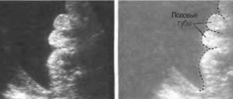
First of all, we are talking about the possibility of visualizing certain facial defects, accurately determining the number of fingers and toes, non-fusion of the spinal cord,

the presence of congenital abnormalities of the skin or genital organs. In all these cases, future parents will be helped by a three-dimensional study. In addition, this study can help in determining the exact gender of the child, because the three-dimensional image allows you to see the whole child or enlarge the image of a specific part of the body. You can also see the child's face in close-up. Agree, wouldn't anyone be interested in the opportunity to receive a photo album with pictures of an unborn child? It is all these possibilities of three-dimensional research that attract future parents.

The three-dimensional ultrasound procedure includes two- and three-dimensional ultrasound examination of the fetus. The patient receives the results of the study - a doctor’s report, a printed color photo and a disk with a digital recording of the 3D ultrasound on the day of the study. In the absence of special indications, two or three sessions of 3D ultrasound during pregnancy will be sufficient.

Traditional ultrasound during pregnancy does not always allow one to accurately determine the sex of the unborn child, as well as identify possible anomalies in its development. Then 3D ultrasound comes to the rescue. Unlike conventional ultrasound, 3D ultrasound data provides additional information that allows you to diagnose many abnormalities in the fetus.

In addition, the combination of two-dimensional and three-dimensional fetal ultrasound is a unique diagnostic method that makes it possible to see the child as realistically as possible: the way he is, right down to his fingers and toes.

The first pictures in 2D mode can be taken when the length of your baby does not exceed 15 mm; at a period of 8 weeks, the head, torso, developing arms and legs can be easily distinguished. From 10 to 16 weeks of pregnancy, the entire fetus can be seen.

Thus, two-dimensional ultrasound makes it possible to characterize the condition of both mother and child. Data from 3D ultrasound of the fetus, which allows one to obtain a three-dimensional image, complement and clarify the image obtained in the traditional way.

Three-dimensional ultrasound is performed according to indications in cases of suspected fetal developmental anomalies or the presence of various neoplasms.

3D also makes it possible to examine individual parts of the body, facial features, which parent the child resembles, and it also provides a psycho-emotional connection between parents and baby. In gynecology, this type of examination is the latest diagnostic method, it allows you to determine the condition of the fetus and identify developmental anomalies in organs and systems, and examine the fetus in a three-dimensional projection (length, height, depth).

The results of the study are issued immediately after the procedure. This type of study is best carried out in the second half of pregnancy.

It is better to combine two-dimensional (2d) research and three-dimensional (3d). The latter only provides additional information to 2d. The optimal period is 25-30 weeks. The power of the ultrasound wave remains the same as in the 2d study.

How safe is 3D ultrasound?

There is an opinion among some parents that ultrasound examination has a negative effect on the fetus, but this is not the case. Three-dimensional ultrasound is recognized as safe and does not harm either you or your baby.

Ultrasound, used in medicine and obstetrics, is absolutely safe for both the expectant mother and the baby. This has been officially confirmed by clinical studies and many years of experience in use during pregnancy.

The safest are modern models of ultrasound scanners that have special gynecological and obstetric programs and calculations. Ultrasound examination of the fetus, including 3D ultrasound, does not have any negative effect on the fetus, and remains one of the safest and most informative methods for monitoring the intrauterine development of a child.

When is it recommended to undergo a 3D ultrasound?

You should try to perform ultrasound during pregnancy within strictly regulated periods - that is, when the diagnostic value of the method is especially high. In addition, the exposure time to ultrasound during a three-dimensional ultrasound of the fetus is only 1% of the time of the entire study. The rest of the time, the sensor receives reflected signals and processes the information.

Therefore, 3D ultrasound during pregnancy is as safe as 2D. Various types of anomalies that interfere with the normal course of pregnancy can also be detected using ultrasound. Among them are the threat of miscarriage, polyhydramnios or oligohydramnios, fetal malnutrition, and entanglement of the baby’s neck with the umbilical cord.

In order to promptly detect certain problems and avoid the development of pathology, ultrasound during pregnancy is recommended to be performed five times:

• the first one must confirm or deny the fact of pregnancy. In this case, an ultrasound is prescribed if there is a delay of five to seven days;

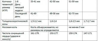
• a second ultrasound should be performed at 11-13 weeks;

• third time - at 16-18 weeks;

• fourth time - at 22-24 weeks;

• last, fifth time - at 32-34 weeks.

It is at all of the above stages that the child develops a risk of developmental defects. It is most optimal to conduct a three-dimensional 3D ultrasound examination from the 25th week to the 30th week of pregnancy.

In what situations is it difficult to see a child?

To the disappointment of future parents, sometimes a situation arises when it is difficult to see the child using an ultrasound. Let's try to figure out why this is happening:

• location of the umbilical cord and placenta;

• presence of scars on the abdomen after operations;

• the activity of the child (the image directly depends on how much he moves. That is, if the child is active, the quality of the frames will be more interesting, and the image itself will be clearer);

• overweight mother;

• incorrect position of the child in the womb;

• the amount of amniotic fluid (the more there is, the better the image on the screen during the ultrasound procedure).

By contacting the Center for Aesthetics and Health, you will have the opportunity to undergo an ultrasound quickly and in comfortable conditions. At your request, in addition to the examination, you can receive a video recording of the ultrasound examination on electronic media.

At your disposal are modern equipment and highly qualified ultrasound diagnostic specialists. You can ask your question or make an appointment with a doctor by calling or ordering a call on the company’s website.

By contacting the Optimus Center for Aesthetics and Health, you will have the opportunity to undergo an ultrasound quickly and in comfortable conditions. At your request, in addition to the examination, you can receive a video recording of the ultrasound examination on electronic media.

At your disposal are modern equipment and highly qualified ultrasound diagnostic specialists. You can ask your question or make an appointment with a doctor by calling or ordering a call on the website.

*As an advertisement

Rating
( 2 ratings, average 4.5 out of 5 )
Did you like the article? Share with friends:
For any suggestions regarding the site: [email protected]
Для любых предложений по сайту: [email protected]