What does ultrasound visualization mean?
Basic information about ultrasound
Obstetricians and gynecologists began to resort to ultrasound diagnostics in working with pregnant women back in the late 60s of the last century.
Over the past time, echography methods have been significantly modernized, and thanks to many years of practice, they have been significantly enriched with useful information that helps specialists make correct diagnoses. Pregnant women are required to undergo three ultrasound examinations, which must be carried out at the end of each trimester. Thanks to the data obtained, doctors can detect any problems with the fetal development process in time, and, therefore, react in time to save the life and health of the future baby. By the way, we cannot fail to mention the fear of some mothers to undergo sonography. Scientists have long been concerned about the impact of ultrasonic waves on a living organism, which is why many large-scale clinical studies have been carried out, which resulted in the following data: high-frequency sound waves do not cause any harm to the human body, even if it is in the mother’s womb.
Difficulty with visualization
It may well happen that when undergoing a routine examination, the results may contain a similar phrase: visualization is difficult. What does visualization on ultrasound mean is difficult? This question will arise for any woman who sees this phrase in a medical report.
In fact, you should immediately put aside any concerns and fears. During the second screening, the placenta is examined to look for cysts and calcium deposits that may complicate its functioning. In this study, the amount of amniotic fluid is also studied, and in the case when the fetus occupies a special position that does not allow defecation.
Source
Errors in ultrasound diagnostics
Having learned about pregnancy, a large number of women rush to get an appointment with a specialist, for an ultrasound, to clarify the timing. But it happens that even if you are confident in conception, if you have no menstruation, the ultrasound machine does not show a positive result. Therefore, the question arises: “Can ultrasound fail to detect fertilization?”
It has long been no secret that an ultrasound machine uses ultrasonic waves to display a picture of the state of internal organs on a monitor. Not confirming pregnancy, probably due to an outdated diagnostic device or its malfunction.
But there are cases that even the highest quality research equipment does not confirm the fact of the birth of new life. The reason for all this is the low level of professionalism of the person sitting at the ultrasound device. Therefore, first, it is recommended to inquire about the skills of a specialist in the field of ultrasound examinations.
In conclusion, I would like to add that when the question arises, “At what stage of pregnancy is visible on an ultrasound?” It is very important not to neglect examinations by specialists and diagnostic tools, since violations of fetal development and maternal health that are detected in a timely manner can be eliminated.
Ultrasound imaging during pregnancy is a phrase familiar to many. Pregnancy is one of many expected events. They prepare for it, select the necessary conditions, monitor unfavorable factors, and look forward to it. This seemingly simple and natural process is not always achievable, and can sometimes require significant time to establish the fact of its completion. Even after everything has already happened and you have become a potential mother, there are still many additional and mandatory parameters necessary for tracking. A visit to the gynecologist during pregnancy becomes one of the traditional actions, and a lot of examinations become the norm of life. The main screening test during pregnancy still remains ultrasound, which provides the most complete and objective information confirmed visually. Doctors often use the term ultrasound imaging during pregnancy. Let's figure out what it is and what it's needed for.
Imaging during pregnancy is, simply put, confirmation that pregnancy has taken place and the fetus has begun to develop. Accordingly, all these signs can be seen with your own eyes on the monitor screen showing what is in your stomach. There are quite clear standards for the timing of ultrasound examinations. In total, if no difficult situations arise, a pregnant woman undergoes an ultrasound scan 3-4 times during the entire period. A larger amount may only be determined by your doctor's recommendations. The first ultrasound is done at 12-13 weeks of pregnancy. At this time it is already possible to see something and talk specifically about the ongoing processes. There are often situations where expectant mothers do their first ultrasound too early and get unreliable results, and then try to independently make various terrible diagnoses for themselves. You should not anticipate the situation and create additional difficulties with your own actions. Ultrasound visualization during pregnancy acquires its maximum information content at 20-24 weeks of gestation. Here it is already possible to accurately identify the sex of the child, measure certain parameters that were previously inaccessible, and identify possible developmental defects. The role of the visual component in this matter is extremely high.
The main goal of the second screening is to examine the placenta for the presence of cysts, and to monitor for excess calcium in the form of various deposits that interfere with the normal functioning of the placenta. The developing child's organs are studied and assessed. A situation may well arise when the position of the fetus is such that sexual characteristics are not identified, and in this case the doctor may use the term visualization is difficult. This phrase should in no way be understood as a diagnosis; it is just a temporary condition due to the location of the fetus. Such situations occur in more than 9% of pregnancies, and cannot be considered dangerous, much less pathological. At the third planned study in 12-14 weeks, you will definitely find out the sex of the child, if it did not work out earlier, and ultrasound visualization during pregnancy will be as accurate as possible.
It is visual examination through ultrasonic examination that gynecologists trust most, and this examination technique has been successfully used for over 50 years. The likelihood of errors and misinterpretation of data is relatively low. If any abnormalities are suspected, you will be prescribed additional tests, so do not worry or be afraid of the unknown. Visit your gynecologist regularly during pregnancy and listen to his recommendations.
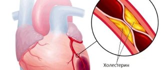
Local thickening of the myometrium: normal or pathological?
Pregnancy is an important period in the life of every woman, however, it is often overshadowed by various troubles and complications. One of these pathological conditions of the expectant mother is thickening of the myometrium, the progression of which can lead to fetal death. That is why it is important to know the reasons for the development of this condition and the symptoms of its manifestation.
Myometrial thickening during pregnancy: normal or pathological?
One of the components of the uterine layer is the myometrium, the thickness of which can vary depending on the day of the menstrual cycle, as well as in the event of pregnancy. Particular importance is attached to identifying the cause of thickening of the muscle layer, which allows timely detection of pathological changes in a woman’s body.
One of the common symptoms that is detected in females is considered to be a local thickening of the muscle layer along the anterior wall of the uterus. However, often the thickness of the uterine wall is prone to changes under the influence of a decrease or increase in the level of hormones in the female body, or under the influence of other factors. It is for this reason that the detection of thickening of the muscular layer of the reproductive organ does not always signal any pathology.
Changes in the myometrium during pregnancy
Doctors diagnosing local thickening of the myometrium when expecting a child indicates that the woman has increased uterine tone. Of course, during the process of childbirth this is considered very important, but in other cases such increased contraction of the reproductive organ is accompanied by unpleasant painful sensations.
Local thickening of the muscle layer during pregnancy is a dangerous pathology that requires increased attention and control.
Hypertonicity of the uterus can result in a disruption of the process of nutrient intake
Source
Forecast
The detection of local thickening of the uterine wall (myometrium) in itself is not necessarily a sign of pathology, but in combination with the above factors, it can pose a serious threat to a woman’s health.
In order for a woman to feel healthy, it is necessary to regularly see a gynecologist for preventive purposes. This should be done at least once every 6 months, even without any complaints. If there are any signs of the disease: pain, discomfort, itching, discharge, bleeding outside of menstruation, then this is definitely a reason to consult a doctor immediately!
PZhS
We went for an ultrasound and wrote to me that “visualization is difficult due to the characteristics of the pancreas.” As I later found out, the subcutaneous fat layer is the subcutaneous fat layer! I’m wondering, is it because of the stitch after the CS or am I fat with my 57 kg?????
Comments
Something very strange, maybe the device is not very good? I was overweight before B, but now I’ve already gained 8 kg, so our PWS is excellent and rich. But everything was perfectly visible. Maybe not the thickness, but some kind of structure that does not allow waves to pass through? Ask your doctor - maybe it’s worth doing it somewhere else?
Damn, I went for an ultrasound... How many times have I stuttered that I will no longer use our free medicine, and yet I ended up going to the clinic for an ultrasound, since I have no money at all... took the child to a friend, went... came...
They did an ultrasound at 25 weeks, in the conclusion it says visualization is difficult, at 30 weeks, the same thing - visualization is difficult, what does this mean???? I read in someone’s diary that this is related to the development of the baby. Who knows what, please write!!!!!!
Girls, hello everyone! Tell me, we went for an ultrasound today to sign up for a joint birth, the term is 36+3, according to the ultrasound everything is fine, but in the paragraph about “the area of the appendages, the walls and the cervix” they wrote that visualization is difficult due to the NVO. I…
cpr 36mm bpr 16.3mm heart rate 170 tvp 1.1mm nasal bone 2.2mm heart 4-cam. the cut is difficult to visualize the heart especially worries me
Girls, your opinion is very interesting - who should we expect - a boy or a girl? My husband really wants a boy, and I want a girl... P.S. in this photo I’m already B Girls, what can I tell you? of course 100%...
I started making appointments for BB with an ultrasound at the same time with my daughter, although now I regret that I didn’t write everything, but only the half. ((((So, today we went to Tula for an ultrasound, the period corresponds to 22-23 weeks,...
We've been waiting for this moment when you find out
Source
Telephone diagram dialogue 601
This article will discuss a comparison of SUVs from the Japanese company Toyota. Thus, it will be necessary to answer the question, which is better: Land Cruiser Prado 150 or Land Cruiser 200?
In 2009, a ready-made version of the Prado 150 SUV was presented at an exhibition in China. Then, for seven years, at factories in Japan, it was refined, modified, and polished. In 2013, a modern, restyled version was presented. The length of the body is 4760 mm, width – 1885.
As for the Toyota Land Cruiser 200 SUV, its brutal design is actually a legacy of the 100 version, which successfully debuted in 1997. The model turned out to be reliable, with high cross-country ability, with good driving characteristics for this standard size of SUV; it is not surprising that it was actively sold all over the world and brought in a decent income. And it was decided to develop the success with the 200 model, which debuted in 2007. Four years later, the first, light restyling followed, and in 2015, the current restyled version was released with an unusual combination of the radiator grille and headlights. The body length of the “two hundredth” is 4760 mm, width – 1970 mm.
The interior space of the Land Cruiser Prado 150 retains family features, while at the same time changes are constantly being made in connection with modern trends. Because of this, the layout of the organs on the front panel was even rearranged. Where the puck of the new Multi-Terrain Select adaptation system to various driving conditions catches your eye. The seats are quite basic and only moderately comfortable.
The main emphasis that Toyota engineers tried to bring to the Land Cruiser 200 is that it looks expensive and graceful, both externally and internally. This is expressed in neon interior lighting that is pleasing to the eye, as well as in the use of the correct geometry
Source
I'm hysterical……….
Comments
In the free one they did it on the belly, but in the paid one they did it this way and that, the baby kept turning away and didn’t show his face)) then with grief he turned sideways and used a sensor on the belly to calculate the size of his nose!
It’s just that my visualization is very difficult in the abdomen, there is a fat layer, adhesions, and we immediately switched to a vaginal ultrasound, I did both at the second screening. But if only for the stomach, I can imagine what they would measure for me there))) So there is no reason to worry, the TVP is normal and the nose is neat.
in general, to get rid of all doubts (about genetic abnormalities), you can undergo non-invasive prenatal genetic diagnostics
You call and clarify, the essence of the method is to find out the child’s karyotype (and the karyotype just shows genetic abnormalities, like all sorts of syndromes)
Lingonberry, of course I don’t understand anything about these meanings, I can only support and wish me great luck!!! Don't be nervous and don't torment yourself with bad thoughts!!! It’s best to have the ultrasound done in another place and calm down!)) Everything is fine with your little one, he (she) just hid his nose and decided to joke with the ultrasound technician)) Everything will be fine! Good luck!!!
Cowberry, what are you doing?.. Everything is fine, the TVP is good, but we didn’t see the nasal bone, because... your baby lies the way he wants, and not the way the uzist is comfortable :) when there are no bones, they write “not visualized”, but for you they wrote “cannot be assessed”
1.6 is a good number, regarding visualization of the nose - find the best screening center, let them take a look there. Have you been tested for risks? In my opinion, there can be no talk of Down syndrome now! Just don't panic, it's bad for you!!
The thickness of the collar space is 1.6 - this is a good indicator. And the ultrasound can be repeated in another week and the nasal bone will definitely be visualized. You shouldn't have worried!
after a caesarean section, the pain goes away for a long time. For some women, doctors do not recommend giving birth on their own, since due to increased blood pressure or severe muscle tension, the woman in labor may experience undesirable consequences.
Some women are afraid of
Baby
Source
Visualization is difficult due to the position of the fetus
During a woman's routine ultrasound examination, her doctor may tell her that imaging is difficult during pregnancy. What does this wording mean? During a normal pregnancy, the second planned ultrasound should be performed between 20 and 24 weeks of pregnancy.
During this examination, parents usually find out the gender of their unborn child. In addition, this ultrasound examination can detect congenital malformations in the development of the fetus. The child’s size is already large enough for the doctor to examine all the organs in detail. In the first trimester, the fetus is still too small for such an examination, and in the third trimester, the placenta may interfere with the examination if it is located on the anterior wall of the uterus.
It must be said that studying the condition of the placenta is also included in the tasks of the second planned ultrasound. The examination is carried out to detect cysts and calcium deposits in the placenta that impede its functionality. The amount of fetal fluid is also assessed. Sometimes the position of the child is such that it is impossible to determine his gender. It is in these cases that doctors say: Visualization is difficult during pregnancy .
You shouldn’t be upset, because you will have another scheduled ultrasound at 32–34 weeks, which will certainly make it possible to find out the gender of the expected baby. Difficulties in determining gender during the third ultrasound are quite rare.
If an ultrasound reveals any problems with the blood supply to the fetus, the doctor should know that measures should be taken to normalize it. Therefore, do not neglect routine examinations.
is it possible to get pregnant if you stop taking pills? Today the word “hormones” sounds terrifying to many. And all because the first generation of oral contraceptives actually had a large number of side effects. The woman's blood pressure increased and her blood pressure increased.
Remember that all doctor's prescriptions are for the benefit of your health and normal intrauterine development of the fetus. A gynecologist may prescribe an additional ultrasound examination for pregnant women over 35 years of age, and
Source
MEN'S
Treatment in MOSCOW
Clinics in MOSCOW
TREATMENT IN SPb
How is the fetus developed at 20 weeks of pregnancy?
That is why the twentieth week is characterized by the baby’s rather erratic movements - on an ultrasound examination you can see how he is trying to test his capabilities:
- flexes and extends limbs;
- moves his fingers and tastes them;
- grabs the umbilical cord;
- sticks out tongue;
- yawns;
- tries to smile and frown.
The baby's activity depends on the established schedule of sleep and wakefulness, certain sounds, eating sweet food, stroking the mother's belly. The structure of the integumentary tissue is the same as that of an adult. However, the skin is still loose and looks more wrinkled - fat cells are just forming under it. Hair appears on the surface of the head, and nail plates appear on the fingers.
The reproductive system is maturing - the girl’s ovaries produce oocytes, the active movement of the testicles towards the exit from the peritoneum confirms the fact that a boy will be born; Using an ultrasound photo, you can accurately determine the baby’s gender. The fetal immune system is already ready to distinguish the molecules of its body from foreign ones, the central nervous system is fully developed. However, his lungs cannot yet transport oxygen into the bloodstream - this is the unpreparedness of the baby for birth.
During pregnancy, visualization is difficult, ultrasound
During your next scheduled ultrasound, your doctor may indicate in the examination results the phrase: visualization is difficult during pregnancy . Let's learn to understand what is behind these words. If the pregnancy is progressing normally, a routine ultrasound examination is carried out at 24 weeks.
It is during this examination that parents are informed of the gender of their unborn baby, and doctors also monitor whether the child has a congenital pathology. The child is already quite large in size, and the doctor can see the organs quite well. The first trimester will not provide such a complete overview; in the second, the placenta is denser in structure, especially if it is located on the anterior walls of the uterus.
During the second ultrasound examination, the doctor examines the condition of the placenta. Be sure to look to see if there is a cyst on it, and if calcium is deposited, which will complicate its functions. During this examination, the amount of amniotic fluid is also judged. So, if the fetus is located in a special way, then its gender cannot be determined. This gives the doctor a turn of phrase such as difficulty visualizing. You should not be upset and take this for a violation; at the third scheduled examination, at the 34th week, an ultrasound will certainly show the sex of your unborn newborn.
Routine examinations should not be neglected, because problems in the blood supply to the fetus noticed in time can probably be corrected. In such a course of events, doctors take measures and eliminate the problem. All doctor’s advice should be administered in a clear prescribed manner; they all serve to support the health of the expectant mother and fetus.
With the help of regular examinations and advice from a gynecologist, you can achieve the harmonious development of your unborn child and maintain excellent health.
If necessary Source
Excess weight requires control
If visualization of the fetus is difficult, this is most often due to the woman being obese. Here are some statistics for you: in the 2nd trimester, overweight women experience poor visualization much more often than those who keep themselves within limits. And there are 25% more of them.
This is why doctors recommend monitoring your weight from the first days of pregnancy. What does it mean for a woman that visualization is difficult due to the PVC? On the abdominal wall of the mother there is a layer of fat, which is highly dense.
Ultrasonic waves cannot overcome it completely; a certain error is created, which necessarily affects the results of the study. By the way, it is possible to determine whether such a pathology is present in a simple way.
You can check how large the subcutaneous fatty tissue is and how it relates to the total body weight:
- take a layer of skin from the navel in your hand;
- managed to do this, then everything is fine with you;
- If you couldn't get a crease, you are overweight.