June 29, 2020 Admin Home page » Ultrasound during pregnancy and childbirth Views: 818
Studying the condition of the fetus in the womb is extremely important at any stage of pregnancy, but there are certain, established deadlines that should be observed so that doctors can have the maximum amount of information about the health of the unborn child. This diagnosis is complex and includes a number of diagnostic measures, including ultrasound.
Ultrasound examination (ultrasound, ultrasonography) is a method of instrumental diagnostics, in which the condition of the fetus and various organs is studied using ultrasonic waves, because ultrasound is performed not only on pregnant women. This method appeared relatively recently, a little more than half a century ago, but now it has become widespread throughout the world, from the poorest to the richest countries. And this comes down to many factors.
Since this article discusses ultrasound at 22 weeks of pregnancy, it is worth covering everything from the point of view of the woman being examined. Thus:
- The first thing that an expectant mother may worry about is safety. Such questions arise not only out of curiosity, but also because of the emergence of responsibility for the child’s life.
Ultrasound diagnostics in this regard is absolutely safe, especially considering the current level of development of ultrasound technologies in medical practice.
This is due to the nature of the waves used in the method. At its core, without going into the specifics of physics, ultrasonic waves are a very loud sound, and the device itself works on a principle similar to echolocation. That is, the device emits waves with certain characteristics that pass through tissues that do not need diagnostics, are reflected from target structures and then recorded and processed by the device. The latter, in turn, analyzes the received data and displays ready-made information on the monitor.
- In addition to the fact that ultrasound at 22 weeks of pregnancy is safe, it is also quite informative. Ultrasound diagnostics can detect not only gross malformations or lack of fetal growth, but also genetic diseases, as well as heart defects. At the same time, to improve the quality and accuracy of the information obtained, in addition to conventional ultrasound during the 22nd week of pregnancy, Dopplerography or 3D ultrasound can also be used.
- When the fact that ultrasound is available almost everywhere was mentioned, the question of price immediately arises. Almost everyone can afford an ultrasound examination, or it can be performed free of charge, subject to the appropriate referral. The machines are available in most clinics, but there are also portable versions of the machine that allow ultrasound to be more common.
When a doctor or anyone else talks about the advantages of a method, then, of course, questions arise about the disadvantages.
- Much in ultrasound diagnostics depends on the specialist and the quality of the equipment. But can an ultrasound be wrong at 21–23 weeks of pregnancy? Here, a lot depends on what kind of defect is present or what the doctor is “looking for” on an ultrasound.
- Sometimes a conventional ultrasound examination may reveal only certain signs that require clarification using other methods. For example, the already mentioned Doppler ultrasound. In addition to diseases, the sex of the child is also important for parents, but sometimes it is also important from a medical point of view. It is extremely important for doctors to know the sex of the child if there is a risk of genetic diseases, such as hemophilia, which is inherited only through the male line.
- At the first screening study (10-14 weeks), it is almost impossible to determine gender using ultrasound. But starting from the 22-23rd week, ultrasound can give a more accurate answer, but even here there are mistakes, as mentioned above - a lot depends on the experience of the specialist and the modernity of the equipment used for diagnosis.
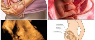
Ultrasound photo of pregnancy 22 weeks
What does an ultrasound show at 22 weeks of pregnancy?
When the date for the examination has been set, which must be determined with your obstetrician-gynecologist, the question arises: what will be examined on the ultrasound? In other words, about an ultrasound at 22 weeks of pregnancy, what do they look at?
- Well, first of all - the fetus itself, namely fetometry. It represents the measurement of parts of the fetus. Measurements of the head, limbs, and abdominal volume are taken. The symmetry and proportionality of development are assessed, and the gestational age is determined.
There are cases that there is some kind of developmental delay. It should be assessed by an obstetrician-gynecologist who is seeing a pregnant woman, because sometimes this can be a variant of the norm.
- Other structures of the child’s body are also examined by ultrasound. Brain, liver, kidneys, intestines, lungs, etc. At 22 weeks of pregnancy, heart defects are often detected using ultrasound. In this case, a repeat ultrasound is prescribed, this time with Doppler ultrasound. It’s worth saying right away that ultrasound can be performed as many times as necessary. In practice, when a pathology is detected for the first time, an ultrasound examination is prescribed by another specialist or even in another clinic to confirm the diagnosis.
- In addition to the fetus, amniotic fluid is also examined at 22 weeks. An ultrasound evaluates, first of all, their number. Both polyhydramnios and oligohydramnios are signs of the presence of various developmental defects. In addition to defects, oligohydramnios may indicate an intrauterine infection, and polyhydramnios may indicate the presence of diabetes mellitus in the mother. Also, polyhydramnios, characterized by an excessively large volume of water, indicates an increased risk of developing umbilical cord entanglement.
- Assessing the condition of the organs on the maternal side is very important. Examination of the umbilical cord and placenta may indicate the need for cesarean section delivery due to the low location of the placenta. Since this increases the risk of bleeding. The degree of maturity of the placenta is also studied; normally, at 22 weeks of pregnancy, it is 0.
- Another indicator is the thickness of the placenta; its decrease indicates sexually transmitted infections. In the umbilical cord, its vessels, their number and the presence of calcifications are studied. In some pregnant women, the number of blood vessels may be extremely small. This indicates a high risk of fetal hypoxia, which is extremely dangerous for the child’s body.
- If isthmic-cervical insufficiency is suspected, it will be necessary to perform a transvaginal ultrasound at 22 weeks of pregnancy and determine the length of the cervix, which is extremely important for diagnosis and treatment.
At 22 weeks of pregnancy, you can perform a three-dimensional ultrasound, which will make it possible to take the very first photos of the baby or, if it is necessary, to carry out additional diagnostics using this method.
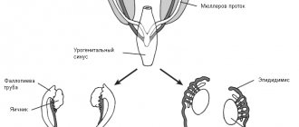
Differentiation by gender
An ultrasound at 22 weeks of pregnancy visualizes the gender of the child in 90% of cases. In the second half of pregnancy, the external genitalia in girls are visualized as parallel lines in the perineum. The angle between the surface of the skin and the genital tubercle is not visible or is less than 30 degrees. In boys, this tubercle is pronounced and looks like a small snail on the monitor.
Determining the sex of the baby at 22 weeks. Photo ultrasound
The doctor may make a mistake or not see the gender of the baby:
- if there are two children in the womb and one “hides” behind the other;
- if the child is excessively mobile and turns away during the examination;
- in the case when the baby covers the genitals with his palm.
These reasons are temporary. During subsequent examinations, it is usually not difficult to differentiate between a boy and a girl.
How does a woman feel?
At week 22, a woman enjoys the cloudless feeling of pregnancy. The toxicity of the first trimester has already passed, and the feeling of heaviness and clumsiness of the last months has not yet arrived. The main impression on a woman at this stage is the increasingly active movement of the baby in the stomach. He is already quite noticeably pushing with his arms and legs. Often the life rhythm of mother and baby does not coincide. A woman may unexpectedly wake up at night from too active movements of the fetus in the womb.
The pregnant woman’s body weight continues to grow, and changes in the outline of her figure are already noticeable to the naked eye. However, the woman’s movements are still light and graceful, she calmly walks, bends, squats, and performs light housework. The weight of her belly does not greatly affect her gait and coordination. But in the back area, unpleasant and sharp pain sensations may unexpectedly arise due to a shift in the center of gravity in the body due to the increased volume of the uterus.
Abundant blood flow to the pelvic organs causes increased sexual desire. During this period, you should not avoid close relationships with your partner. The sensations obtained during sex at 22 weeks of pregnancy become especially vivid and memorable. Restrictions may be set by a doctor if intimacy could lead to an unplanned birth. The reason for this is complications such as placenta previa, isthmic-cervical insufficiency, etc.
The skin of the abdomen and thighs gradually stretches, which is why the first traces of stretch marks may appear on it. Due to the pressure that the uterus puts on the bladder, a woman’s desire to go to the toilet appears more and more often. In addition, she may experience severe swelling of the arms and legs, and the volume of the foot may increase slightly.
Burning and itching in the anus are a sign of hemorrhoids. This unpleasant disease often becomes a companion to pregnant women. Displacement of the walls of the abdominal cavity and insufficient outflow of blood from the venous arteries leads to their enlargement. Unpleasant sensations may increase if a pregnant woman suffers from constipation. In other cases, hemorrhoids do not cause any particular problems, but they must be treated.
What happens in a woman's body
At week 22, the total increase in a woman’s body weight compared to the initial period averages 5-8 kilograms. The uterus can be easily felt and is located 20 mm above the navel. The abdomen increases in size gradually.
The woman’s emotional state has improved significantly due to the fact that her hormonal levels have returned to normal and become stable. The body produces special joy hormones that allow a woman not to notice the hardships of bearing a child.
The color of the skin on the face, the condition of the hair and nails improves. An increase in estrogen levels in the blood can cause spider veins to appear on the skin. Fatigue, irritability and drowsiness are not typical for this period. A healthy appetite and a constant desire to eat something tasty indicate that the body needs an increased supply of nutrients.
At this stage of pregnancy, the total blood volume in a woman’s body actively increases. Plasma cells, responsible for transporting nutrients throughout the body, begin to divide rapidly. The consistency of the blood becomes thin, and there is a serious risk of developing anemia. After standing or sitting for a long time, swelling and the first signs of varicose veins may appear.
How is an ultrasound performed at 22 weeks?
How the diagnosis will be carried out and preparation for it depends on the method by which the diagnosis will be carried out. Most often this is a transabdominal method. It is performed this way only if there are no indications for transvaginal.
- Transabdominal ultrasound is performed through the anterior abdominal wall. Preparation for this study is not required; it can be carried out almost immediately after visiting an obstetrician-gynecologist, who sets the exact date of the study. The examination is performed as follows: the pregnant woman comes at the appointed time and lies down on the couch, then exposes the anterior abdominal wall. The doctor applies a special gel that improves the conductivity of ultrasound waves and begins the examination.
- If the diagnosis is performed using the transvaginal method, then no preparation is required either, but before the diagnosis itself, in case of severe flatulence, you may need to take carminatives, such as espumisan. The woman also lies down on the couch, but she needs to bend her legs at the knee and hip joints, after which the doctor inserts a special sensor into the vagina. To improve conductivity, personal hygiene and reduce trauma, a special condom is placed on the sensor. It is also lubricated with a special gel. The duration of the diagnosis is approximately 15-30 minutes.
After an ultrasound is performed at 21–23 weeks of pregnancy, it may need to be repeated, often more than twice. This is especially true in cases where the issue of the need for artificial termination of pregnancy is being decided.
Ultrasound photo 22 weeks
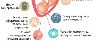
How does a baby's body develop?
By week 22, the formation of the baby’s brain is almost complete, and the nervous system begins to function.
The baby is already consciously exploring the boundaries of her small world, pushing off from the abdominal wall of her tummy, waving her arms, trying to feel her face and tummy, actively pulling the umbilical cord and sucking her finger. It is by the 22nd week, in the case of a multiple pregnancy, that twins begin to become aware of each other’s presence and begin to actively study not only themselves, but also their brother or sister. Of course, the baby sleeps most of the day, but he already adjusts his activity schedule to his mother’s schedule, that is, at night he mostly sleeps and prefers to wake up with his mother. Most often, the baby falls asleep during the daytime during the period of active movement of the mother, since smooth movements in the amniotic fluid have a rocking effect.
During the day, daylight seeps through the abdominal wall in the form of a pinkish glow, which the baby can already distinguish. He blinks, winces, tries to cover his eyes with his hands, and then turns his butt, trying to avoid a new and as yet unfamiliar manifestation of the outside world. By the way, as experts who conduct ultrasound note, it is girls who turn their butts most often; boys, even in the womb, are not shy about appearing in all their glory, also in their first photo.
At this stage of pregnancy, the baby actively reacts to sounds coming from outside, can calm down when the mother’s voice sounds affectionate, or become more active when exposed to extraneous noises. The baby is already reacting to the mother’s palm being placed on her tummy; when she sees such an obstacle, she strives to respond with a kick.
The baby’s skeleton continues to form; now each bone contains bone marrow, which produces red blood cells and deposits calcium. The spine is fully formed, even intervertebral discs can be clearly seen on ultrasound. Sweat glands begin to form, and the liver is actively involved in its work.
The liver processes “indirect” bilirubin, that is, the breakdown products of hemoglobin and neutralizes its action with enzymes, thus converting it into “direct” bilirubin, which is excreted through the placenta. If the liver is not yet able to form a sufficient amount of enzymes by the time the baby is born, the newborn baby will have a yellowish skin color, which is called physiological jaundice.
Interpretation of ultrasound results and norms at 22 weeks of pregnancy
The data obtained must be transferred to an obstetrician-gynecologist who is sufficiently knowledgeable about the mother, father and immediate relatives of the unborn child. This is important for assessing the development of dangerous conditions, if any. Also, when a doctor evaluates the development of the fetus, he must take into account the constitutional characteristics of the parents, because those parameters that may be normal for some will be low for others and vice versa. The average ultrasound readings for 22 weeks of pregnancy are shown in the table below.
| Index | Meaning |
| Height, cm | 28 |
| Weight, g | 430 — 500 |
| BDP (Biparetal size) mm. | 48 — 60 |
| Fronto-occipital size, mm. | 64 — 76 |
| Head circumference, mm. | 178 — 212 |
| Abdominal girth, mm | 148 — 190 |
| Femur length, mm. | 35 — 43 |
| Length of shin bones, mm. | 31 — 39 |
| Length of humerus, mm. | 31 — 39 |
| Length of forearm bones, mm. | 26 — 34 |
| IR (resistance index) of placental vessels | average 0.51 (acceptable values from 0.36 to 0.69) |
| SDO (systole-diastolic ratio) in the umbilical cord | 3,87 — 3,95 |
| IR (index of resistance) in the umbilical cord arteries | average 0.73 (acceptable values from 0.61 to 0.83) |
Ultrasound diagnostics
At 22 weeks, the pregnancy calendar does not provide for an ultrasound diagnostic procedure. A routine ultrasound is performed at 17-20 weeks as part of the II screening examination. Reasons for performing an ultrasound at this time:
- the expectant mother did not have time to undergo screening within the specified time frame;
- questionable data from screening II, which requires monitoring the condition of the fetus or its life support systems.
During this period, the doctor pays special attention to the proportionality of the baby’s skeletal bones. At this time, the CTR (coccygeal-parietal size) on ultrasound loses its leading significance. The state of amniotic fluid (quantitative data, transparency) is also assessed and compared with the norm. Thus, using ultrasound, it is determined:
- norm;
- high and low water levels.
Next, the functional state of the umbilical cord and placenta, as well as their structure, is assessed. Ultrasound assesses the presence/absence of damage to the placenta, foreign inclusions and the degree of its maturity. The number of umbilical cord vessels and indicators of the quality of blood flow through them are determined.
At 20-22 weeks, not only the fetus is examined, but also the mother’s reproductive system - especially the uterus and placenta. At this time, the doctor can diagnose “polyhydramnios” or “oligohydramnios” and check the functioning of the uterus-placenta-fetus ligament.
At the 22nd week of pregnancy, the doctor carefully examines the fetal brain, its structure, including the cerebellum, comparing them with the norm. The ventricles of the brain are examined to exclude hydrocephalus. With normal brain development, their size is no more than 10 mm. Structurally, the nervous system and brain of the fetus have practically formed, so during this period an ultrasound specialist can evaluate the structure of the brain, compare the data obtained with standard parameters and give an answer regarding the normal development of the baby’s brain structures.
Next, the spinal column and the spinal cord in its canal are subject to evaluation. The structure and integrity of these formations is determined. If necessary, a 3D ultrasound scan can be performed at 22 weeks.
What to do if you miss an ultrasound in the second trimester of pregnancy and more
Sometimes there are situations in which a woman skips an ultrasound examination. Of course, you should not skip the diagnosis, since at each stage a different pathology can be detected.
- Thus, at the first screening, gross disorders and genetic diseases are detected.
- On the second - mostly organ ones. Of course, even in the second one you can find signs of genetic pathology, but this has its own characteristics.
- At the third ultrasound, genetic pathology is also more difficult to detect. But the timeliness of detection of the disease is closely related to artificial termination of pregnancy.
The earlier the disease is detected, the sooner the pregnancy can be terminated, which means the consequences for the woman’s physical and mental health will be less.
When a pregnant woman misses an ultrasound for any reason, she should contact her doctor immediately. He will set a date or, if it is too late, the study will simply be missed. To avoid such situations, you should discuss all aspects in advance with the doctor prescribing the examination.
What happens to the mother's body
Pregnancy at 22 weeks is not yet too burdensome and restrictive for the mother, the tummy is relatively small, but nagging pain in the back is already felt due to a shift in the center of gravity, and the legs also begin to swell. Mom is still actively moving, but there are already moments of sudden fatigue.
By week 22, the blood volume in the mother’s body increases, the number of red and white blood cells grows unevenly, this period is characterized by the thinnest blood throughout pregnancy. There is a danger of developing anemia; you should carefully monitor all indicators during tests, as well as the intake of sufficient iron into the body.
The mother begins to gain weight sharply, which can subsequently affect the course of labor, and therefore doctors recommend drinking more fluids and plant foods. The amount of discharge increases at this stage of pregnancy, the color and density of which directly depend on possible infections in the amniotic fluid. The gums begin to bleed, and nasal congestion is possible, which is considered normal and can be regulated by taking vitamin complexes. Heartburn wakes up, as well as a sudden appetite, and varicose veins may develop.
And yet, the mother is happy, the baby moves, responds to her touch, voice, and reacts to rhythmic sounds and movements. In the photo, he looks like a little man, recognizes the voices of his dad and perhaps his older brother or sister, and also affectionately strokes his mother’s tummy from the inside.