Small, childish, infantile - all these are designations of the same structural feature of an important female organ. Uterine hypoplasia is a pathological condition characterized by its unusually small size. It is believed that the chances of pregnancy and a successful birth in this condition are reduced. Despite this, it is possible to become pregnant with hypoplasia, and the likelihood of conception is determined by the individual characteristics of the patient.
When you hear the diagnosis of “infantile uterus,” you don’t need to give up. Modern treatment methods and assisted reproductive technologies achieve the patient’s main goal – the birth of a child.
Characteristics of the “adult” uterus
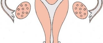
The uterus is rightfully considered the main organ in the female body; it performs a hormonal function; in its absence, menstruation and, importantly, pregnancy are impossible. During the process of childbirth, the uterus actively helps the birth of a child, which is due to the developed muscle layer and its contractions (contractions).
The size of the fruit container depends on the age of the fair sex. A newborn girl has a uterus about 3 cm long, with the cervix in relation to the body of the uterus as 3/1. The angle between the cervix and the body is not pronounced, and the weight of the uterus is about 4 grams.
During childhood, which lasts up to 8 years and ends at the start of puberty, the size of the uterus undergoes changes. In the first year of life, its length is 2.5 cm and its weight is 2.3 grams. By the age of 4, the weight of the uterus increases to 2.8 grams, and by the girl’s sixth birthday, the weight of the uterus is equal to its weight at birth. The ratio of the cervix and uterus also changes: by the end of the first year it is early 2/1, by the age of four – 1.7/1, and at 8 years – 1.4/1. If a newborn’s uterus is located in the abdominal cavity, then by the age of 4 it descends into the pelvis.
The size of the uterus of a woman of childbearing age depends on the presence/absence of a history of pregnancies:
- there were no pregnancies - length 4.5 cm +/- 3 mm, width 4.6 cm +/- 4 mm, thickness (antero-posterior size) 3.4 cm +/- 1 mm;
- there were pregnancies, but were interrupted (abortions, miscarriages) - length 5.3 cm +/- 3 mm, width 5.0 cm +/- 5 mm, thickness 3.7 +/- 1 mm;
- women who have given birth - length 5.8 cm +/- 3 mm, width 5.4 cm +/- 6 mm, thickness 4.0 +/- 2 mm.
As soon as a woman becomes pregnant, the uterus begins to grow rapidly (hypertrophy and stretching of muscle fibers), its length at birth reaches 32 - 33 cm, and its weight is 1.5 kg. After the birth of a child, the organ undergoes involutive changes and gradually returns to normal size, but remains somewhat heavier and larger than before the “non-pregnant state”.
Normally, the length of the cervix corresponds to approximately a third of the uterine length and is 28–37 mm. The thickness of the neck reaches 29 – 53 mm. The shape of the cervix also depends on the presence of a history of childbirth. In nulliparous women it is conical, and in women who have given birth it is cylindrical.
Underdeveloped uterus and its degrees
Hypoplasia of the uterus is a uterus when it is underdeveloped, that is, by the end of puberty it does not reach the size corresponding to normal indicators, and the organ itself is formed correctly, has a body, fundus, cervix and fallopian tubes. A “small” uterus is often found in combination with underdevelopment of the appendages, vagina and external genitalia, that is, it acts as one of the symptoms of genital infantilism.
Degrees of uterine underdevelopment:
- I degree - the dimensions of the uterus in length, measured with a probe, do not reach 3.5 cm, moreover, most of the length belongs to the cervix - such a uterus is called rudimentary or embryonic;
- II degree - measured with a probe, the length of the uterus is within the range of 3.5 - 5.5 cm, and the proportion of the cervix and body is 3/1 - such underdevelopment is called the infantile or childish uterus.
- III degree - the length of the uterine cavity according to the probe is 5 - 7 cm and the ratio of the cervix to the uterus is within the normal range - 1/3, a slight lag in the size of the uterus from the norm is called a hypoplastic or teenage uterus.
Causes of uterine underdevelopment
This disease can be congenital, that is, caused by factors affecting the maternal body during gestation, or acquired.
A small congenital uterus is one of the pathognomic signs of genital infantilism, the causes of which are:
- chromosomal abnormalities and genetic diseases;
- occupational hazards to the mother during pregnancy;
- bad habits (smoking and alcohol);
- fetoplacental insufficiency and intrauterine growth retardation;
- past infections;
- taking medications.
Acquired uterine hypoplasia develops against the background of:
- dysregulation of the hypothalamic-pituitary system (infectious or toxic origin, trauma);
- tumor-like formations of the pituitary gland, hypothalamus;
- severe infections;
- chronic extragenital pathology (heart defects, kidney and liver diseases, respiratory organs);
- endocrine diseases - diabetes (see symptoms of diabetes), thyroid diseases (read about symptoms of thyroid diseases);
- autoimmune processes;
- dishormonal disorders after severe childhood infections (mumps, rubella) or ovarian cysts and tumors;
- underdevelopment of the ovaries (in this case, hypoplasia of the uterus and ovaries occurs);
- weight deficiency (starvation, poor and insufficient nutrition, diets for weight loss);
- hypopolyvitaminosis;
- mental disorders (depression, neuroses), stress;
- surgery on the ovaries (significant damage to the glandular tissue of the ovary or removal of the ovaries);
- toxic factors (drug and alcohol use, smoking);
- excessive physical activity, professional sports;
- frequent colds;
- mental stress;
- hereditary predisposition.
Is it possible to get pregnant with a small uterus?
The question of whether it is possible to get pregnant with a small uterus worries many women who have been given a similar diagnosis. Since hypoplasia is a developmental pathology, there are methods for correcting anomalies.
Depending on the degree of pathology, the potential to cure the disease is determined. Stage I hypoplasia, unfortunately, can only be treated in rare cases, and it is rarely possible to become pregnant with it. The causes of the embryonic state of the genital organ may be hidden in genetic disorders during intrauterine development or ovarian surgery in childhood.
The uterus in grade II can be corrected, but the approach must be comprehensive. Grade III often does not require radical measures. In these cases, doctors prescribe hormonal drugs as the basis of treatment, which accelerate the process of maturation of the reproductive system. As an auxiliary therapy, the doctor may prescribe a number of vitamins, including folic acid. Treatment begins six months before the expected conception, although often the sexual organ reaches normal levels within a few months.
The best remedy for all diseases is a healthy lifestyle - proper nutrition, exercise to improve blood circulation and giving up bad habits. These measures should help in trying to get pregnant with III degree of genital infantility.
It is quite possible to become pregnant and carry a child with a child’s uterus, but during pregnancy a woman may face a number of difficulties:
- the manifestation of toxicosis is much stronger;
- improper implantation of a fertilized egg due to inelasticity of the endometrium;
- fading of pregnancy or slow development of the fetus;
- complications during childbirth due to insufficient muscle tissue strength;
- in particularly difficult cases, surgical intervention may be required.
Clinical manifestations
The leading symptom of this pathology is menstrual irregularity. Girls with grade 1 uterine hypoplasia have amenorrhea or extremely rare and scanty bleeding. In grades 2 and 3 of the disease, patients complain of late onset of menstruation (after the 16th birthday), their irregularity (long intervals), scanty or, on the contrary, heavy bleeding. As a rule, menstruation is very painful, accompanied by headaches, lethargy, nausea and even fainting. Algomenorrhea is explained by three factors.
- Firstly, the reduced elasticity of the uterus on the eve of menstruation and the flow of blood to the organ reacts with pain impulses.
- Secondly, blood and particles of the uterine mucosa pass through a cervical canal that is too long and narrow with difficulty, which is aggravated by hyperanteflexion of the uterus (uterine inflection).
- Thirdly, impaired innervation of the organ leads to discoordinated contractions, which leads to the sending of pain impulses to the brain and the occurrence of pain.
A general examination allows you to identify a girl/girl’s retardation in physical development. Patients, as a rule, are thin and thin-boned, short in stature, they have a narrow pelvis and shoulders, the mammary glands are underdeveloped, and there is little hair growth in the armpits and pubis.
During the examination on the gynecological chair, underdevelopment of the labia, retraction of the perineum, a narrow and short vagina, the clitoris not covered by the labia, a long and conical cervix are revealed, while the body of the uterus is small, flattened and quite dense, there is a significant bend of the uterus anteriorly (hyperanteflexia ).
Women of puberty age complain of the absence of pregnancies or their spontaneous termination, often in the early stages, decreased or complete absence of libido (with a rudimentary uterus), and anorgasmia.
How to diagnose a baby's uterus?
Often, girls with uterine infantilism experience a late onset of menstruation—at 16 years of age or even later. The nature of menstrual flow differs from the norm - it is either scanty or, on the contrary, abundant. The menstrual cycle is usually irregular, libido is low or absent, and anorgasmia may occur. Very often, such women have other characteristic signs of general physical underdevelopment: miniature forms, small breasts, insufficiently formed labia and clitoris, narrow pelvis, and a thin, almost childish voice.
Often, many people do not even suspect that they have a child’s uterus. And only when faced with the problem of conceiving and/or bearing a child can a woman hear about uterine infantilism for the first time.
But it also happens that a doctor gives a disappointing verdict to a young girl who is not planning a pregnancy during a manual (that is, using her hands and fingers) examination. Under no circumstances should the diagnosis of “baby uterus” made in this way be accepted as final and reliable. Practice shows that doctors quite often make mistakes here: firstly, this organ cannot be palpated through the abdominal cavity so well as to trust the doctor’s assumption one hundred percent; and secondly, the physiological characteristics of each individual woman are of no small importance - in small, puny, miniature women, the uterus is often small, but completely developed and normal for their composition.
That is why the preliminary diagnosis of “baby uterus”, made in the gynecologist’s chair, should always be rechecked in the ultrasound room, and the study must be carried out using an intravaginal sensor. Only such a diagnostic method can be considered highly accurate: it determines the condition of the reproductive organ and its ability to bear a fetus.
But even if an intravaginal ultrasound confirms the gynecologist’s assumption, in no case should you give up and lose hope. Modern methods of treating infertility in women can solve even this problem.
Complications
Women with this disease often develop the following complications:
- infertility, both primary and secondary (more about the causes of infertility in women);
- habitual miscarriage;
- inflammatory processes of the cervix and uterus (cervicitis and endometritis), due to the low resistance of the reproductive system to infections);
- complicated course of labor (discoordination and weakness of labor forces);
- severe early toxicosis;
- premature birth;
- tubal pregnancy (due to tortuosity and elongation of the fallopian tubes);
- obstruction of the fallopian tubes (read about the treatment of tubal obstruction);
- early postpartum bleeding.
Diagnostics
Diagnosis of the disease begins with the collection of complaints and anamnesis, in which diseases and factors predisposing to underdevelopment of the uterus are identified. After a general and gynecological examination and identification of characteristic signs of sexual and general infantilism, additional research methods are prescribed to confirm the diagnosis of “baby uterus”:
- carrying out functional diagnostic tests (cervical mucus tension, rectal temperature measurement, “pupil” symptom) allows us to determine anovulation;
- ultrasound examination of the internal genital organs (length and width of the uterus, cervical length, open internal os, long and tortuous fallopian tubes, hyperanteflexia);
- determination of hormonal status (testosterone and estradiol, prolactin and progesterone, follicle-stimulating and luteinizing hormones, thyroid hormones, ketosteroids);
- measuring the size of the pelvis (a decrease in indicators indicates a delay in sexual development);
- determination of bone age using an x-ray of the hand (lag from biological age by 1 – 4 years);
- hysterosalpingography helps to differentiate degrees 2 and 3 of the disease, tortuosity/obstruction of the tubes, long cervical canal;
- magnetic resonance imaging of the brain;
- radiography of the skull (condition of the sella turcica);
- if necessary, diagnostic laparoscopy;
- determination of sex chromatin and karyotype in complex cases.
Treatment of pathology
Treatment of the disease in adolescents first begins with correcting the diet, which should contain the norms of carbohydrates, proteins, fats and is rich in vitamins and microelements. Particular attention must be paid to the psycho-emotional state of the child (exclude stress and unnerving situations).
For uterine hypoplasia, treatment is based on hormone therapy (both replacement and stimulating therapy can be used). Hormonal drugs (estrogens and progestins) are prescribed in a cyclic mode with a break for menstruation in courses of 3 to 4 months and at intervals of 3 months. Stimulating treatment with hormones for stage 2–3 disease allows not only to regulate the menstrual cycle, but also to increase the size of the uterus. In case of stage 1 pathology, treatment with hormones has a replacement goal, which helps restore the cycle.
Also, teenage girls are prescribed courses of cyclic vitamin therapy (groups B, E, A, and C).
Additional treatments
Of the additional methods, together with the main treatment, physiotherapy is widely used:
- electroreflexotherapy (acupuncture, electropuncture);
- endonasal electrophoresis with thiamine (stimulates the activity of the hypothalamus-pituitary gland region, increases the production of FSH and LH);
- collar according to Shcherbak;
- paraffin treatment;
- ozokerite therapy;
- electrical stimulation of exocervix receptors;
- abdominal decompression;
- UHF therapy;
- laser and magnetic therapy;
- inductothermy.
Balneotherapy and sanatorium-resort treatment (mud treatment, baths with sea water and sea bathing) are effective. Gynecological massage and physical therapy classes are also prescribed (but not for adolescents).
How is pregnancy going?
Pregnancy with uterine hypoplasia may be accompanied by various abnormalities. The first problem is miscarriage. It usually occurs as a result of a hormone imbalance. Various disturbances in the functioning of the ovaries are provoked by insufficient release of progesterone in the second phase, and it is this hormone that is responsible for maintaining pregnancy and maintaining the muscle layer in a relaxed state. The interruption occurs already in the second week. However, with timely initiation of hormonal therapy, the likelihood of a threat occurring during pregnancy is reduced.
Another problem that a small uterus during pregnancy can lead to is cervical insufficiency. Premature expansion of the cervical canal threatens miscarriage in the early stages or premature birth in the later stages. With timely diagnosis, all these problems can be corrected using modern safe techniques.
Pathology of the uterus can cause the fallopian tubes to acquire a tortuous structure. In this case, the fertilized egg may not have time to descend into the cavity of the reproductive organ at the right time. As a result, an ectopic pregnancy will occur, threatening the patient’s life. If this condition is detected, a woman needs emergency medical care.
During childbirth, the baby’s uterus may also not perform well, so gynecologists sensibly assess the possibility of natural delivery, and if necessary, prescribe a cesarean section. The most common difficulties that arise as a result of the natural birth of a child are the absence or weakness of labor, difficulty opening the cervix, and unproductive contractions.
For the child, the danger lies in the occurrence of hypoxia, oligohydramnios, and breech presentation.
Forecast
The prognosis of the disease depends on the severity of the pathology and strict adherence to the doctor’s recommendations. When asked: “Is it possible to get pregnant with a child’s uterus,” a positive answer will be given only for grades 3 and 2 of the disease. The prognosis for pregnancy with stage 3 pathology is favorable, since the treatment carried out relatively easily brings the size of the uterus to normal, which allows you to conceive and carry a pregnancy. With stage 2 disease, treatment is long and does not always result in pregnancy or its successful completion. Pregnancy with a rudimentary uterus is impossible.
But in the case of normally functioning ovaries and their production of full-fledged eggs (if it is impossible to carry a pregnancy to term), there is the option of IVF with subsequent gestation of the embryo by a surrogate mother (even with stage 1 pathology).
Author:
Sozinova Anna Vladimirovna obstetrician-gynecologist
Is it possible to get pregnant with uterine hypoplasia?
A baby's uterus and pregnancy are not mutually exclusive. However, the chances of a quick and successful conception are low. The likelihood of pregnancy is determined by the degree of underdevelopment of the reproductive organ, which can be determined during an ultrasound scan or tomography.
- Hypoplasia of the uterus of the 1st degree (embryonic form, aplasia) leaves no chance for successful conception and pregnancy. In this condition, the cavity of the reproductive organ is practically absent, the cervix is disproportionate.
- Uterine hypoplasia of the 2nd degree (infantilism) is likely to perform reproductive function after treatment. The prognosis of therapy is individual, as is the chance of conception.
- Hypoplasia of the 3rd degree (teenage uterus) is the most favorable form of the pathological process for conception. The less pronounced the distortion in the size of the reproductive organ, the higher the chance of pregnancy without prior treatment. When using modern methods of correction, the need for which is determined by a doctor, it has a good prognosis.
With a child's uterus, if it is not the 1st degree of pathology, you can get pregnant. But after implantation of the fertilized egg, problems may begin. The first thing women encounter is corpus luteum deficiency. An ectopic pregnancy can happen to any woman, and if the uterus is small, then the likelihood of this pathology increases. Therefore, before planning, it is necessary to be examined and follow the doctor’s recommendations, and if pregnancy occurs, be under the supervision of a gynecologist.