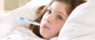
Duchenne myopathy is a congenital pathology that causes constantly developing weakness in the muscles. The disease begins to develop in childhood. Sometimes parents do not even suspect that the child is sick: they do not notice that it is difficult for the baby to run, climb stairs and even stand. Patients with this diagnosis should undergo frequent examinations. This type of myopathy affects mainly boys; it is very rare in girls (but they are carriers).
- Etiology
- Symptoms
- Diagnostics
- Treatment
- Possible complications
- Prevention
Duchenne myopathy is a serious disorder that affects the muscles of the shoulders, torso, and hips. Hands and fingers are not affected. Problems arise when walking and running. Muscle weakness develops gradually. In childhood, when the pathology begins to develop, the symptoms are weak. As the child ages, the manifestation of symptoms increases, and the quality of life deteriorates. The pathology is diagnosed in one boy out of 3,500 births.
Pathogenesis of the disease: in the muscles of every person there is a special protein called dystrophin. People with myopathy have very little protein, which causes their muscles to weaken over time. The cause of the development of the disease is a genetic predisposition: an abnormal gene is passed on from the older generation to the younger. The cause of the disease may be a gene mutation that occurs during puberty. Then healthy parents may have a child with a pathology.
The probability that a carrier mother will pass on the defective gene to her son is 50%. If a woman gives birth to a daughter, the girl is just as likely to become a carrier.
Causes of myopathy
Primary myopathy is a genetically determined disease.
The direct mechanism that triggers the chain of pathological reactions and leads to metabolic disorders in muscle cells is not yet clear. According to the current theory of defective membranes, the primary pathology lies in the plasma membrane of muscle fibers, which leads to uncontrolled penetration of calcium, which activates enzymes (proteases) that destroy muscle tissue. Often, manifestations of myopathy arise (and existing ones also intensify) under the influence of unfavorable provoking factors, which include:
- infectious diseases;
- intoxication;
- physical and psycho-emotional stress.
Most myopathies are of hereditary origin, some of them are associated with the X chromosome.
Acquired forms of myopathy are caused by diseases that cause nerve damage, metabolic disorders, toxic effects, intense inflammatory or tumor processes.
The leading cause of congenital muscular dystrophy is genetically determined defects (mutations) in various genes, which causes disruption of the functioning of mitochondria and ion channels of myofibrils, the process of synthesis of proteins/enzymes involved in the regulation of muscle tissue metabolism, which causes disruption of the structure of muscle fibers (atrophy and muscle paresis), proliferation of connective tissue and (fatty degeneration of muscle fibers), infiltration by lymphocytes.
Inheritance of the disease (defective gene) can occur dominantly, recessively and sex-linked (X chromosome). The role of triggers that launch the pathological process is often external/internal factors - infectious diseases (frequent acute respiratory infections, chronic tonsillitis, salmonellosis, bacterial pneumonia, pyelonephritis, etc.), physical overexertion, severe injuries, intoxications of various origins, nutritional dystrophy.
The leading causes of acquired myopathies are most often endocrine disorders (Cushing's disease, hyperparathyroidism, hyperaldosteronism), tumor processes, chronic intoxications (occupational hazards, substance abuse, chronic alcoholism, drug addiction), vitamin deficiencies, malabsorption, severe chronic diseases (COPD, chronic renal failure, chronic liver/heart failure.
Etiology of the disorder
According to the international classification of diseases ICD-10, Duchenne-Becker muscular dystrophy is coded G71.
One third of cases of the disease are associated with a spontaneously occurring mutation, while the remaining cases of pathology account for the inheritance of the X chromosome carrying the pathological gene.
Cases of gonadal mosaicism are associated with 20% of cases of Duchenne muscular dystrophy. Average life expectancy is less than 20 years. An increase in the level of the enzyme creatine phosphokinase (CPK) is found in 2/3 of women who are carriers of the pathological gene. Most of them do not have any manifestations of the disease. Myopathy almost always affects boys, since the disease is linked to the X chromosome and the type of inheritance is recessive.
One of the genes in the structure of the human genome was given the name of a neurologist, after whom the deviation was named. Duchenne muscular dystrophy can be influenced by various factors:
- incest;
- genetic predisposition, for example, if a relative has Duchenne myopathy;
- improper synthesis of muscle fibers, accelerated spread and replacement by fatty tissue, connective fibers;
- hereditary forms of Duchenne syndrome, most often transmitted from the mother;
- genome mutation during formation during pregnancy;
- abnormalities in chromosomal structures of unknown origin;
- severe disturbances in the development of dystrophin;
- pathological changes in biochemistry in the blood.
Duchenne myopathy also develops in connective tissue diseases that are not directly related to genetic abnormalities.
Possible complications
The most serious complication is a shortened life span. Due to the rapid development of the disease, the muscles weaken and serious heart diseases appear. The functioning of the respiratory system is disrupted.
If Duchenne-Becker muscular dystrophy is detected in the early stages of development, and the method of therapy is chosen correctly, the patient can live up to 30 years.
In parallel with this disease, complications also develop:
- osteoporosis;
- diseases of the spine and joints;
- problems with the gastrointestinal tract;
- disturbances in the functioning of the respiratory system;
- cardiovascular diseases.
Symptoms of muscular dystrophy
The first symptoms appear between the ages of three and seven years. Parents usually notice a swaying gait and hyperlordosis. There are several main criteria that suggest Duchenne myopathy. These include:
- Muscle weakness that appears suddenly and begins in the lower extremities.
- Hyperlordosis is a pronounced anterior curvature of the spine, which is especially noticeable when walking.
- Hypertrophy of weakened muscles.
- Poor muscle response to electrical stimulation in later stages of the disease.
Over time, all of these symptoms progress. Despite damage to muscle structures, bladder and intestinal dysfunction are not observed. By the age of twelve, most patients cannot walk independently and are in a wheelchair. Becker myopathy is very similar to Duchenne dystrophy, since the same locus of the X chromosome is affected, as a result of which dystrophin synthesis is affected. The difference between Becker myopathy is the onset of the disease, usually after three years of life or even in adolescence.
Duchenne muscular dystrophy is a pathology that affects not only skeletal muscles. Dystrophin is also found in the myocardium, brain tissue and smooth muscle. Late stage of the disease is associated with severe heart failure and respiratory disorders - the main causes of death in patients.
Types of myopia
Ophthalmologists distinguish several types of disease, which appear for different reasons, have different classifications and different methods of correction.
| View | Characteristic |
| Congenital | Diagnosed at birth. Appears due to abnormal development of the eye. The rarest form. |
| High | Classified from -6.25 dioprics. |
| Combination | A weak degree in which natural refraction does not occur. |
| False (pseudomyopia, spasmodic) | Temporary visual impairment caused by a spasm of the ciliary muscle. |
| Transitional | Myopia caused by chronic diseases or taking certain medications. |
| Night (twilight) | Myopia that appears in low light. |
| Axial | It is caused by the optical length of the axis of the eye to a large extent. |
| Complicated | Caused by anatomical changes in the eye. May cause absolute blindness. |
| Progressive | Gradual, irreversible loss of vision due to stretching of the back of the eye. |
| Refractive (optical) | Appears due to the large refractive effect on the optical system of the eye. |
Forecast
Unfortunately, the prognosis is unfavorable, since the mortality rate for this pathology is 100%. If there is a family history of a close relative who is a carrier of the pathological gene, male children are at higher risk of developing cardiomyopathy with progressive heart failure between the ages of 20 and 40 years. Research is being conducted on the use of stem cells for the treatment of muscular dystrophies.
The prognosis depends on the form of the disease. It is unfavorable in relation to pseudohypertrophic myopathy, which quickly leads to severe disability and early death. The prognosis for myopathies that debut later, at the end of adolescence, is more favorable.
The prognosis for hereditary congenital myopathies is generally unfavorable, however, it is largely determined by the form of myopathy, the severity of the course, the rate of development and the degree of degeneration of skeletal muscles, as well as the involvement of the respiratory and cardiac muscles in the process. The most unfavorable process is in Duchenne muscular dystrophy, in which patients die before the age of 20-25 years. Late developing forms of myopathy (after 20-30 years) are more benign and have a gentle nature.
The patient can lead a more or less normal lifestyle and even get a profession that corresponds to his ability to move, depending on the level of muscle damage (manual/mental labor). However, with other forms of myopathies, the risk of developing disability remains high. The prognosis of acquired myopathies, provided that the disease that caused the myopathy is successfully treated, is more favorable.
Classification
According to the official classification, low myopia is divided into the same types and forms as high myopia. For reasons of occurrence it can be:
- false - caused by a spasm of the visual muscles responsible for accommodation, changes in the shape of the lens;
- true - caused by congenital or acquired changes in the size of the eyeball (axial myopia), thickening of the cornea or lens (refractive myopia).
It is believed that mild false myopia can be cured fairly quickly. To do this, doctors will have to remove the muscle spasm and prevent its reappearance. The causes of true myopia cannot be eliminated using conservative methods, so treatment requires more time and money.
Based on the nature of development, low myopia is divided into 4 types:
- Stationary. The mildest and most stable form, which practically does not progress, even if observed for years. Does not require serious therapy.
- Progressive. A more serious form of the disease, which is accompanied by a gradual decrease in vision up to 1 diopter per year.
- Transitional. A form that progresses when taking certain medications. In some cases, concomitant diseases can provoke a sharp deterioration in vision.
- Malignant. The most aggressive and rapidly progressing disease. In ophthalmology, cases have been recorded when vision deteriorated to 1 diopter per day, and within a month after its discovery the person received disability.
Twilight myopia has been classified as a separate subspecies. At any degree, myopia of this form manifests itself only in conditions of lack of light. During the day and in well-lit rooms, signs of visual impairment are not observed.
Diagnostic methods
A child with Duchenne-Becker myopathy up to 2-3 years of age may be no different from other children. However, there are a number of signs that are worth paying attention to.
A number of studies of myopathies have noted that speech and motor delays were more common in children with dystrophin deficiency. IQ levels may be one standard deviation below the population average.
30% of children with mutations in the gene encoding the synthesis of dystrophin had difficulties with learning and acquiring new skills, obsessive-compulsive disorders, attention deficit disorder, and mental retardation. Children with Duchenne and Becker dystrophies have more difficulty with verbal skills.
Diagnosis of Duchenne myopathy includes:
- Study of the level of CK, which exceeds the reference values by 50-100 times.
- Molecular diagnostics - a study of the gene located in the Xq21 locus and responsible for the synthesis of dystrophin, allows you to confirm or refute the diagnosis.
If a patient has a combination of high CK levels and muscle weakness, this is highly likely to suspect Duchenne-Becker myopathy.
Of the instrumental methods used:
- plain chest x-ray. The study allows us to conclude how severe scoliosis is;
- electromyography: the method is used for the purpose of differential diagnosis with spinal muscular atrophy;
- electrocardiography – to identify sinus arrhythmias;
- ultrasound examination of the heart - often determines the small size of the ventricles of the heart and a longer diastole;
- Holter monitoring determines the presence of paroxysmal arrhythmias.
If molecular diagnostics do not reveal a dystrophin gene mutation, a muscle tissue biopsy is recommended. Characteristic histological changes are given below:
- muscle fibers with a pronounced degenerative process and necrosis;
- proliferation of connective tissue;
- the appearance of adipose tissue in significant quantities.
The dystrophin protein isolated from the muscle is also analyzed to determine its molecular weight.
Prevention
Prevention of hereditary myopathies comes down to a thorough collection of family history, especially in couples who had relatives diagnosed with hereditary myopathy. Such couples need medical-genetic consultation (preferably at the pregnancy planning stage) with an assessment of the likelihood of having a child diagnosed with myopathy.
Today, Duchenne muscular dystrophy is already determined in the prenatal period, for which it is necessary to study the amniotic fluid and fetal cells for the presence of mutations and their presence is a contraindication to childbearing, however, this issue is resolved by medical genetic consultation doctors and parents individually in each case.
The basis for the prevention of acquired forms of myopathies is the timely treatment of endocrine/infectious diseases, the elimination of the effects of toxic substances on the body, and the correction of metabolic disorders.
It is not possible to prevent the occurrence of congenital myopathy. Identifying the defective gene can help people with a family history of hereditary forms of the disease plan pregnancy.
Preventive measures are effective for acquired myopathies. They include avoiding self-medication, avoiding alcohol abuse, timely detection and treatment of thyroid diseases, and prevention of intoxication.
Treatment
There is still no standard method for treating Duchenne dystrophy, although scientific experiments are being carried out by scientists from many countries, in particular Israel, Great Britain and the USA. Currently, doctors can only offer symptomatic treatment with medications and non-drug treatments.
To improve metabolic processes in muscles, anabolic steroids are used - Methandienone, Nandrolone Decaonate. Actoprotectors give good results, increasing the body’s resistance to stress without increasing the absorption of heat and oxygen.
The most effective drug from this group for muscle atrophy is ethylthiobenzimidazole. To improve the transmission of neuromuscular impulses, Neostigmine is prescribed.
Physiotherapeutic procedures, therapeutic exercises and massage are used as remedies against the development of contractures and further loss of motor activity.
It is very important to monitor respiratory function and blood gas composition. If vital capacity (VC) falls below 41%, artificial ventilation (ALV) at night is indicated. Further, the duration of ventilation increases in accordance with the decrease in vital capacity.
Massage is an important component of treatment; it is done selectively, with a predominant effect on the affected muscles
In the initial stages, mechanical ventilation can be performed using an S-shaped air duct or a Ruben bag. Severe cases require tracheostomy and mechanical ventilation by connecting the device to a tracheostomy tube. Hospitals have portable ventilators that are battery-powered and can be attached to wheelchairs.