Agnosia is a pathological condition associated with a violation of various types of perception, subject to the preservation of consciousness and sensitivity. The reason for this is damage to the secondary parts of the cerebral cortex, which are responsible for the analysis and synthesis of information.
There are different types of agnosia depending on the analyzer in the regulation of whose activity disturbances occurred (auditory agnosia, tactile agnosia, visual agnosia, olfactory agnosia, spatial agnosia and others).
This condition is diagnosed during a neurological examination.
Treatment of agnosia consists of treating the underlying disease.
Causes of agnosia
Agnosia is caused by damage to the projection-associative parts of the cerebral cortex, which are part of the cortical level of the analytical systems.
In this case, a person retains elementary sensitivity, but loses the ability to analyze and synthesize information received from the analyzer, which results in a violation of one or another type of perception.
Damage to the cerebral cortex can result from: cerebrovascular accident, Alzheimer's disease, toxic encephalopathy, subacute sclerosing panencephalitis.
What causes visual agnosia?
It is believed that visual agnosia occurs when areas of the cerebral cortex are damaged in its parietal and occipital-parietal lobes. It is here that information responsible for associations with objects is analyzed and synthesized. The parietal cortex stores all sensory information from the visual system, as well as tactile and spatial associations, sensory data from the skin about temperature, touch, and pain.
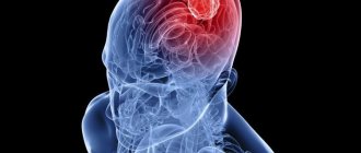
Visual images are formed in the brain, its damage causes optical agnosia
Lesions in these parts of the brain may be caused by:
- Stroke. Brain damage occurs due to disruption of its blood supply as a result of ischemia, thrombosis, arterial embolism, or hemorrhage. Nervous tissue, not receiving oxygen, begins to die, and gradually parts of the brain lose their functions.
- Neurological disorders and tumor processes. Visual agnosia occurs when there is massive damage to the occipital and parieto-occipital regions of the brain.
- Dementia. Neurodegenerative processes that occur in the brain with age lead to loss of cognitive functions and memory. The most common cause of dementia is Alzheimer's disease.
Other possible reasons:
- hereditary predisposition;
- meningeal infection;
- mechanical damage to the occipital lobes due to traumatic brain injury;
- carbon monoxide poisoning;
- recovery after long-term blindness.
Types of agnosia
The following types of agnosia are distinguished:
Visual agnosia - occurs when the occipital cortex is damaged. In this case, the person does not lose visual acuity, but at the same time cannot recognize objects or distinguish the characteristics of objects.
Visual agnosia, in turn, is divided into:
- object - there is a violation of object recognition while maintaining vision function. Patients describe individual signs of objects, but cannot determine which object is in front of them;
- simultaneous - there is a functional narrowing of the visual field to one object. Patients perceive only one semantic unit at a time;
- agnosia for faces (prosopagnosia) – the process of recognizing familiar faces is disrupted. Patients distinguish the face as a whole object and its parts, but cannot say who is in front of them;
- color agnosia, that is, the inability to determine whether a color belongs to a specific object or to select the same colors;
- agnosia caused by optomotor disorders, that is, the inability to direct the gaze in the required direction while maintaining the function of eye movement. The patient finds it difficult to fix his gaze on a given object, it is difficult for him to read;
- weakness of optical concepts - the inability to represent an object, as well as describe its properties.
Auditory agnosia - develops with damage to the temporal cortex. If the left side is affected, there is a violation of the discrimination of speech sounds, which leads to speech disorder. If the right side is affected, the patient does not recognize familiar noises and sounds, or amusia develops, when the ear for music disappears. In this regard, they distinguish:
- simple auditory agnosia - lack of ability to identify simple sounds and noises - rustling paper, knocking, gurgling, clinking of coins;
- auditory-verbal agnosia - lack of ability to recognize speech, which is perceived by the patient as a set of sounds unfamiliar to him;
- Tonal agnosia is the inability to distinguish the expressive aspects of the voice. Patients do not perceive the tone, timbre, or emotional coloring of the voice. But at the same time they understand speech.
Tactile agnosia develops when the central gyrus located in the posterior part of the cortex is damaged. Tactile agnosia is manifested by a lack of recognition of objects by touch (with eyes closed) or a lack of recognition of the texture of an object. In this regard, a distinction is made between object tactile agnosia and tactile texture agnosia.
Spatial agnosia is the inability to determine various spatial parameters. Stands out:
- violation of topographic orientation - inability to navigate in a familiar place. The patient cannot find his home, is lost in his apartment, but his memory is preserved;
- Depth agnosia – the inability to correctly localize objects in space, to determine parameters closer or further;
- unilateral spatial agnosia - a disorder in which loss of one half of space occurs, usually the left;
- impaired stereoscopic vision;
Somatoagnosia is a violation of recognition of parts of one’s body, the inability to assess their location relative to each other. These include: anosognosia (lack of awareness of one’s own illness) and autotopagnosia (failure to recognize individual parts of the body and impaired perception of them in space).
Impaired perception of movement and time - the patient has an incorrect perception of the movement of objects and the passage of time.
Olfactory, gustatory, and finger agnosia are also distinguished.
How to recognize
Most cases of this disease occur in older people who have experienced some degree of brain damage. But signs of visual agnosia can occur at any age.
To understand how everything looks from the first person, imagine the following: you see two circles and a crossbar between them and you cannot even imagine what it is and what it is used for. But a healthy person, when looking at this object, easily determines that these are glasses. He immediately “knows” that they fit on his face and help improve his vision.
Drawing of a person with simultaneous visual agnosia
Depending on where exactly the brain damage occurred in the occipital lobes, the characteristics of visual agnosia vary.
Optic nerve atrophy
- With object agnosia, there is no holistic perception of the object, but its individual parts can be identified. A person lists the characteristics of an object, but cannot find out what it is.
- Agnosia for faces (prosopagnosia) occurs when there is damage in the right hemisphere. A person cannot recognize people he knows by face or in a photo, and in severe cases, he cannot recognize himself.
- Simultaneous agnosia does not allow the patient to look at all objects that are in the field of vision. The violation manifests itself in misses and disorientation. In this state, a person cannot draw anything within a given area (for example, in a circle) or outline a figure.
- Visuospatial agnosia. This type is also called one-sided, or left-sided, agnosia and is characterized by “omission” of the left half of space and even one’s body. The lesion is located on the right side of the brain, and the synthesis of images from the two hemispheres into one does not occur. It manifests itself in the fact that a person “does not see” the left side of the text or drawing; when asked to draw an object, he also draws only its right side from memory.
- With agnosia for symbols (letters), the area of damage is determined at the border of the temporal and occipital lobes. The patient can copy letters and numbers, but cannot recognize and name them, and accordingly the reading skill is lost.
- Agnosia by color. It is characterized by the fact that, in principle, the patient can name the color if it is presented separately on a card, but when asked to name the color of a certain object (for example, strawberries), he experiences difficulty. Variants of the disorder: amnesia regarding the names of shades, inability to imagine a color (imagine it), cortical blindness (complete lack of color discrimination).
- Space agnosia with damage to the area in the upper occipital of the brain. There are varying degrees of disturbance in the perception of the coordinate system. The patient is not oriented in the concepts of left-right, up-down, cardinal directions, or the distance of objects, but normal recognition of objects is maintained. Spatial agnosia is often combined with impaired coordination of movement; accordingly, the ability to write and read suffers. The following types of visual agnosia regarding spatial perception are also possible: macropsia (objects seem larger) and micropsia (smaller than they really are), polymelia (perception of false limbs).
Diagnosis of agnosia
The diagnosis of agnosia is made based on medical history (trauma, stroke, tumor) and the clinical picture of the disease. Special tests are also performed to determine the type of agnosia.
The patient is asked to identify simple objects using various senses. If the doctor suspects denial of half of space, then he asks the patient to identify paralyzed parts of his body, or objects in different parts of space.
Conducting a neuropsychological examination helps determine the presence of more complex types of agnosia.
Brain imaging methods (MRI or CT) are also used to establish the nature of central lesions (hemorrhage, infarction, massive intracranial process) and to identify areas of cortical atrophy.
To identify primary disorders of certain types of sensitivity, a physical examination is performed.
Agnosia is...
Agnosia (from the Greek agnosia, where a is a negative particle, gnosis is knowledge) is a violation of gnosis, one of the types of higher nervous activity of a person. Characterized by impaired recognition of environmental stimuli (light, sound, touch) while maintaining the normal functioning of the analyzers. This term was first encountered in the works of the German physiologist G. Munch in 1881.
Agnosia develops when the secondary projection-associative zones of the analyzers located in the cortex and subcortical structures of the brain are damaged. It is not a separate nosological entity, but a symptom of many diseases of the nervous system, in which focal lesions of the brain occur.
Agnosia develops in women and men equally often. In children it is diagnosed from the age of 7 - 8 years, when the corresponding parts of the central nervous system have already been formed. About 1 - 1.5% of the world's population suffers from this pathology.
The basis for the formation of gnosis
Gnosis is the highest mental function of the brain, manifested by the ability to analyze and compare information received from the outside world with one’s own experience. This type of activity of the central nervous system allows you to recognize objects, phenomena, parts of your body, etc.
This function of our brain develops due to the formation in the cerebral cortex of three groups of projection-associative fields, which process information coming from the external environment at different levels. Primary fields perceive stimuli coming from peripheral analyzers. Secondary – analyze and summarize this information. Then, in the tertiary zones, synthesis occurs, the unification of images and the development of a reaction to stimuli, taking into account the experience accumulated throughout life.
Disruption of the functioning of neurons in the secondary cortical zones leads to the loss of the ability to recognize external stimuli, identify them and compare them into holistic images. At the same time, the analyzers (visual, olfactory, tactile, etc.) remain intact.
Impaired visual recognition (visual anosia)
Previous11Next
Gnostic disorders (agnosia) are a violation of the recognition of stimuli (objects of the surrounding world) related to one or another modality.
Visual agnosia
occur with damage to the secondary projection zones of the cortex (18 and 19 Brodmann areas), localized in the parieto-occipital region of the cerebral cortex. With visual agnosia, there is a disorder of perception and recognition of previously familiar objects while consciousness and intelligence are preserved. Mental pathology is described by the phrase: “It sees, but does not understand.” Elementary visual functions in gnostic disorders are preserved (visual acuity, visual fields, color perception are not impaired). Children with visual agnosia see objects, touch them, hear sounds, but cannot understand what they mean. Adult patients lose the ability to recognize even familiar stimuli perceived by the senses.
In the clinic of local brain lesions, different types of visual gnosis disorders are distinguished, which indicates a high functional differentiation of the visual cortex. The specific designation for visual agnosia depends on whether it refers to an object, space, movement, color, or symbol. Accordingly, the following types of visual agnosia are distinguished:
– object agnosia;
– agnosia for colors;
– agnosia for faces;
– finger agnosia.
Visual agnosia was first described by G. Munch in 1881, observing a dog with an affected occipital region, in which elementary visual functions were preserved (she saw surrounding objects = did not bump into them, but she “did not understand” their meaning). The term “agnosia” was proposed by S. Freud.
The following types of visual agnosia have diagnostic significance in neuropsychological practice: object, facial, optical-spatial, color, letter, color, simultaneous and symbolic.
Object visual agnosia is characterized by a violation of the recognition of individual objects and images or a violation of the integrity of the perception of an object with the possible recognition of its individual features or parts. It is based on defects in recognizing the shape and contours of an object. Most often, this disorder occurs with bilateral lesions of the temporo-occipital regions of the brain, but it can also be caused by unilateral lesions of the right or left hemispheres. Bilateral lesions cause severe disturbances in objective visual gnosis; patients do not recognize even simple images of everyday objects and confuse similar images.
The impossibility of visual identification of an object can manifest itself as a listing of individual fragments of an object or its image (fragmentation), or the isolation of only individual features of an object that are insufficient for its complete identification. For example, recognizing the image of “glasses” as a “bicycle”, since there are two circles connected by crossbars; identification of a “key” as a “knife” or “spoon” based on the selected features “metal” and “long”.
Object agnosia can have varying degrees of severity - from maximum (difficulty in recognizing real objects) to minimal (difficulty recognizing contour images in noisy conditions or when superimposed on each other). As a rule, the presence of extensive object agnosia indicates bilateral damage to the occipital region. Thus, with severe damage, the perception of real objects is impaired: the patient does not recognize previously familiar objects or can only describe their individual features. To facilitate identification, he tries to feel the object, and if it is food, then taste it. Often, to identify an object, the patient names randomly selected features by brute force and thus tries to “recognize” the object. A less severe gnostic visual disorder is manifested by the fact that the patient recognizes real objects, but does not recognize schematic images, inverted or superimposed drawings.
In mild cases, only the time of tachistoscopic recognition increases when a stimulus is presented in a dosed manner (normally, the time sufficient to recognize a stimulus is 0.01 sec; with visual gnostic disorders, the recognition time can increase to 1 sec or more).
With unilateral lesions of the occipital region of the brain, differences in the structure of visual object agnosia can be seen. Damage to the left hemisphere is manifested to a greater extent by a violation of the perception of objects by the type of listing of individual details. The pathological process in the right hemisphere leads to the virtual absence of the act of identification. In this case, the patient can evaluate the visually presented object according to its significant characteristics, answering the researcher’s questions about the relationship of this object to “living - inanimate”, “dangerous - non-dangerous”, “warm - cold”, “big - small”, etc.
Diagnosis of subject visual gnosis[5].
1. Recognition of depicted objects (methodology of A. R. Luria). 16 pictures are presented, which depict externally similar objects, for example, a snake - a belt, a cap - a plate, a table lamp - a mushroom, etc.
2. Recognition of objects with missing features (unfinished pictures).
3. Poppelreiter's test - identification of superimposed images.
4. Recognition of disguised (hidden) figures.
5. Folding pictures from parts.
6. Correlating an object with a form.
Facial agnosia (prosopagnosia) often develops with right-sided lesions of the secondary and tertiary projection areas of the visual cortex. Often prosopagnosia manifests itself as an independent gnostic defect. Face agnosia is a selective gnostic disorder, manifested in difficulties in recognizing familiar faces. In some cases, with a gross manifestation of the defect, patients do not recognize their loved ones, photographs from a family album, cannot imagine or describe a familiar face, evaluate people by random signs (moles, hairstyles, etc.), as well as by voice, gestures. In rare cases, patients with such disorders find it difficult to evaluate facial expressions expressing a particular emotion, and also see distorted grimaces. Agnosia on faces
caused by a lesion in the temporo-parietal-occipital regions of the right hemisphere.
| Prosopognosia is characterized by difficulty or inability to distinguish familiar faces |
Manifestations of prosopagnosia vary in severity. Thus, a mild degree is characterized by difficulty distinguishing familiar faces and recognizing facial expressions. In severe cases, the patient does not recognize previously familiar people, does not identify their gender, age, and uses auxiliary signs for identification: voice, gestures, gait, etc. In some cases, he does not even recognize himself in the mirror. Violations of moderate severity are characterized by difficulty in identification from a photograph or schematic image. The patient cannot combine the front and profile.
There are apperceptive and associative facial agnosia. Apperceptive
characterized by a violation of gender and age identification.
With associative
prosopognosia, the analysis and synthesis of perceptual information is available (the patient recognizes gender, age, determines the degree of similarity of faces), but associative connections are impaired. Thus, the patient cannot provide any information about the person he sees, while the emotional reaction to this person is preserved.
Diagnosis of facial gnosis [H4] [6] .
1. Recognition of familiar faces. Presentation of photographs of outstanding domestic writers.
2. Identification of unfamiliar persons. The subject is presented one by one with standards - three images of faces unfamiliar to him - and is asked, looking at them, to find identical ones in a set of 20 images. If completed successfully, the task becomes more complicated: the photograph is presented for 10 seconds, and the search is carried out from memory.
2. Emotional identification of portraits. The patient is presented with images of faces (photographs or diagrammatic images) that reflect certain emotional states: joy, pleasure, surprise, fear, anger, grief, contempt, pride - and is asked to identify them.
Optical-spatial agnosia is characterized by the fact that the patient, while retaining the recognition of individual objects, loses the ability to navigate in a familiar space. With less severe lesions, orientation in real space is preserved, but there is a loss of the ability to distinguish between “right and left,” impaired orientation in geographic maps, in the position of clock hands, and loss of the ability to mentally rotate an object by 90–180°.
Optical-spatial agnosia occurs when there is predominant damage to the superior parietal and parieto-occipital parts of the cortex of the left or right hemispheres of the brain, due to which the complex interaction of several analyzer systems (visual, auditory, tactile, vestibular) occurs. Optical-spatial agnosia is especially severe in cases of symmetrical bilateral lesions.
Features of the functioning of the visual areas of the brain make it possible to recognize such spatial characteristics of visual images as size, distance, direction, and relative position of objects. These characteristics of objects are learned in ontogenesis due to associative connections between various modalities: tactile, auditory and visual. Such disorders vary in form depending on the location of the lesion and, above all, on the side of the brain in which it is located. During the period of mastering the corresponding types of activity, the cause of such disorders can be not only lesions, but also various dysfunctions in the maturation of brain structures.
Left hemisphere lesions or dysfunctions lead to disturbances in spatial orientation activity. They are characterized by insufficient discrete-logical analysis of optical-spatial objects. In these cases the following are violated:
1) schematic ideas about the spatial relationships of objects of reality (inability to rotate a figure in space, orientate oneself in a geographical map, a clock, spatial games, etc.);
2) various types of constructive activities, drawing;
3) body diagrams (autotopagnosia);
4) naming and understanding words denoting spatial relationships: prepositions with spatial meaning on, in, under, above
etc., adverbs such as
far, on the side, below
, etc., on the basis of which agrammatism, characteristic of patients with semantic aphasia, often arises;
5) identification and naming of fingers (finger agnosia);
6) writing and reading (based on a defect in analytical-synthetic, simultaneous actions for sound-letter analysis of the composition of a word).
Right hemisphere lesions with optical-spatial agnosia and dysfunction are observed much more often than left hemisphere lesions. They cause a lack of holistic perception of the spatial situation:
1) simultaneous agnosia, in which there is an inability to assess the meaning of a plot picture due to the fragmentation of the perception of the spatial situation, although recognition of individual objects, as a rule, remains intact;
2) impaired recognition of a familiar spatial situation, inability to reproduce it from memory;
3) a violation of the body diagram (autotopagnosia), when orientation in the location of body parts is difficult because they are perceived as distorted in size, disproportionate.
The listed situations are especially pronounced on the contralateral (opposite to the lesion on the left half of the body).
As a consequence of a violation of the body diagram and weakening of visual control, difficulties arise in constructing movement in space, i.e. apraktoagnosia,
consisting in the disintegration of established everyday skills, for example, dressing (apraxia of dressing), the ability to draw, perform professional actions, etc.
The most striking and characteristic symptom of optical-spatial disorders that occur when the right hemisphere of the brain is damaged is unilateral spatial agnosia. With it, the phenomenon of ignoring the left half of space, as well as visual, auditory, and tactile stimuli emanating from the left half of space, appears.
| Optical-spatial agnosia is characterized by the patient losing the ability to navigate in a familiar space |
Diagnostics of optical-spatial gnosis [H5] [7] .
1. Draw a floor plan.
The study can begin with asking the patient to orient himself in the real space in which he is currently located (draw a plan of the department, tell how to get from this office to the exit; indicate which of the visible objects is closer or further).
2. Determination of parts of the world: the experimenter places a dot on the picture. The subject is asked to determine the direction of the world.
3. "Blind compasses." The figure shows only one part of the world, and not in a typical place. Task: identify the direction indicated by the arrow.
4. Line orientation test by A. Benton. The subject needs to be shown in the drawings which of the set of lines corresponds to the lines exhibited as stimulus material (they need to be correlated by angles of inclination). Instead of recognition, the option of sketching lines is possible.
5. Drawing figures
The experiment usually begins with asking the subject to draw a “house” and a “cube” on his own. In case of inadequacy of the drawing, it is proposed to copy the same object from the sample.
6. Drawing “House – tree – man.” When drawing in patients with optical-spatial agnosia, the overall scheme of the drawing may disintegrate, the spatial arrangement of parts of the drawing may be disturbed, and proportions may be disturbed.
7. Test with rotation of figures.
The subject is offered a form with rows of drawn squares, inside each of which there are one or two graphic marks located in a certain way in the space of the square. The leftmost square is a sample, in relation to which you need to mentally rotate the subsequent squares 90° clockwise and then select the one that fully matches the sample.
8. Braid cubes. The subject is offered a series of increasingly complex patterns of two-color patterns that must be laid out from the upper faces of the cubes. The cubes have two opposite faces painted red, two – white, and two more – diagonally divided into white and red triangles.
Agnosia for colors
or
color agnosia
, is caused by damage or dysfunction of the temporo-occipital regions of both the left, dominant, and right, subdominant hemispheres. Color perception is preserved, i.e. the patient distinguishes colors, names them correctly, but has difficulty correlating the color with a specific object: he cannot remember what color an orange, carrot is, or cannot name objects of a certain specific color. In addition, there is evidence that when the right hemisphere is damaged, difficulties may arise in differentiating mixed colors (brown, purple, orange, pastel colors). Color agnosia of the dominant type, like other types of visual agnosia, is characterized by a violation of abstractness and generalization in perception. Patients with color agnosia cannot select shades of color into a single color scheme - a selective disorder accompanied by forgetting the names of colors.
| With color agnosia, the patient experiences difficulty relating a color to a specific object. |
Previous11Next
Date added: 2017-05-02; views: 7214;
Similar articles:
Diagnosis and treatment of deviation
Letter agnosia is a condition in which a person is unable to correctly identify letters using one or more senses. To make a correct diagnosis, the doctor must take into account all clinical manifestations.
Often, during the diagnostic process, a neuropsychological study is carried out, brain imaging methods are used, for example, computed tomography or MRI, which help to establish the causes of the formation of pathology. The prognosis for the curability of the disease depends on the severity of the damage, the neglect of the disease, the nature of its course and the age group of the person.
There are no specific methods for organizing the treatment process of agnosia. Usually, the main task of treating pathology is to treat the underlying disease, which has resulted in damage to a specific area of the brain and, as a consequence, the development of agnosia. In order to compensate for the symptoms of the disease, the help of neuropsychologists, speech therapists and occupational therapy specialists is often required.
In medical practice, the treatment process for agnosia usually lasts about three months, and in particularly advanced cases up to twelve months. But, as a rule, with the usual development of the disease, three months is quite enough for the patient to fully recover. The success of the treatment depends to a large extent on the age of the person, on the nature of the damage that provoked agnosia, and on the severity of this damage. Even if the slightest deviation is detected, it is strongly recommended to visit a specialist and undergo a thorough examination in order to prevent serious consequences.
Rate this article:
(votes: 5 , average: 4.80 out of 5)
Loading...
Related posts:
- The concept of autopagnosia and its treatment
- Tactile agnosia, its types and treatment
- Characteristic features of the manifestation of simultaneous agnosia
- Features of the manifestation of visual agnosia
- Facial agnosia, its symptoms and subsequent treatment
- The treatment process for color agnosia
Optical-spatial pathology
Characterized by a disorder in determining the parameters of space. Here are the types of this violation:
- Agnosia of depth. A person cannot correctly localize objects in three spatial coordinates. Especially in depth. Reason: disturbances in the parieto-occipital region (middle sections, as a rule).
- Stereoscopic pathology. Inability to see a three-dimensional image, in other words. Reason: disruption of the left hemisphere.
- Spatial one-sided pathology. A person simply does not perceive what is happening on the left side of him. Cause: damage to the parietal lobe, contralateral side of the prolapse.
- Violation of topographic orientation. With this pathology, a person retains his memory, but loses the ability to navigate. He can get lost in his yard, get lost in the city where he has lived all his life. Cause: damage to the parieto-occipital region.
Having one of the pathologies is not just inconvenient, but dangerous. If, for example, a person is diagnosed with optical-spatial visual agnosia, the signs of which were discussed above, then he can easily get lost even in his own apartment. Therefore, the disease must be dealt with. But how?
Mechanism of disease development
Simultaneous agnosia develops with right-sided or bilateral damage to the occipital-parietal region of the brain. The process of perceiving visual information consists in the ability to process only one operational unit of this information, which is currently the object of the patient’s attention.
So, for example, if a patient is given the task of placing a point in the center of a circle, then the patient will not be able to complete the task, since in this case it is necessary to perceive and interrelate all three objects at once (the boundaries of the circle, its center, the tip of a pencil). In this case, the patient “sees” only one object out of three.
It is worth noting that simultaneous agnosia is not always so clearly expressed. Most often, difficulties are observed in recognizing and reproducing a complex of any objects, when some details are lost and elements “fall out”. This is noted when drawing and reading independently. Also, a disorder of eye movements, which is called “gaze ataxia,” may occur.
Often, a violation of letter identification is closely related to the recognition of numbers, while difficulties with their writing, as a rule, do not arise.
A severe manifestation of simultaneous agnosia is Balint's syndrome, in which it is very difficult for patients to keep their gaze in a certain direction, fixing it on some object.
There is another manifestation of this disease - fragmented perception, in which the patient sees only a part or detail of an object, and not the entire object as a whole. For example, if such a patient is shown a table lamp and asked what he sees, the patient may answer that he sees, for example, an ashtray, since he sees the lower part of the table lamp.
Some patients report that objects that the patient was just looking at suddenly suddenly disappear. What is happening can be explained by the fact that the patient’s gaze does not return to its starting point. In the same way, for patients with this form of agnosia, the perception of moving objects is extremely difficult, and distraction of attention further aggravates the picture.
Description of the disease
Agnosia is a pathological condition that is formed as a result of damage to the cortex in the brain, as well as nearby subcortical structures. If the brain damage is asymmetrical, then unilateral agnosia may appear.
Facial agnosia usually develops due to damage to the right hemisphere of the brain, usually in the posterior or middle regions. The strength of the pathology manifestation varies, ranging from impaired recognition and memory of faces during experimental tests to the inability to recognize one’s own reflection in the mirror. Selective impairment of face memory may also develop.
The face, as a visual object, has a certain specificity compared to objects. The possibility of impaired perception of faces is confirmed by data on the difficulties of playing chess in people with damage to the right hemisphere of the brain.
In addition, the perception of a human face contains a certain individuality of the perceiver, who captures something of his own in the facial features, completely subjective even when examining portraits of famous personalities. The specificity of the person in question lies in its uniqueness, which expresses the individuality of the sample in relation to the original perceived by a person.
A special form of this pathology includes a violation of a person’s recognition of his own body. This process often occurs when a large part of the right hemisphere of the brain is affected. In this case, a person ceases to recognize his own limbs and may experience the presence of several arms or legs. At the same time, the patient realizes the absurdity of the current situation. This group of deviations may include phantom pain and the feeling of having a missing limb or the inability of a person to realize that he has abnormal vision, hearing, or the presence of paralysis.
Finger agnosia
Finger agnosia is considered a special form of autotopagnosia. With this form, a person loses the ability to recognize and show specified fingers, both on his own hand and on the hand of another person.
The patient is not able to show the finger on his hand that the doctor asks, especially. If at the same time the position of the hand changes sharply. Most often, recognition errors are present for the second, third and fourth fingers of the right and left hands. This form of the disease usually develops when the left side of the parietal lobe, its angular gyrus, is damaged.


