Systemic lupus erythematosus ( SLE
) is a connective tissue disease that occurs with damage to various body systems caused by circulating immune complexes and autoantibodies. Systemic lupus erythematosus is characterized by typical lesions of the joints, skin, serous membranes, central nervous system and internal organs with disruption of their function. The prevalence of the disease is 20–50 cases per 100 thousand people. It occurs mainly in women of reproductive age (90% of cases).
Etiology
The etiology of the disease is currently unknown. Lupus is characterized by impaired immune response, manifested by increased activation of T-lymphocytes and B-lymphocytes, which is caused by genetic predisposition and the influence of unfavorable environmental factors.
The role of genetic factors is confirmed by a large number of familial cases of the disease (10–15%) and higher concordance in identical twins (up to 50%).
Unfavorable environmental factors include viruses, including retroviruses, toxic substances and drugs. But there is no convincing evidence of the importance of these factors in the pathogenesis of lupus, with the exception of ultraviolet radiation, which provokes an exacerbation of the disease.
Hormones involved in the development of immunological tolerance play a significant role in pathogenesis. In women with systemic lupus erythematosus, an excess of estrogens and prolactin was detected, causing polyclonal activation of B lymphocytes. Therefore, women of childbearing age get sick 8–9 times more often than men.
Etiology and pathogenesis of systemic lupus erythematosus
The leading hypothesis for the development of systemic lupus erythematosus is genetic predisposition and the emergence of stimulating factors during life. This complex interaction may explain the different clinical manifestations of the disease in each individual.
- SLE has a fairly modest recurrence rate within families—only 8% of patients have at least one first-degree relative with the disease.
- Symptoms occur in 24% of identical twins and approximately 2% in twins.
- If the mother has signs of the disease, then the probability of relapse in her son will be 1:120, and in her daughter - 1:40.
Genetic factors cause the so-called true systemic lupus erythematosus . In addition, there is a false course of the disease due to the influence of certain medications, this is drug-induced lupus erythematosus . This type of disorder is reversible and occurs due to exposure to chemical components of drugs due to long-term use of drugs.
The clinical picture of drug-induced lupus erythematosus disappears immediately after discontinuation of the drug causing the pathological disorders. And there are about 38 drugs known to cause symptoms of lupus. Which ones are the most common?
- Procainamide.
- Isoniazid.
- Hydralazine.
- Quinidine.
- Phenytoin.
Potential stimulating factors influencing the development of systemic lupus erythematosus include a number of conditions.
- Professional or household activities associated with large quantities of silicone dust.
- Smoking tobacco.
- The use of estrogens in therapeutic regimens in postmenopausal women.
- Exposure to light stimulates rapid symptoms on the surface of the skin. Ultraviolet radiation increases the activity of keratinocytes, which leads not only to excessive expression of substances on the surface of epithelial cells, but also affects the secretion of cytokines, which increase the production of autoantibodies.
- Some anecdotal studies suggest an interaction between vitamin D levels and lupus symptoms. Hypovitaminosis stimulates the production of autoantibodies in healthy people and also leads to B-cell hyperactivity and interferon-alpha activity in patients with SLE.
At the cellular level, the pathogenetic characteristics of lupus erythematosus are attributed to disorders of apoptosis - programmed cell death. This is when outdated or damaged cells are carefully disposed of by one’s own body, which is a physiologically normal process of life. A significant role in the functioning of physiological apoptosis is played by antibodies of the immune system, which absorb, digest and utilize unnecessary cellular substances.
In SLE, the immune system produces antibodies programmed to destroy healthy cells, or more precisely, to destroy protein formations in their cell nucleus.
Normally, the immune system is quite sensitive to antigen-presenting cellular structures, which, as a “friend or foe” signal, make it possible to distinguish between foreign substances and native cells. Viral and bacterial pathogenic agents do not contain antigen-presenting structures familiar to the antibodies of the immune system, which causes the latter to attack the foreign antigen. This is the basis of immunity.
Gene disorders in SLE may not manifest themselves for quite a long time. But the formation of abnormal DNA in antigen-presenting structures can occur, which provokes an attack on them by their own immune bodies under a certain set of circumstances - under the influence of viruses, bacteria, ultraviolet radiation, and medications.
Pathogenesis
The pathogenetic basis of systemic lupus erythematosus is the constant production of autoantibodies and immune complexes. In patients, high-affinity autoantibodies are produced by individual clones of B lymphocytes (polyclonal immune activation).
The production of autoantibodies is also stimulated by T lymphocytes (CD4, CD8 and T lymphocytes that express neither CD4 nor CD8). Activation of B and T lymphocytes is a consequence of apoptosis (programmed death) of cells of various tissues, violations of immunological tolerance and the removal of immune complexes by the reticuloendothelial system, proliferation that controls the immune response of cells. Antigens that are important in the immunopathogenesis of systemic lupus erythematosus are components of one's own cells (nucleosomes, ribonucleoproteins, surface antigens of lymphocytes and erythrocytes).
The resulting autoantibodies have a damaging effect on tissue as a result of specific binding to antigens or, having a positive charge, are able to bind to negatively charged structures. Thus, autoantibodies to erythrocytes cause hemolytic anemia, to platelets - thrombocytopenia, to lymphocytes - leukopenia and dysfunction of T-lymphocytes. Autoantibodies to DNA react with the glomerular basement membrane. Autoantibodies to the ribosomal P protein damage the tissues of the meninges, while antineuronal antibodies damage neurons.
The resulting autoantigen–autoantibody complexes circulate in the blood, activate complement and become fixed in the capillaries and renal tubules, leading to tissue damage.
Lupus - causes of the disease
What is lupus erythematosus? The main version is disturbances in the functioning of the immune system, as a result of which healthy cells of the body perceive each other as foreign and begin to fight among themselves. The disease lupus, the causes of which are still not fully understood, is now widespread. There is a safe type of the disease - medicinal, which appears while taking medications and goes away after they are discontinued. Can be transmitted from mother to child at the genetic level.
Pathological anatomy
For systemic lupus erythematosus, typical changes characterizing damage to connective tissue are the accumulation in it of amorphous masses of nuclear substance, stained with hematoxylin in a purple-blue color. Foci of fibrinoid necrosis formed by immune complexes consisting of DNA, antibodies to DNA and complement are also detected.
Leather.
Skin lesions at the onset of the disease are manifested by nonspecific lymphocytic infiltration. Subsequently, deposition of Ig and complement occurs, degeneration of the basal layer of the epidermis and necrosis of the dermoepidermal junction develop. Infiltrates of lymphocytes, macrophages and plasma cells around the skin appendages and vessels of the upper layers of the dermis are characteristic. Open damage to the walls of small vessels is often detected.
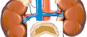
Kidneys.
Deposition of immune complexes in the mesangium and basement membrane of the glomeruli causes the development of glomerulonephritis. Ig deposition only in the mesangium leads to the development of the most commonly detected mesangial nephritis, which is characterized by a mild course and does not lead to renal failure. Deposition of immune complexes in the basement membrane worsens the prognosis. When glomerular segments are involved, focal proliferative glomerulonephritis develops in more than 50% of glomeruli, which can progress to the development of diffuse proliferative glomerulonephritis, characterized by cellular proliferation of most glomerular segments in more than 50% of glomeruli. Membranous nephritis, which affects peripheral capillary loops without glomerular cell proliferation, is rare. The prognosis for this form is relatively favorable.
To assess the activity of the process and its chronicity, a kidney biopsy is performed. High inflammatory activity is characterized by glomerular necrosis, epithelial crescents, necrotizing vasculitis and interstitial infiltrates. Glomerulosclerosis, fibrous crescents and tubular atrophy are signs of irreversible kidney damage.
Central nervous system.
All parts of the brain, meninges, spinal cord, cranial and spinal nerves are affected. The morphological substrate of these lesions is perivascular inflammatory changes in small vessels, microinfarctions and microhemorrhages.
Classification
The classification of systemic lupus erythematosus used in Russia was developed by V. A. Nasonova (1972–1986). The classification presents the nature of the course of the disease (acute, subacute and chronic), phases and degree of disease activity in accordance with the severity of clinical symptoms and the level of laboratory parameters in a given period of time (0 - remission, I - minimal, II - moderate, III - high) and clinical and morphological characteristics of lesions.
The acute course is characterized by febrile fever, fatigue, general weakness, and severe articular syndrome such as polyarthritis. Moreover, in most cases, the patient remembers the day the disease began. In the coming months, a polysyndromic picture develops with the involvement of vital organs in the pathological process.
In the subacute course, the onset of the disease is not so pronounced; over the course of 1-1.5 years from the first symptoms of the disease, a polysystemic system is formed, there is a tendency to progress when, during the next exacerbation, new organs or systems are involved in the pathological process.
In a chronic course over several years, there is one syndrome (monosyndromic) or several syndromes or symptoms that cannot be combined into systemic lupus erythematosus. Exacerbation is replaced by spontaneous remissions; in some cases, glucocorticoid therapy is not required.
What is lupus
Libman-Sachs disease is an autoimmune disease that affects connective tissue and the cardiovascular system. One of the recognizable symptoms is the appearance of red spots on the cheekbones, cheeks and bridge of the nose, which look like butterfly wings, and in addition, patients complain of weakness, fatigue, depression, and fever.
Lupus disease - what is it? The reasons for its appearance and development are still not fully understood. It has been proven that this is a genetic disease that can be inherited. The course of the disease alternates between acute periods and remissions, when it does not manifest itself. In most cases, the cardiovascular system, joints, kidneys, nervous system suffer, and changes in blood composition occur. There are two forms of the disease:
- discoid (only the skin suffers);
- systemic (damage to internal organs).
Symptoms
Systemic lupus erythematosus mainly affects women aged 20–30 years. The onset of the disease has various options. It is possible to damage one system with subsequent spread to others, but it is also possible to damage several systems at once. The main general symptoms of systemic lupus erythematosus are weakness, fatigue, loss of appetite, weight loss.
- In acute cases, the disease develops within two to three days. Patients experience fever accompanied by chills, acute polyarthritis, polyserositis, and the “butterfly” symptom.
- In the chronic course of the disease for a long time, the main syndromes are polyarthritis, less commonly polyserositis, and Raynaud's syndrome. In the 5th–10th year of the disease, nephritis, pneumonitis, and polyneuritis appear. The chronic course is the most favorable.
Musculoskeletal system.
The most common signs are arthralgia and myalgia, which are found in 85–90% of patients at the onset of the disease. Subsequently, most of them develop arthritis. Usually the small joints of the hands, wrist and ankle joints are affected, and less commonly the knee joints. The joints are swollen, and fusiform thickening of the fingers may develop. X-ray examination reveals epiphyseal osteoporosis, mainly of the joints of the hands and wrists, and in chronic polyarthritis - thinning of the subchondria plates. Joint deformity and erosion of articular surfaces are usually absent. Most patients develop persistent myalgia, myositis, and muscle atrophy, especially pronounced on the back of the hands. The causes of muscle damage can be inflammation and side effects of medications (glucocorticoids, aminoquinoline derivatives).
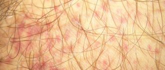
Leather.
Skin lesions are a characteristic symptom of systemic lupus erythematosus. The most typical is butterfly erythema (persistent redness of the skin with a cyanotic tint on the cheeks, bridge of the nose, often spreading to the ears and chin). Redness increases with insolation and exposure to wind and cold. In some patients, nonspecific exudative erythema is detected on the skin of the extremities and chest, similar to the décolleté type. Approximately 20% of patients develop discoid erythema on the face, chest, and limbs. Diffuse or focal hair loss on the scalp appears relatively early, which can grow back as the condition improves. Concomitant skin vasculitis, manifested by hemorrhagic rash, infarction of the nail folds, ulcers and gangrene of the fingers, is of diagnostic importance. Skin rashes are often combined with ulcers on the oral mucosa, angular stomatitis and damage to the red border of the lips.
Kidneys.
Kidney damage (lupus glomerulonephritis) develops in half of the patients. In the early stages, some patients are diagnosed with nephrotic syndrome, characterized by proteinuria, hematuria and cylindruria. Kidney function is usually normal. Diffuse proliferative glomerulonephritis quickly leads to deterioration of renal function, requiring immediate initiation of active treatment with high doses of glucocorticoids and cytostatics. It is advisable to perform a kidney biopsy before starting treatment. If therapy is ineffective and the serum creatinine level increases to more than 270 µmol/l (3 mg/%), this indicates sclerosis of a significant part of the glomeruli, which should be confirmed by a kidney biopsy. Such patients undergo hemodialysis or kidney transplantation. There is also an increased risk of developing severe glomerulonephritis in patients with persistent changes in urine, high titers of antibodies to native DNA and low levels of complement in the serum.
Nervous system.
Affects 55–60% of patients. The most common manifestations are symptoms of asthenovegetative syndrome: weakness, adynamia, headache, irritability, sleep disturbances. As the disease progresses, epileptic seizures, polyneuritis, and decreased tendon reflexes are possible. Changes in the emotional sphere, depressed mood, memory and intellectual impairments, and psychosis are less common. Some patients develop extrapyramidal and cerebellar disorders, damage to the hypothalamus with impaired ADH secretion, serous meningitis, intracranial hypertension, optic neuritis and sensorimotor neuropathy. On the EEG, 70% of patients show a generalized slowing of the rhythm or focal changes, which can be detected using CT and angiography. MRI can detect changes in the brain in most patients, but they are nonspecific. Lumbar puncture is indicated for patients with neurological symptoms. In the cerebrospinal fluid, half of the patients have an increased level of protein, a third have an increased number of lymphocytes, and in some patients immunoglobulins and antibodies to neurons are detected.
The cardiovascular system.
Damage to the cardiovascular system is characteristic of systemic lupus erythematosus. Most often, exudative pericarditis occurs, the rapid progression of which can lead to cardiac tamponade. Myocarditis may develop, accompanied by rhythm disturbances, blockades and heart failure. A characteristic symptom of lupus is aseptic thromboendocarditis Libman-Sachs, sometimes leading to embolism of the blood vessels of the brain, spleen and kidneys, aortic or mitral heart defects. Various vascular lesions develop (Raynaud's syndrome, thrombosis of arteries and veins), accompanied by severe complications. Vascular lesions are a consequence of vasculitis. But there is evidence indicating the possibility of thrombosis without inflammation as a result of the action of antiphospholipid antibodies (lupus anticoagulant, antibodies to cardiolipin). Some patients develop atherosclerotic vascular lesions against the background of hyperlipoproteinemia, which is a consequence of long-term glucocorticoid therapy.
Respiratory system.
Respiratory organ lesions usually develop in the 2nd–3rd year of the disease, most often dry or exudative pleurisy. Sometimes lupus pneumonitis develops, characterized by shortness of breath, cough, and fever. X-ray examination reveals pulmonary interstitial infiltrates and subsegmental atelectasis. Infiltrates in the early stages are treatable with glucocorticoids. But it is possible to develop pulmonary fibrosis, the treatment of which is ineffective. In addition to pneumonitis, secondary bacterial pneumonia, candidiasis, and tuberculosis can develop. Very rare complications include pulmonary hypertension and adult respiratory distress syndrome.
Gastrointestinal tract.
Lesions of the gastrointestinal tract can manifest as lupus peritonitis and vasculitis of the mesenteric vessels, accompanied by cramping abdominal pain, nausea, vomiting and diarrhea. Sometimes small intestinal pseudoabstruction syndrome develops due to swelling of its wall. Glucocorticoids are effective for these gastrointestinal disorders. In case of intestinal perforation, urgent surgery is necessary. In some patients, aminotransferase activity increases without significant liver damage, which decreases as the exacerbation subsides.
Eyes.
Eye damage is one of the severe lesions of systemic lupus erythematosus. It is possible to develop choroiditis, leading to blindness within a few days, conjunctivitis, episcleritis, and optic neuritis. In the fundus of the eye, ophthalmoscopy reveals a narrowing of the artery and white lesions near them.
Antiphospholipid syndrome.
This syndrome develops in approximately 20–30% of patients and is characterized by the appearance of lupus anticoagulant and antibodies to cardiolipin. The presence of lupus anticoagulant is confirmed by prolongation of the aPTT and its normalization in the presence of phospholipids. Sometimes, antibodies-inhibitors of coagulation factors VIII or IX are found in the serum of patients. Clinical manifestations of antiphospholipid syndrome are recurrent thrombosis, most often of the cerebral arteries with a clinical picture of ischemic microstrokes and deep veins of the lower extremities, hepatic and portal veins. In some patients, the heart valves are affected (from small thickenings of the valves to severe mitral and aortic defects). Sometimes thrombosis of the coronary arteries develops with the development of myocardial infarction. In women, antiphospholipid syndrome is manifested by repeated spontaneous abortions, intrauterine fetal death at any stage of pregnancy and preeclampsia. With thrombocytopenia and hypoprothrombinemia, combined with a lupus anticoagulant, patients may experience bleeding. Another cause of bleeding may be antibodies - inhibitors of clotting factors.
Symptoms of discoid lupus erythematosus
- Plaques, redness on the skin of the face, upper body.
- Atrophy, scars with dilated vessels at the site of lesions.
- Scarring alopecia of the scalp.
- Skin itching in the area of the rash.
- Ulcerations on the oral mucosa (in 15% of cases).
- In the common form, there is damage to joints, muscles, and deterioration in general well-being.
Dermoscopy reveals 3 stages:
- Erythema: pink-red scaly plaques on the face (butterfly-shaped), chest, back; along the edges of the lesions - telangiectasia, areas of hyper- and depigmentation.
- Follicular hyperkeratosis: plaques have small asbestos scales, when removed, pointed spines are revealed (symptom of “ladies’ heels”); painful removal of scales (Besnier-Meshchersky symptom).
- Cicatricial atrophy: three zones in the lesions - cicatricial (in the center), infiltrative-hyperkeratotic and erythematous (peripheral) atrophy.
Diagnostics
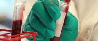
A general blood test reveals hypochromic, less often hemolytic, anemia, leukopenia and thrombocytopenia. ESR in most patients weakly correlates with disease activity. In the urine of lupus glomerulonephritis, proteinuria, hematuria, and casts, including granular ones, are found with high activity. It is mandatory for patients with kidney damage to study serum protein levels. To assess the activity of the process and its chronicity, a kidney biopsy is performed.
Immunological studies can detect autoantibodies characteristic of systemic lupus erythematosus. Antinuclear antibodies, determined using immunofluorescent methods using human epithelial cells (cell lines WIL-2 and HEp-2), are detected in 95% of patients. Antinuclear antibodies, when interacting with components of the nuclei of epithelial cells, form fluorescent immune complexes that have diffuse, homogeneous or ring staining. But they are not specific for lupus erythematosus, as they are found in the serum of healthy people, especially older people, with other autoimmune diseases, viral infections and chronic inflammation.
More specific for lupus are antibodies to native DNA and the Sm antigen, which consists of proteins associated with small nuclear ribonucleoproteins (SNPs) U1, U2, U4/6 and U9. In Sjogren's syndrome, subacute cutaneous lupus erythematosus and in elderly patients with SLE, antibodies to Ro/SS-A are detected, which can cause glomerulonephritis. If antibodies to the La/SS-B antigen are detected simultaneously with these antibodies, this indicates a low risk of glomerulonephritis. The degree of activity of lupus erythematosus is determined by clinical and laboratory characteristics.
Diagnosis of systemic lupus erythematosus in typical cases, in which characteristic skin syndromes, polyarthritis and serositis are identified, is not difficult. Laboratory tests of immune status confirm the diagnosis and help exclude other diseases. The criteria of the American Rheumatological Association are used to diagnose lupus erythematosus. The presence of 4 out of 11 criteria confirms the diagnosis.
Differential diagnosis:
- Differential diagnosis is carried out with rheumatoid arthritis, in which extra-articular manifestations (serositis, carditis) are possible. Unlike systemic lupus erythematosus, rheumatoid arthritis exhibits persistent articular syndrome, rapid development of erosive-destructive changes in small joints, and less pronounced systemicity.
- Great difficulties arise when carrying out a differential diagnosis with mixed connective tissue disease, which has signs of several connective tissue diseases (systemic lupus erythematosus, dermatomyositis, scleroderma, inflammatory muscle lesions). Patients with this pathology usually do not develop diffuse glomerulonephritis and central nervous system damage. With long-term observation, the disease transforms into systemic scleroderma or systemic lupus erythematosus.
- Systemic lupus erythematosus must also be differentiated from drug-induced lupus syndrome, which can develop with long-term use of certain drugs (penicillamine, phenytoin, hydralazine, procainamide, etc.). Drug-induced lupus syndrome is characterized by polyserositis, arthritis, and the appearance of antibodies to histones.
- Antiphospholipid syndrome is often a consequence of lupus erythematosus and is characterized by venous and arterial thrombosis, prolongation of prothrombin time, false-positive serological reactions to syphilis and positive antiphospholipid tests.
Treatment
Treatment of systemic lupus erythematosus is ineffective. It is possible to achieve remission with intensive therapy, but it is usually short-lived and is replaced by exacerbation. In the complex of therapeutic measures, it is necessary to explain to patients the features of the course of the disease, recommendations for reducing psycho-emotional stress and insolation, and diet.
Food should not contain large amounts of fat, which are replaced by polyunsaturated fatty acids. Regular intake of vitamins and calcium is recommended. Treatment of concomitant infections is necessary, including vaccination. It is not recommended to take oral contraceptives with high estrogen content.
Drug treatment depends on the degree of activity and severity of clinical manifestations of systemic lupus erythematosus.
In mild forms (I degree of activity), salicylates and nonsteroidal anti-inflammatory drugs (NSAIDs) are used to treat arthralgia, myalgia, arthritis and moderate serositis. But NSAIDs in patients can increase serum aminotransferase activity, cause serous meningitis and impaired renal function.
For skin lesions and increased fatigue, antimalarial drugs (chloroquine, hydroxychloroquine) are prescribed. In the first 3 months, hydroxychloroquine is prescribed at a dose of 400 mg/day. When skin manifestations decrease, the dose is reduced to 200 mg/day. Side effects (retinopathy, rash, myopathy, neuropathy) are rare. But due to the increased risk of retinopathy, patients should be examined by an ophthalmologist at least once a year. Prescribing glucocorticoids for mild forms of lupus erythematosus is not advisable.
With moderate activity (polyserositis, exacerbation of arthritis, urinary syndrome, anemia, thrombocytopenia), patients are prescribed oral glucocorticoids in moderate doses (≤35 mg/day) for 2–4 weeks with a gradual reduction in the dose to the minimum maintenance level.
With high activity and severe complications (myocarditis, cardiac tamponade, coronary vasculitis, rapidly progressive glomerulonephritis, severe neurological, pulmonary, gastrointestinal and hematological disorders) that threaten life, patients are prescribed glucocorticoids in high doses (equivalent to 1–2 mg/kg/day ). Glucocorticoids are recommended to be taken 2 times a day for 4–10 weeks until the symptoms of the disease subside. Then the dose is gradually reduced to prevent relapse of the disease. Some patients are recommended to take prednisolone or methylprednisolone in the morning every other day.
In cases of exacerbation of lupus erythematosus, including diffuse proliferative glomerulonephritis, pulse therapy with methylprednisolone 1000 mg IV drip for 3–5 days is effective, followed by a transition to lower doses of glucocorticoids. Maintenance doses of drugs are 5–10 mg/day.
Long-term treatment with large doses of glucocorticoids requires constant monitoring of the development of side effects (Cushing's syndrome, arterial hypertension, infections, osteoporosis, aseptic bone necrosis, diabetes mellitus, adrenal insufficiency, etc.) and their prevention. Side effects are less likely to develop when taking prednisolone at a dose of 10–15 mg/day in the morning. For infectious complications, anti-inflammatory therapy is carried out. To prevent osteoporosis, patients are prescribed calcium supplements (1 g/day in terms of calcium). When daily calcium excretion is below 120 mg, ergocalciferol (vitamin D2) 1–5 mg orally or intramuscularly 1–2 times a week is prescribed under monitoring the level of calcium in the blood.
Cytostatic drugs in combination with glucocorticoids can reduce disease activity, the frequency of exacerbations and the dose of glucocorticoids. Cytostatic drugs are prescribed for lupus glomerulonephritis, complicated by renal failure, and severe damage to the central nervous system. The most effective for the treatment of lupus erythematosus with damage to the kidneys and central nervous system is cyclophosphamide, which is administered orally at a dose of 1.5–2.5 mg/kg/day intravenously daily. Pulse therapy with cyclophosphamide is possible at a dose of 10–15 mg/kg IV once every 4 weeks for at least 6 months, then every 3 months for 2 years. The most common side effects of cyclophosphamide include hemorrhagic cystitis and hematopoietic depression.
Less toxic cytostatics, but also less effective, are azathioprine (used at a dose of 2–3 mg/kg/day daily) and methotrexate, which is prescribed orally and intramuscularly at a dose of up to 15 mg/week. These drugs are used in patients resistant to glucocorticoids, or as a component of complex maintenance therapy for remission achieved with cyclophosphamide. Treatment with cytostatics is continued for several months and after the exacerbation subsides, the dose is gradually reduced.
In case of antiphospholipid syndrome, for the prevention of arterial and venous thrombosis, acetylsalicylic acid is prescribed at a dose of 75 mg/day orally or the indirect anticoagulant warfarin in high doses in long courses (under INR control). The INR (international normalized ratio), an important indicator of blood clotting, was introduced into clinical practice to standardize the results of the prothrombin time test. Normal INR values in healthy people range from 0.8 to 1.3. With an INR above 5.0 there is a high risk of bleeding, with an INR below 0.5 there is a high risk of thrombus formation. When treating with warfarin, the target INR level should be 2.0-3.0. It usually takes 10-14 days to stabilize the INR and select the required dose of warfarin. After this, the patient needs to donate blood again every 2-4 weeks and, if necessary, adjust the dose.
It is advisable to prescribe antimalarial drugs with antithrombotic and antihyperlipidemic activity. For mental disorders, psychotropic drugs are used.
In seriously ill patients with complications accompanied by progressive dysfunction of organs and life-threatening, plasmapheresis is indicated in combination with therapy with glucocorticoids and cyclophosphamide. With the development of chronic renal failure, program hemodialysis should be performed.
Prevention
There are currently no special preventive measures against discoid lupus erythematosus for the reason that the occurrence of this disease remains a mystery to official medicine. People who have managed to recover from this type of lupus cannot be completely sure that it will not manifest itself again, so they need to carry out preventive courses of treatment every six months, in spring and autumn, in which they need to take one tablet of antimalarial drugs per day, then their number is increased to three per week.
Also, such people are not recommended to be in the sun and frost - temperature changes can trigger an outbreak of the disease. Avoid air conditioning and strong winds.
If possible, then refuse all types of vaccinations and surgical interventions. Try not to injure the skin. Use photoprotective creams and eat foods rich in nicotinic acid.
Physical exercise and long walks in the fresh air will be very useful. There should be a nutritious diet with a minimum amount of sugar and salt, and a rejection of bad habits.
Happiness and health to you!