The most modern means of determining pregnancy is ultrasound examination. Of course, today pregnancy can be determined using pharmacy test strips, which are widely used for these purposes due to their accessibility and information content. In addition, the pregnant uterus can be examined by a gynecologist without much difficulty.
However, only after receiving the result of the examination is the fact of pregnancy confirmed. Based on this, most women remain perplexed when there are difficulties with this method. Our article will tell you whether an ultrasound may not show pregnancy, and for what reasons this situation occurs.
Methods for determining pregnancy
There are several methods that provide relatively reliable information:
- Test;
- hCG;
- Ultrasound.
The instructions for most tests state that they can show the presence/absence of pregnancy already on the first day after a missed period. But it is worth considering a very important point - the reliability of the results. It may depend on the following factors:
- Product quality (tests from some companies receive only negative reviews from women);
- Gestational age. The test can be carried out at any stage, but in the early period the error may occur more often;
- Time of the diagnostic method. It is advisable to perform the procedure in the morning;
- Follow the instructions for using the test.
The test shows the correct result only after a week or two, and not immediately after sexual intercourse. This is explained by the fact that the hormone indicating the birth of a new life (hCG) begins to be produced after the fertilized egg reaches its intended place in the uterus.
At an early stage, the second stripe on the test is very faintly colored (it is barely visible). A positive test result is almost 100% true, but a negative one can be unreliable very often.
HCG, or in full translation - human chorionic gonadotropin, is a hormone that indicates the presence of a fertilized egg in a woman’s uterus. HCG must be checked over time for maximum reliability of information. During pregnancy, the hormone level exceeds 1000 IU/l.
Many women consider ultrasound to be the most accurate diagnostic method. Pregnancy can be recognized after 10 days of delay. But even this method is not flawless and may not see the fertilized egg under certain conditions. Therefore, in some cases, it is recommended to undergo a comprehensive examination, including a repeat (transvaginal) ultrasound and hCG.
Ultrasound diagnostics of the nasal fibroid
Ultrasound diagnosis of ectopic pregnancy can be carried out in several ways:
- Transvaginal. This technique involves the introduction of special equipment directly into the uterine cavity, which allows the condition of the organs of the reproductive system to be determined with maximum accuracy. This method is the most informative, therefore, in the early stages of pregnancy it can be used to diagnose intrauterine fibroids.
- Transabdominal. This type of ultrasound examination is characterized by the use of equipment with sensors that are placed on the outside of the abdominal cavity. In comparison with a transvaginal examination, a transabdominal examination does not detect the UMV in the early stages, as well as physiological pregnancy.
Ultrasound to determine pregnancy
Ultrasound diagnostics is considered the most accurate and most popular method that can diagnose conception in its early stages. Thanks to an ultrasound machine, the doctor determines the presence of a fertilized egg in the uterus and its location. But in some cases, the test shows pregnancy, but the ultrasound does not.
There are cases when the test shows a positive result, but an ultrasound examination does not show the fertilized egg in the uterus. Why might this be so? There are several reasons why the embryo is not visible, we will consider them further. According to statistics, cases where the gestational sac is not visible on an ultrasound despite a positive test result are quite common. One of the reasons may be incorrect device data.
What measures to take in case of ectopic pregnancy?
Thanks to modern techniques, it is now possible to diagnose VMB in a relatively short period of time. But, unfortunately, in most cases, ectopic pregnancy is detected only at 7-9 weeks, when there is already a danger to the woman’s health. And one of the worst cases is the admission of a patient with an already ruptured pipe, which is more likely to lead to the most dire consequences due to peritonitis and large blood loss.
That is why it is very important to identify the pathological condition as early as possible. If an embryo is detected that grows outside the uterine cavity, the following development options are possible:
- During the operation, the doctor cuts the tube, removes the fertilized egg from it and applies stitches.
- If necessary, resection is performed - removing the already torn part of the pipe and then connecting it to the adjacent sections.
- In the case when a complete rupture of the fallopian tube occurs, it is completely cut out and only one tube remains functional. But there is a chance for natural conception. You can read this in one of our previous publications.
If ectopic pregnancy is diagnosed at a short term, drug, non-surgical therapy is possible. For such purposes, a drug such as Methotrexate can be used, under the influence of which the fertilized egg dissolves. In this case, the body independently removes the fertilized egg from the reproductive system. In addition, drug therapy can be prescribed after surgery in order to completely cleanse the reproductive organs of the remnants of the fertilized egg.
The most dangerous option is delaying treatment. In any case, a rupture during the IMB will lead to a large loss of blood. Next comes hemorrhagic shock, followed by death.
Recommended period for determining pregnancy by ultrasound
To avoid unreliable results when undergoing an ultrasound examination, you should not rush to carry it out. To obtain the most accurate result, experts recommend doing the examination on days 20-22, starting with the delay. During this period, an ultrasound will not only determine the presence of a fertilized egg, but will also record the first contractions of its heart.
There are cases when the fertilized egg is in the uterus, but it does not develop. This pathology is called anembryogenesis. In this case, the embryo is missing. There is only a shell. Doctors can use ultrasound to determine this pathology.
What is ultrasound
It is advisable that the ultrasound be performed by a qualified professional who could detect a deviation in the child’s development in a timely manner. If it so happens that the test showed pregnancy, but the fetal egg was not seen on the ultrasound, such a doctor must understand how to proceed further.
During the examination, the radiologist applies a small amount of a special gel to the pregnant woman's abdominal area and moves the transmitter from side to side, capturing the best images of the fetus. When preparing for an ultrasound early in pregnancy, you should drink several glasses of water before the procedure. A full bladder helps to capture better images of the fetus.
When ultrasound waves pass through the uterus of a pregnant woman, if there is a fetus, they detect it and are reflected from the embryo and the woman's tissues, like an echo. These echoes are turned into an image on a screen that shows the position of the fetus in the uterus and its movements.
Hard tissue, such as bone, produces stronger echoes, which appear white on the ultrasound screen. When an ultrasound detects a gestational sac, the softer tissues appear in shades of grey. Amniotic and other fluids are black on the screen because ultrasound passes through them with almost no reflection. When interpreting the images obtained during the examination, the doctor deciphers these images, visible as transitions of halftones of white and black.
If the baby is deep in the pelvic area or the pregnant woman is overweight, the woman is offered a transvaginal ultrasound. This test is performed by inserting a lubricated probe into the vagina and performing an internal ultrasound. The method is used when the fetus is too small to be seen with conventional ultrasound. A transvaginal ultrasound can detect the embryo or gestational sac up to a week earlier than a regular scan.
When pregnancy is not visible on ultrasound
An ultrasound can detect pregnancy no later than a test or hCG. Why is there no pregnancy on ultrasound? In order for the fetus to be visible in the womb, the following conditions must be met:
- Not early stage. The period should be long enough for the examining specialist to distinguish a small embryo from the polyps present on the mucous membranes of the uterus;
- No swelling on the uterine mucosa. Since due to the presence of any inflammatory process in this area, swelling of the organ is provoked. As a result, a small fertilized egg may not be noticed;
- Good modern equipment. Due to incorrect equipment readings, specialists cannot detect pregnancy, even if it is normal (even if there are no pathologies);
- The equipment has an ultrasonic scanner with the highest possible resolution;
- Determination of the shape of the uterus. Some women have a standard uterus shape, while others do not. Because of this physiological feature, the device cannot see the fertilized egg. He is simply not in the field of view of the device;
- An ultrasound examination should be performed by a highly specialized sonologist at an obstetrics and gynecology clinic who specializes in examining pregnant women. The doctor’s qualifications are an important factor influencing the reliability of the diagnosis. It is the doctor who interprets the picture provided by the technology. The same picture can be interpreted in different ways.
It is in these cases that the embryo in the uterus is not visible, although the test shows a positive result, but the ultrasound examination does not. To confirm the presence of pregnancy, the doctor may prescribe an additional ultrasound after 1.5-2 weeks. Only after several examinations can you find out whether there is a pregnancy or not.
Using ultrasound, you can determine the presence of a fertilized egg within 10 days from the moment of delay. A more informative examination can be carried out using a vaginal sensor.
uziprosto.ru
Delayed menstruation, deterioration in health, nausea, changes in taste and olfactory sensations are signs that many women experience in connection with pregnancy. To verify the onset of this physiological state, some people purchase a special test at the pharmacy. After passing it, appropriate conclusions are drawn.
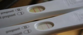
Finding out about pregnancy using a test is quite simple. You just need to see how many stripes appeared after passing it. If there are two of them, then the result means that pregnancy has occurred. If one line appears on the indicator, this means that conception has not occurred.
Pregnancy test
The presence of two lines on the test is a reason to visit a gynecologist to register with the antenatal clinic. Doctors send all patients for an ultrasound to confirm pregnancy. However, in some people who test positive, the fertilized sac is not visualized. When is pregnancy not detected by ultrasound?
Why is the fertilized egg not visualized on a scan with a positive test?
There are many reasons why an ultrasound examination does not indicate that pregnancy has occurred. One of them is an expired or defective test. You can avoid purchasing expired dough. The expiration date is indicated on the packaging, so you should always pay attention to it. But it is impossible to know whether the test is defective. To avoid a false positive result, it is recommended to purchase several different tests.
Ectopic pregnancy on ultrasound
Another possible reason for the absence of a fertilized sac may be an ectopic pregnancy. With this complication, during an ultrasound examination, sonologists detect free fluid behind the uterus and a heterogeneous mass formation in the area of one of the fallopian tubes and the ovary, but do not detect a fertilized egg in the uterus. In order to confirm an ectopic pregnancy, women are prescribed a transvaginal ultrasound and a blood test to determine the level of hCG (human chorionic gonadotropin).
Pregnancy may not be detected by ultrasound if it is done too early. You should go to the gynecologist no earlier than 10 days after the delay. It is also worth noting that a sonologist can perform a transvaginal or abdominal scan:
1. The first type of study is considered more informative, because the sensor is inserted into the vagina. During transvaginal scanning, the gestational sac is visualized at an early stage.
2. During an abdominal ultrasound, the specialist places the sensor on the anterior wall of the abdomen. This study allows you to determine the onset of pregnancy a little later than transvaginal pregnancy.
Abdominal scan
A blood test shows pregnancy, but a scan does not show a fetus
Human chorionic gonadotropin is a hormone that is produced in the human body. The hCG norm in women is 0−5 mU/ml. When pregnancy occurs, this value increases significantly. In the first hours after fertilization, the level of the hormone in the blood increases slightly. By the 5th–7th week, the rate of human chorionic gonadotropin increases several thousand times.
A blood test for hCG is one of the ways to determine pregnancy. However, there are cases when the results reveal a significant increase in hormone levels, and ultrasound does not confirm pregnancy. This is possible if women have serious health problems:
1. Chorionic carcinomas. This is a rare cancer that occurs due to malignant transformation of the chorionic epithelium.
2. Tumors in the ovaries, uterus, kidneys, gastrointestinal tract, lungs. Neoplasms arising in the human body are a pressing problem. They are detected in many women, regardless of age. The appearance of tumors causes hormonal imbalance. That is why a blood test sometimes indicates that pregnancy has occurred, and ultrasound results indicate the absence of a fertilized egg in the uterus.
3. Hydatidiform mole. This term refers to an anomaly in the formation of the fertilized egg. The embryo does not develop. The chorionic villi grow in the form of bubbles filled with liquid. A hydatidiform mole is formed as a result of fertilization of a defective egg.
Chorionic carcinoma
Scanning is one of the methods for determining pregnancy
In obstetric and gynecological practice, among the existing diagnostic methods (computed tomography, x-ray examination), ultrasound takes the leading position.
It is used not only to diagnose various diseases, but also to determine pregnancy. During an ultrasound, the doctor performs the following actions:
- visualizes the fertilized egg in the uterine cavity (if it is detected, then the assumption about the presence of pregnancy is confirmed);
- determines the number of embryonic objects;
- assesses the structure and size of the fetal egg (the specialist uses the information obtained to calculate the gestational age);
- studies the condition of the uterine cavity, examines the ovaries.
Ultrasound is used to determine pregnancy due to its absolute safety. This diagnostic method does not have a negative effect on the female body and fetus. Another advantage of ultrasound is its high resolution. Carrying out a transabdominal, transvaginal ultrasound allows you to form a clear ultrasound picture. However, there are cases when this diagnostic method indicates that there is no pregnancy, although the test and blood test results for hCG indicate the opposite.
When pregnancy is not visible on scan
The detection of a fertilized egg is influenced by many factors:
1. Pathological changes. With inflammation occurring in the uterine cavity, swelling of the mucous membranes occurs. Because of this, the sonologist faces difficulties in detecting pregnancy.
2. Features of the structure of the uterus. In some women this organ has a normal shape, in others it does not. Because of this, specialists sometimes do not detect the fertilized egg in the second group of patients, although in fact it is present in the uterus.
3. Professionalism of the doctor. Ultrasound examinations should be performed by a sonologist. An important factor influencing the reliability of the results is the experience of the specialist.
4. Equipment. Experts obtain false results as a result of using old technology. It is desirable that scanners with the highest possible resolution be used, but, unfortunately, not every medical institution is equipped with modern equipment.
There may be no pregnancy at all. Positive tests and blood test results are sometimes explained by the use of hormonal drugs. If any treatment is being carried out, the specialist should be informed about this before undergoing an ultrasound examination. The doctor will take this information into account when interpreting the results.
What to do if the scan does not show pregnancy
Many women begin to worry and worry when the test turns out to be positive, and during an ultrasound examination specialists discover that there is no fertilized egg. You need to be prepared for any results, because in the early stages, methods for determining pregnancy cannot guarantee receiving the correct information.
To avoid finding yourself in such situations, women are advised to:
1. Before visiting a sonologist, perform pregnancy tests several times. There should be intervals of several days between them. The first test may indicate the presence of pregnancy, and subsequent tests may indicate its absence. Incorrect results are possible when tests are defective or corrupted.
2. Go through ultrasound several times. Ultrasound examinations are harmless to the adult human body and the fetus, so they can be performed repeatedly. If you receive a negative result, you can come back for scans at other medical institutions. A transvaginal ultrasound examination is recommended.
3. If the results are questionable, you must have your blood tested to determine the hCG level. If the concentration of the hormone is within normal limits, and the scan does not reveal the presence of a fertilized egg, then the positive pregnancy test is not taken into account by specialists. Most likely, the woman used a defective or damaged test. If the level of human chorionic gonadotropin is elevated, and the ultrasound indicates that there is no fertilized sac, then the specialist may have made a mistake in interpreting the scan results.
In conclusion, it is worth noting that the test is a simple and informative way to determine pregnancy. It can be done at home. However, you should not rely on the results. There are other highly informative ways to determine pregnancy. Women are recommended to make final decisions only after undergoing comprehensive examinations.
Why does ultrasound not detect pregnancy with a positive test?
There are cases when, when there is a delay, a woman takes a pregnancy test and gets a positive result. At the same time, when she comes for an ultrasound examination, she is told that there is no pregnancy. What is the reason and which diagnostic method can you trust: test or ultrasound?
Pregnancy is not visible on ultrasound in the following cases:
- Early examination. Ultrasound examination will be able to recognize the fertilized egg starting from the 5th day of the delay (transvaginal method) or from the 10th day (abdominal method);
- Development of ectopic pregnancy. It is determined during examination much later. To confirm the diagnosis, you can check the level of human chorionic gonadotropin in the blood (test values should be 1000 IU/L or more). After 2 weeks it is necessary to retake this analysis. HCG levels during pregnancy should fluctuate in the range of 5-30 thousand IU/l at 4-5 weeks;
- The reason for the appearance of a second line on the test may not only be pregnancy. There may be another source in the body that produces chorionic gonadotropin: hydatidiform mole - pathology of pregnancy, liver tumors. To confirm/refute the diagnosis, you need to determine hCG over time, and you can also undergo a repeat ultrasound.
There are the following types of ultrasound examination of the pelvic organs:
- Transrectal - done through the rectum. This procedure is performed on girls who are not sexually active.
- Transvaginal – performed through the vagina in early pregnancy. Provides the most accurate result.
- Transabdominal – carried out through the anterior abdominal wall (lower abdomen).
- Combined – a combination of vaginal and transabdominal ultrasound methods. Prescribed for a complete diagnosis.
- 3D and 4D ultrasound - with the help of these two types of studies you can get a realistic image of the child. And 4D will also allow you to watch his movements and even facial expressions in real time. Indicated to clarify the condition of the fetus.
Methods for confirming pregnancy after a delay
Pregnancy is always “unexpected,” even when the couple has a stable sex life.
Young girls who mark the days of their cycle and expected ovulation are also mistaken, noting with amazement the delay (menstruation not occurring). But conceiving is quite physiological, even if it is protected using not entirely reliable methods. Sperm that enters the female body before ovulation “knows how to wait.” That’s why women’s forums are filled with topics like “I took contraception, why is there a delay, the test is positive” or “An ultrasound doesn’t show pregnancy, but I feel like I’m pregnant...”
It is important to look not only at the absence of menstruation, but also to monitor the state of your body. For example, during menstruation, many people have a tummy tug and a little swelling, which is noticeable in tight clothes. The breasts swell, become more sensitive, and the nipples are painful to touch. Lethargy and apathy, mood swings, want to sleep - it’s time to get tested. But it happens that the test is positive, but there is no pregnancy according to ultrasound, what next?
Attention: You cannot rely entirely on one of the methods of confirming conception. Symptoms of “pregnancy” can provoke diseases or pathologies, tests often lie, and ultrasound does not “see”!
Feelings must be confirmed by something more obvious:
- A longer delay than the due date of the next menstruation.
- Ultra sensitive test systems (take again).
- Increased basal temperature.
- Blood test for hCG.
- Modern ultrasound.
Many women also report that they intuitively received confirmation. And this despite the fact that the ultrasound did not show pregnancy, and the test was positive, but the line was weak. Over time, this was confirmed by the birth of a child, but in some strange way an understanding of the accomplished fact came. But intuition is not considered as reliable confirmation, only medical tests and examination results.
The fruit is not visible, what next?
The embryo may not be visible on ultrasound until the sixth week of pregnancy. To see it, you need to wait this time. But you should know that if the gestational age was determined incorrectly (the doctor decided that the pregnancy occurred earlier), the fetus may not be detected. In this case, the answer to the question of whether an ultrasound scan can fail to detect pregnancy is positive.
You should also know that the first signs of the initial development of the embryo, based on how the fertilized egg looks, become noticeable on an ultrasound no earlier than the eighth week of pregnancy.
If the fetus or gestational sac is not visible on ultrasound, you can try to do the analysis in another laboratory equipped with more powerful and modern ultrasound equipment, since ultrasound errors during pregnancy are possible in the early stages. Another option is to come back for another test in a few days. If there is a pregnancy, the fetus will by this time be fixed in the uterus and begin to grow.
If pregnancy is not to blame for the increase in hCG, additional examination is necessary. There is a high probability that a tumor or other pathology is developing in the body.
If a couple is trying to conceive a child, they greet every delay with anticipation. But a delay in menstruation does not yet indicate pregnancy. The reason for this may be a hormonal imbalance in the body. Therefore, to determine conception, you can use one of several diagnostic methods: test, ultrasound, hCG.