Why is the embryo not visible on ultrasound?
It happens that a woman who saw the long-awaited two lines on the test comes to the doctor and hears: “The fertilized egg is empty, the embryo is not visible on the ultrasound.” This phenomenon is called anembryonic pregnancy.
If a pregnant woman is diagnosed with anembryonia, this means that with an increase in the level of hCG in the blood, there is no embryo in the fertilized egg. It is difficult to say exactly what week specialists will be able to see the embryo on an ultrasound. This period ranges from 5 to 9 weeks, depending on certain factors:
- Features of the body of each specific woman.
- The correctness of calculating the period from the date of conception.
- What kind of pregnancy is it? With each subsequent pregnancy, the likelihood of detecting an embryo earlier increases significantly.
On average, it has been determined that visualization of the embryo is possible at 7 weeks from the date of conception, with an active and ongoing increase in the level of hCG in the blood. However, even if at this time specialists did not see the embryo in the fertilized egg, you need to panic only if the growth of the hCG level has stopped or has started to decline. This picture indicates that the pregnancy is frozen. However, it doesn’t hurt to make sure of this once again, so it’s worth double-checking everything with another doctor or doing a transvaginal ultrasound.
A woman should consult a doctor if, several weeks after the growth of hCG levels has stopped, the embryo is not visible in the fertilized egg, even when examined transvaginally, while the pregnancy is approaching nine weeks. Stopping the growth of the embryo and the beginning of its decomposition may be accompanied by the following accompanying symptoms:
- Unreasonable jump in body temperature.
- The appearance of nausea and vomiting.
- Constant weakness, muscle pain.
- Lower abdominal pain.
- The appearance of discharge with blood impurities or bleeding.
You should not delay your visit to the doctor and put off the curettage procedure. The decomposition of the embryo can threaten a woman with serious health problems.
How much can you trust ultrasound?
Ladies who really want to get pregnant want to know about a miracle happening in their lives as early as possible, literally in the first days of the delay. Of course, you can always buy a pregnancy test. But even ultra-sensitive tests can only accurately give a specific answer to the question “has pregnancy occurred?” They are able to answer yes or no, but more precise methods will be needed to set the deadline.
During a manual examination of a pregnant woman, an obstetrician-gynecologist may notice that the uterus is loose and slightly enlarged. But this phenomenon is also observed before menstruation. To accurately clarify the situation, you will need an ultrasound examination.
Ultrasound can accurately determine pregnancy when the level of hCG (human chorionic gonadotropin) in a woman’s blood exceeds one thousand units. In this case, the doctor is already able to see the fertilized egg in the uterine cavity (in case of multiple pregnancy - two or three fertilized eggs). The longer the period, the more complete the picture the study shows - the doctor will note the presence of not only the fertilized egg, but also the yolk sac in it, or even see the embryo and its heartbeat, and will be able to measure the CTE of the embryo (the distance from the tailbone to the crown).
If your menstrual cycle is more than 30 days, the gestational age according to ultrasound and menstruation may differ significantly.
The following situation often arises: a woman comes for an ultrasound, the doctor asks about the date of her last period, conducts a study, and then announces that the pregnancy most likely turned out to be frozen - the embryo and its heartbeat are not visualized, a fetal egg is visible, the size of which is smaller than it should be be. The doctor may be wrong if a woman had late ovulation. For a significant proportion of women, the menstrual cycle is not the standard 28 days, but 33, or even 35-40. This means that conception did not occur on days 14-16 of the cycle, but a week or two later, and the woman simply came for the ultrasound too early, so the fetus was not seen. Most likely, the pregnancy will not be frozen and the fetus is developing normally, it’s just worth repeating the study in a couple of weeks to make sure there is no mistake. It is also possible to explain the situation when pregnancy is not visible at all on an ultrasound: probably, the level of hCG in the blood is less than that at which pregnancy can be seen.
At what age should an embryo be visible on ultrasound?
While waiting for the birth of a baby, a woman asks the question: at what time will the embryo be examined by ultrasound? During diagnosis at a period of 5-6 weeks, the fertilized egg is about seven millimeters in diameter. At this stage, in most cases, the doctor has already visualized the embryo. Around this time, you can also hear his heart beating.
If you have a regular menstrual cycle, an embryo should be visible at the end of the sixth week. If an embryo is not visible on ultrasound, it is recommended to undergo a repeat examination in a week to exclude all possible abnormalities.
There are also cases when the fertilized egg is located outside the uterus. During an ultrasound, the egg is not visible well enough or is not visible at all. In this case, the heartbeat is heard outside the walls of the uterus.
Ectopic pregnancy
Another reason why pregnancy may not be seen on an ultrasound is an ectopic pregnancy. This condition is extremely dangerous. According to the results of the ultrasound, the embryo will not be visible in the uterine cavity, and the heartbeat will not be heard. When the diagnosis shows a false fertilized egg, we can talk about an ectopic pregnancy. It is worth noting that the embryo and pregnancy are still present. But this is an abnormal condition when the pregnancy is terminated on its own or specialists are forced to do so.
It is imperative to monitor your condition, since an ultrasound can help detect an ectopic pregnancy in the early stages, when you can get by with gentler methods of terminating it. Delays in treatment can be fatal for a woman. Modern high-quality equipment is capable of fairly accurate predictions in combination with other types of analysis.
What to do if the fetus is not visible on ultrasound and what could this mean?
There are situations that during an ultrasound, the embryo is not visualized inside the fertilized egg, and sometimes the fertilized egg itself is not visualized. First of all, you need to try not to panic. There may be no pregnancy at all, or there was an error in calculating its duration, so it is still difficult to diagnose. If a frozen pregnancy is not definitely confirmed, there is no need to rush into cleaning. First, it is better to undergo an ultrasound again, in another clinic. It may be necessary to conduct one or more studies. The best option is when the level of hCG in the blood is monitored in parallel with the diagnosis. If pregnancy develops without deviations, then its level increases. This helps specialists exclude a possible frozen pregnancy.
If an ultrasound does not show an embryo in the fertilized egg, what does this mean?
Very often, a fertilized egg without an embryo is diagnosed in the uterine cavity in young and healthy girls. Why is the fetus not visible on an ultrasound, and is it possible to avoid a frozen pregnancy?
There are a huge number of reasons for this phenomenon. This can be caused by infections of various etiologies, exposure to toxic substances, etc. You can minimize the possibility that an embryo will not be visible on an ultrasound by planning your pregnancy in advance in order to accurately calculate the gestational age. Also, you need to undergo examinations and cure all existing infections before planning to conceive a baby. This is especially important for women who are planning a pregnancy over the age of 35. This category has a significantly higher risk of developing chromosomal abnormalities in the fetus.
The absence of an embryo in the fertilized egg often does not give the woman any symptoms during pregnancy. Blood discharge may appear if a miscarriage occurs. Even a gynecologist during an examination will not be able to say for sure whether there is an embryo in the fertilized egg or whether it is empty. The diagnosis of anembryonia can only be made by a doctor who performed an ultrasound examination no earlier than 5-6 weeks. If the gestational age is calculated from the first day of the last menstruation, then the doctor will be able to visualize the embryo using ultrasound at 1-2 weeks of delay.
It is extremely rare for a patient to be given an incorrect diagnosis after an ultrasound, so if there is no embryo in the fertilized egg, it is necessary to check the result a week later using other equipment if there are any doubts about the professionalism of the doctor or the quality of the ultrasound machine. An error cannot be ruled out for other reasons: a short period of pregnancy or late ovulation, excess weight of the woman, etc.
Outdated equipment and doctor’s qualifications
When coming for an ultrasound examination, make sure that the installed device is a modern generation device. They are the ones who can give a clear picture and demonstrate in detail what is actually happening in the uterus of a pregnant woman. But even the most modern device does not guarantee that pregnancy will be visible at the first visit.
A good device and a poorly qualified specialist will give the same result - you may not see the pregnancy. Even if the embryo is still too small, an experienced specialist will be able to note something unusual in the uterine cavity and make a conclusion about a possible happy state. Therefore, before going for ultrasound diagnostics, you should inquire about the reputation of the person who will perform the ultrasound procedure.
Why can't you see an embryo on an ultrasound?
If a pregnancy test shows two lines, but the embryo is not visualized on an ultrasound, the reason for this may be:
- Incorrect calculation of gestational age from the moment of conception. The embryo may not be visible because the woman conducts the examination too early.
- Ultrasound diagnostics were carried out on an old device or the specialist did not have the proper level of qualifications.
- The examination was done through the abdomen and not transvaginally.
- A pregnant woman had a miscarriage, but she did not pay attention to it (she confused it with the beginning of her period), while the level of hCG in the blood had not yet decreased to its previous value.
If an ultrasound does not show an embryo in the ovum, do not immediately panic. For a number of reasons, the diagnosis of anembryonia can be made incorrectly, so it is necessary to monitor the level of hCG in the blood and undergo diagnostics again.
Quite a large number of couples have been trying to conceive a child for several years. Only a few can boast of quick results. But when the test still shows those coveted two lines, the woman, of course, rushes to the gynecologist to accurately check her interesting position. But what a surprise it is when an ultrasound does not show pregnancy, but the test does.
The main methods for determining pregnancy are:
- hCG analysis
If a woman decides to determine pregnancy using a test, she should remember that the results may be unreliable. Although tests always say that they show pregnancy almost on the first day immediately after a missed period, the result may be erroneous. In this case, many women immediately despair. But a negative test result can also be false. Especially if it was done in the first days after a missed period. Perhaps the test came at a time when the level of hCG in the blood was still too low. A false result may be influenced by factors such as:
- poor quality test
- Diagnostic errors occur much more often if they are too early
- It is recommended to perform this procedure in the morning
- unclear adherence to the attached instructions for use
It has been proven that the test can reliably show any result only a few days after conception itself, since the hCG hormone begins to be rapidly produced immediately after the fertilized egg takes its place directly in the uterine space. HCG is a hormone that accurately indicates the presence of a fetus in a woman’s uterus. During pregnancy, the level of this specific hormone should exceed 1000 IU/l. With each subsequent week, the level of this hormone increases, which allows us to judge the normal course of the process.
Most women consider ultrasound diagnostics to be the most reliable method for accurately determining pregnancy. But this method is also not flawless. There are cases when the fertilized egg itself is not visualized in any way under certain circumstances. Therefore, it is better to undergo the most comprehensive gynecological examination to ensure the accuracy of the result. After all, if several tests were carried out and they all showed a positive result, then most likely the ultrasound was either carried out incorrectly or its interpretation was unreliable.
Results in 2-4 weeks
As for the timing of pregnancy itself, as mentioned above, it is possible to determine quite accurately that an embryo is present in the uterus only by the end of the second - beginning of the third week. The optimal period is four weeks, since during this period the ultrasound specialist, of course, will not yet see the embryo in all details, but will note the presence of a yellow sac. It should measure between two and three millimeters. A week later, diagnostics will show a very small embryo.
Starting from a four-week period, you can almost say one hundred percent whether the pregnancy is uterine or ectopic. An ultrasound technician will be able to tell exactly where the embryo is. Of course, in certain cases, even at four weeks it is not always possible to determine the presence of an embryo and pregnancy. Then the observing gynecologist will recommend undergoing a repeat ultrasound examination in a week or two, as well as donating blood for hCG.
If you do not agree with the results of the ultrasound examination, then it is better not to get upset and not give up. The examination can be carried out after some time. You will also have time to donate blood to determine a special hormone that is produced by the body of a pregnant woman. An examination in a gynecological chair will also clarify the issue of determining early pregnancy.
If pregnancy cannot be detected by ultrasound examination, but the tests show a positive result, try to exclude all factors that would allow the test to show a false positive result.
Ultrasound performed to determine pregnancy
Ultrasound is the most common method to determine pregnancy. The doctor can see the presence and exact location of the fertilized egg directly in the uterine cavity. But there are also cases when a test, and even more than one, already shows the presence of a developing pregnancy, but an ultrasound does not yet. According to statistics, such cases occur very often. One of the most common causes is faulty appliances. After all, if the test shows only a positive result several times, then most likely the woman is still pregnant. The reason for a negative ultrasound result may lie in several other aspects: doctor error, abnormal location or structure of the uterus, ectopic pregnancy. If there is even the slightest doubt about the reliability of the ultrasound examination, it is better to contact another specialist or double-check the result on another device.
To avoid unreliable results, you should not rush through the procedure. Experts recommend doing an ultrasound no earlier than the 22nd day after a missed period. During this period, you can not only see the fertilized egg, but also hear the fetal heartbeat. At this time, it is possible to identify such a pathology as anembryogenesis. In this case, the fertilized egg is in the uterus, but it does not develop. There is no embryo, but only its shell. This pathology can only be determined using ultrasound. Moreover, such situations are not so rare. In this case, the test may still show a positive result, which misleads not only the woman, but also the gynecologist.
Does an ultrasound always show pregnancy?
Many women barely receive a positive test result to determine pregnancy. They immediately rush to the ultrasound room to find out the duration of their joyful state. But here they can expect great disappointment - they may not be able to see the pregnancy on an ultrasound! Yes, this is upsetting and upsetting - after all, several tests immediately confirmed that the woman was expecting a baby. We need to figure out why ultrasound does not show the presence of an embryo.
First, let's clarify that an ultrasound examination is a method that uses ultrasonic waves to image what is happening inside the body. The waves convey on the monitor what they hit and what they bounce off. In this regard, pregnancy may not be seen on an ultrasound in several cases. The first of them is a poor, outdated diagnostic device that has insufficient power.
When is pregnancy not visible on an ultrasound?
An ultrasound may not show pregnancy if:
- carried out in the early stages. In order for a doctor to be able to distinguish an embryo from, for example, a polyp, the gestational age must be long enough. Indeed, in the early stages, the embryo looks like a small dot and has no special distinctive features
- if there is swelling on the uterine mucosa. As a result of inflammation, swelling appears, due to which the fertilized egg is not visible. In this case, the entire uterine cavity is closed by edematous tissue. No matter at what angle and in what projection the examination is carried out, without eliminating the swelling it is simply impossible to obtain a reliable result of an ultrasound examination
- the equipment is in poor condition. Pregnancy may not be noticed due to unreliable instrument readings.
- no ultrasound scanner
- the shape of the uterus itself is incorrectly determined
- The qualifications of the doctor are of great importance
In all of the above cases, an ultrasound may not show pregnancy, although the test already shows a positive result. In this case, the doctor may prescribe an additional ultrasound examination after 1.5-2 weeks. Only after several examinations carried out in a row will it be possible to draw a conclusion about whether there is a pregnancy or not. An even more informative study can be carried out using a special vaginal sensor.
Type of examination
Do not forget that it is important to choose the right method for conducting ultrasound research. There are two main methods - transvaginal and transabdominal. The transabdominal method involves performing the procedure through the abdominal wall. This option is not suitable for diagnosing early pregnancy due to the large distance from the uterus.
A transvaginal ultrasound is performed by placing a transducer in the vagina. This method is the most suitable, since the sensor is located as close as possible to the uterus and allows you to examine in detail what and where is in the uterine cavity. The correctly chosen method allows you to get a more accurate answer.
It is necessary to properly prepare for a transvaginal medical procedure. The sensor will not be able to display a picture if a full bladder interferes. By the way, this will cause considerable discomfort to the woman. An empty bladder is the main condition for this type of diagnosis.
Women believe that pregnancy can be diagnosed using an ultrasound immediately after a positive test. Although the test results may be affected by various other factors that distort the result. Therefore, an ultrasound will not show the presence of an embryo. Pregnancy may not be visible on an ultrasound if there is a miscarriage in the early stages. The woman will still consider herself pregnant, and the test will confirm this.
Why is pregnancy not visible on ultrasound?
Usually, when a test has been done and it shows pregnancy, the woman immediately goes for an ultrasound to confirm the result. But the ultrasound specialist tells her that there is simply no pregnancy. Why is this happening? Which method should you trust more: a test or an ultrasound?
Pregnancy is not diagnosed in the following cases:
- short gestational age, and therefore early research
- ectopic pregnancy, which can be determined much later and for confirmation it is necessary to undergo an hCG test
- The reason for the appearance of 2 lines on the test is not only pregnancy. This could be a pathology such as a hydatidiform mole or a tumor located in the liver. In order to confirm the diagnosis, it is necessary to be tested for the presence of the hCG hormone and undergo a re-examination, including an ultrasound.
What to do if ultrasound does not give a reliable result?
First of all, you should not despair without checking again, even if you first saw a negative result. There are different options here: either the fertilized egg is really missing or the diagnostician made a global mistake. The doctor may assume a frozen pregnancy, as well as the fact that the fertilized egg has not reached the uterine cavity, as happens with the ectopic form. Without making sure of this, you should never immediately agree to cleansing, even if everything suggests that there is still no pregnancy. After all, it may be that the fertilized egg was simply not noticed. Such a step could lead to a full-fledged abortion.
Ultrasound cannot be considered the most informative method of modern diagnostics. To accurately confirm pregnancy, it is necessary to do a mandatory blood test for hCG. Such a study will be the most truthful. In any case, even if the results are too different, it is better to recheck them in another laboratory or on another device.
Thus, situations where the test showed pregnancy several times, but the ultrasound did not, happen quite often. Why this happens can be explained by many factors. Before making any radical decisions, you need to weigh the pros and cons, undergo all the necessary studies, undergo an examination by a gynecologist, and donate blood for hCG. Only with the entire list of confirmatory tests in hand will it be possible to reliably say whether the test and ultrasound results were correct or whether the diagnostician made a mistake.
The most modern means of determining pregnancy is ultrasound examination. Of course, today pregnancy can be determined using pharmacy test strips, which are widely used for these purposes due to their accessibility and information content. In addition, the pregnant uterus can be examined by a gynecologist without much difficulty.
However, only after receiving the result of the examination is the fact of pregnancy confirmed. Based on this, most women remain perplexed when there are difficulties with this method. Our article will tell you whether an ultrasound may not show pregnancy, and for what reasons this situation occurs.
Encyclopedia of Ultrasound and MRI
Pregnancy is a special new state of the female body when an embryo begins to develop in her reproductive organs. Every woman has to deal with this condition sooner or later. When it comes to our body, we look for a way out and solve medical problems. But when it comes to the lives of two, many women panic.
Such sensitive and exciting issues require precision and confidence. Unfortunately, not everything goes smoothly in our lives and we have to face difficulties. One of these difficulties is determining pregnancy. At the present stage of development of medicine, there are many accessible and safe methods for determining pregnancy: diagnostic test strips for the presence of human chorionic gonadotropin in the urine, a blood test for the same hormone, ultrasound of the pelvic organs. The difficulty is that these methods may not always be reliable, and you should never rely on just one of them.
To determine pregnancy, as well as to monitor the progress of pregnancy, two ultrasound techniques are used:
- Transabdominal (trans lat. – through; abdomen lat. – belly) – the sensor is applied directly to the mother’s abdomen at the projection site of the uterus. Normally, the projection of the uterus is located under the pubic symphysis, but during pregnancy the uterus enlarges and begins to protrude more and more above the pubic symphysis. The guided ultrasound is reflected from tissues of different densities and, returning back to the sensor, provides an image of the organs in the abdominal cavity. In this case, it shows the condition of the uterus, making it possible to see whether it is enlarged and to view the embryo in its lumen.
- Transvaginal (trans lat. - through; vagina lat. - vagina) - the sensor is inserted through the vagina at a shallow distance, ultrasound passes through the cervix, allowing you to immediately examine the organ cavity, bypassing its walls. This ultrasound method is more accurate, it allows you to examine the lumen of the uterus in a physiological state; other organs and cavities do not interfere with the sound.
Are errors possible when determining pregnancy by ultrasound?
Answering the question whether an ultrasound may not show pregnancy in the early stages, doctors say: “Yes.” Of course, today ultrasound diagnostics is the most informative method, but you cannot be completely sure of the result.
Causes of errors
The resulting picture directly depends on the specialist’s qualifications and experience working with pregnant women. There are cases when pregnancy is mistaken for fibroids or the doctor conducting the examination does not see anything, although the fetus is more than 2 months old. In addition, the cause of this error is outdated equipment, which at an early stage is not able to detect the presence of a fertilized egg.
If a woman manages to become pregnant and is less than 10 days late, then there is no point in undergoing an ultrasound examination. Although it is often possible with modern equipment to detect a pregnant uterus even at an earlier delay. If the test shows pregnancy, but the ultrasound does not, then this may be preceded by a special anatomical structure of the uterus, which does not allow early diagnosis.
In addition, there are other reasons why the doctor may not notice the fertilized egg. These include:
- pregnancy is too small to be noticed during the study;
- the fertilized egg is not yet in the uterine cavity, during the examination it is located in the area of the fallopian tubes;
- the woman made a mistake in the calculations, which is why the ovulation process is delayed;
- due to early implantation of the egg, an ectopic pregnancy developed. It is usually difficult to diagnose at an early stage. To recognize it, it is advisable to use an intravaginal sensor;
- Inflammation of the uterus often causes errors. This is due to the swelling of the cavity, against which it is quite difficult to see a small fertilized egg.
In the early stages, the fertilized egg can be easily confused with uterine fibroids
Advice: if there is a slight delay, you should not rush to an ultrasound scan, because due to the short period of time, the doctor may not detect the pregnancy, and in addition, inaccurate information often causes unnecessary worry.
To whom and why is ultrasound recommended during early pregnancy?
Typically, gynecologists prescribe early ultrasound diagnosis to a certain circle of women. These include:
- Women with problems that appeared during the previous pregnancy;
- There is a threat of miscarriage;
- The patient's complaints include periodic stretching, pain in the lower abdomen;
- Suspected ectopic pregnancy;
- Bloody discharge.
If doctors have not identified such suspicions in expectant mothers, then an ultrasound examination may not be prescribed so early.
The expectant mother will have to undergo a mandatory scheduled ultrasound scan in the first trimester as prescribed by the gynecologist. Experts prescribe it during the 11-12th week of pregnancy. The main goals of diagnosis are to confirm the presence of a fertilized egg, as well as to assess the compliance of fetal development with the established stage.
Can a test show pregnancy but an ultrasound cannot?
Typically, determining pregnancy begins with a test at home, which today differs in its quality. However, despite its information content, errors may occur.
If a test performed several times shows a positive result, but the diagnosis does not, then most likely you should trust the test, since it reacts to the level of hCG released during pregnancy.
This hormone appears in a woman’s urine 2–3 days after conception. However, one should not ignore the fact that this hormone increases in the presence of diseases, such as tumors.
In order to understand the current situation, you should donate blood to determine the exact amount of the hormone. Advice: when an ultrasound does not detect the presence of pregnancy, but the uterus is enlarged, then it is better to do a transvaginal examination, which can determine the fetus already from the 5th day of the menstrual cycle delay.
Error with irregular menstrual cycle
Some girls suffer from a “floating” menstrual period. This is not a disease, but a simple feature of the body (or a consequence of a minor hormonal imbalance).
With a “floating” schedule, menstruation does not occur every 28 days or every 35 days, but each time at a new time, for example:
- after 28 days;
- then after 30 days;
- after – in 25 days.
Cycle disruption can occur due to lack or excess of nutrition, with vitamin deficiency. If failures occur continuously, it is recommended to consult a doctor.
With “floating” menstruation, even the patient herself cannot say exactly when the day of conception occurred. Therefore, a girl should not count down the days: it is better to trust an ultrasound examination specialist. If the expectant mother nevertheless calculates the period on her own, it may differ from that announced by the doctor by 5-10 days. This is not a critical difference.
What to do when an ultrasound does not detect pregnancy?
It happens that a woman shows obvious signs of pregnancy, the test shows a positive result, but the ultrasound waves do not see anything. In this case, first of all, you need to calm down and try to guess the approximate date of ovulation. In addition, you can visit an experienced gynecologist who, without diagnostics, can assume the presence of a fertilized egg by examination.
Pregnancy tests may show erroneous results
If the test previously showed positive results, which later became negative, then it is urgent to take a blood test to detect hormones and do a transvaginal ultrasound to rule out ectopic pregnancy. This course of events requires prompt intervention, since it carries great risks for the life and health of the woman.
Usually, when the diagnosis does not show the presence of a fertilized egg, the reason is the presence of a short term. You should calm down, listen to your body’s signals and repeat the examination in 2 weeks.
To avoid disappointment at the doctor's office, it is best to do several different tests, preferably 1-2 days apart. The simplest test strips deteriorate if stored incorrectly and often erroneously show a positive result. But if several tests confirm pregnancy, it is worth visiting a gynecologist who, based on indirect evidence, will confirm or refute the test result and prescribe all the necessary procedures, including ultrasound. Although advertisements for tests say that the strips can be used after the first day of a missed period, doctors recommend not trusting the advertisements and waiting at least 2-3 days. During this time, the hCG hormone, which begins to be produced from the moment the embryo attaches, reaches the required concentration in the urine. Already a week after attachment, the hormone level is high enough to conduct a pregnancy test.
The test packages are marked with 10, 20, 25, 30 mIU/ml. This indicates the sensitivity of the test, that is, the level of hormone in the urine required for the strip to “work.” The lower the number, the lower the hormone level the strip is designed for. The most sensitive ones, with an index of 10, can be used 10-12 days after sexual intercourse. A strip with an index of 20 and 25, which can be seen in any pharmacy, will show an accurate result only after a few days of missed period.
On sensitive ultrasound machines, the fertilized egg is visible already in the fourth or fifth week of pregnancy. But you won’t be able to really see anything, because the embryo at this stage is very small, only a few millimeters in length. Therefore, doctors recommend ultrasound diagnostics after the sixth week, when the fertilized egg is about 1 cm in size and will be visible even on an old two-dimensional device.
When choosing a place for diagnostics, it is worth reading the reviews about it and the qualifications of the doctors. The correct choice of a medical institution is very important, since it is recommended that all studies be done by the same doctor and on the same machine. This recommendation is given to patients because different machines may be calibrated differently and test results may vary slightly. This is undesirable because sometimes a difference of 1 mm is fatal and the doctor may make an incorrect diagnosis due to this difference. This is especially important in the early stages, when the embryo is still very small. The qualifications of the sonologist who will do the ultrasound are no less important: an experienced specialist will be able to see the pregnancy even with the help of an old, low-power device.
But, if there are doubts about the doctor’s competence or the reputation of the clinic, then it is better to change the medical institution.
It is necessary to properly prepare for an ultrasound, especially in the early stages. If the procedure is performed abdominally, that is, through the abdominal wall, then an hour before the procedure you will need to drink 2 glasses of water. This will fill the bladder and raise the uterus, allowing the doctor to see the organ as clearly as possible. For transvaginal ultrasound, no special preparation is required, other than hygiene. The transvaginal method of examination is more accurate, since there are no several layers of skin and fatty tissue between the ultrasound sensor and the uterus, and it is used to confirm pregnancy in the earliest stages. In addition, if the patient is severely obese, or the uterus is not positioned correctly, an abdominal examination will not show anything. Most often, in the early stages, an abdominal ultrasound is performed first, and then a transvaginal ultrasound in order to examine the fertilized egg more accurately. In later stages of pregnancy there is no need to drink water; the embryo is already large enough and is clearly visible.
If the due date has already passed, pregnancy is confirmed by indirect signs and a blood test, but it is still not visible on an ultrasound; there may be a danger of an ectopic embryo. This is a serious complication that most often leads to miscarriage and serves as a reason to terminate the pregnancy. With this pathology, the fertilized egg will not be visible on an ultrasound. The diagnosis of ectopic pregnancy is made only after several tests, including a blood test and a repeat ultrasound after a week. And even if the tests show an increased level of hCG, and the embryo is not visible on the ultrasound, the doctor will prescribe laparoscopic surgery to confirm the diagnosis.
But more often than not, everything is not so bad. Women, delighted with the positive test result, rush to an ultrasound, but in vain. Since the gestational age is determined by indirect signs, and the woman rarely knows exactly which day the countdown is from, the gestational age may be determined incorrectly, which is why the ultrasound does not show anything.
Almost always, after a second examination, a week later, the embryo becomes visible and the doctor can confirm pregnancy with 100% probability. If the study shows nothing, you should simply repeat it a week later and contact the antenatal clinic.
When a desired pregnancy occurs, all expectant mothers want to reliably make sure that the fertilized egg is attached to the wall of the uterus and the formation of the unborn baby is happening normally. The most reliable and convenient way to confirm a positive pregnancy test is an ultrasound examination.
Despite the fact that a high-precision test strip, readily available in pharmacies, indicates the onset of pregnancy, and a qualified obstetrician-gynecologist is able to recognize the symptoms of a “pregnant uterus,” only the final ultrasound data confirms the fact of gestation. That is why, in the case when a woman believes that she managed to get pregnant, but the ultrasound does not show the fertilized egg, future parents are perplexed.
In connection with this phenomenon, they have a question: can a diagnostician not see a pregnancy on an ultrasound? In our article, we want to provide information about at what period of delay in menstruation it is possible to confirm the completion of the conception process, when an ultrasound scanner will allow the doctor to see the embryo, and whether it is possible not to see pregnancy on an ultrasound.
How are expectant mothers examined?
If the pregnancy test is positive, this can be confirmed by ultrasound examination - diagnosis is carried out in a commercial center or in a antenatal clinic. It is important to know that equipment with a high level of resolution and functionality, as well as the qualifications of a specialist, plays an important role in obtaining reliable examination results.
Up to 9 obstetric weeks, two methods are used to examine pregnant women:
- Transabdominal – through the area of the anterior abdominal wall.
- Transvaginal - using a transducer that is inserted into the vagina.
Up to 5 weeks, the formed fertilized egg is very small - its size is only about two millimeters. The transvaginal method is considered an effective method for diagnosing the embryonic period - its high-frequency sensor makes it possible to get as close as possible to the uterine cavity and transmit the smallest dimensions of the examined organs to the monitor screen.
The technique of examining the expectant mother using high-frequency waves is non-invasive and absolutely harmless - it allows the doctor to safely monitor the development of the fetus
During the entire gestation period, a woman undergoes at least three ultrasound scans. The examination session is short-term; the doctor tries not to hold the sensor in one place for a long time, especially during the formation of the most important organs and systems of the unborn baby.
What do they look for in an ultrasound scan?
The main purpose of performing an ultrasound scan in the embryonic period is to confirm pregnancy; this issue is especially relevant in the case of in vitro fertilization. The diagnostician has several tasks:
- Confirmation of fixation of the fertilized egg in the uterus.
- Exclusion of the presence of a neoplasm in the uterine cavity, which can “masquerade” as pregnancy.
- Assessment of embryo viability.
- Rule out ectopic pregnancy.
- Determining the presence of a second fetus.
- Study of the localization of the placenta and embryo.
- Clarification of gestation dates.
In gynecological practice, there is one important point that all expectant mothers should know: the doctor measures the duration of the pregnancy period in obstetric weeks - from the first day of the last menstruation. That is why the difference between the actual and obstetric time of conception of a child is two weeks. In a woman of reproductive age with a normal menstrual cycle, recognition of pregnancy during transvaginal examination occurs no later than five weeks. If the cycle is irregular, determining the exact period based on menstruation is difficult.
How to determine the gestational age with an irregular cycle?
Some women suffer from hormonal imbalances and, as a result, irregular cycles. With pathologies such as polycystic disease, multifollicular ovaries, and excessive amounts of male sex hormones, menstruation may not occur for several months. However, ovulation with such disorders still occurs from time to time, which means that such women have almost the same chance of getting pregnant as others.
If pregnancy is not desirable for you, the use of contraception is mandatory, even if you suffer from irregular cycles and have not had a period for several months.
Often, women with irregular cycles do not consider it necessary to use protection, believing that they will definitely not be able to get pregnant. In this case, you can detect pregnancy in the third or even fourth month, because the absence of menstruation for several months becomes a kind of norm. Correct calculation of the obstetric term in such a situation is practically impossible, therefore, when determining the term, the doctor can rely solely on ultrasound data. If the fetus is not too large or too small, ultrasound can determine the gestational age of the fetus to within 2-3 weeks. The date of birth will also be determined using an unconventional method - by counting 40 weeks from the first day of the last menstruation, and relying on ultrasound data.
At what time is the embryo not visible on ultrasound?
Signs of a viable pregnancy are the following factors, which are recorded by an ultrasound scanner:
- the presence of discernible outlines of the embryo in the egg;
- listening to the fetal heartbeat;
- recording the slightest movements of the embryo.
For each woman, the period of bearing a child proceeds individually and it is very difficult to say exactly how long it should take for the doctor to be able to examine the fetus in the form of a point and hear the rhythm of its heart.
In obstetric practice, there are certain normative terms for conducting ultrasound diagnostics for pregnant women. It is taken into account that transvaginal scanning allows one to study changes occurring earlier than transabdominal scanning. So that our readers can evaluate the quality of these methods, we provide a comparative table.
The contractions of the heart muscle of the unborn baby begin between 3 and 4 weeks and can only be detected using a transducer (a special narrow vaginal sensor). It happens that the ultrasound doctor cannot see anything in the fertilized egg and recommends coming for an examination in 7-14 days.
It is the frequency of contractions of the heart muscle of the embryo that will allow the doctor to clarify the gestational age:
- at 5 obstetric weeks, the heart rate is up to 85 beats/min;
- in 6 – from 102 to 126;
- in 7 – from 127 to 149;
- in 8 – from 150 to 172;
- at 9 – 175.
If at 7 obstetric weeks no embryonic parameters are observed in the fertilized egg and no heart rhythm is heard, a preliminary diagnosis of anembryonia is made - the absence of an embryo in the fertilized egg. However, in this case, the woman is recommended to come for an additional ultrasound after another 7 days.
Frozen pregnancy and ultrasound error
If there is an erroneous determination of the SB on ultrasound, it means that there has been overdiagnosis, poor qualifications of the specialist or low quality of the equipment.
The main reasons for incorrect diagnosis on ultrasound are:
- Human factor. Sometimes even a specialist with extensive experience makes mistakes. There is always a possibility that the doctor incorrectly assessed the condition of the fetal egg and missed important points. Such medical errors usually occur in the first trimester.
- Incorrect calculation of gestational age. If a woman has hormonal disorders or chronic diseases, calculating the exact duration of pregnancy is not always possible. If you do not take this important point into account, the doctor may mistakenly diagnose ST.
- Fetal hypoxia. In conditions of oxygen deficiency, the fetus slows down its development, moves little, and its heartbeat is barely perceptible. If the fetus is in this state, the ultrasound will show a picture reminiscent of ST.
- Technical problems. Any technology can fail. The monitor or sensor sometimes does not work correctly and incorrect data may be transmitted. But the specialist will be able to notice that the problem is precisely the breakdown of the device, so he will refer the woman for an ultrasound scan at another clinic.
Treatment for 3D involves removing the dead fetus from the uterus. They do this using various methods. Depending on the situation, the woman is given an abortion, labor is induced, and abortifacient drugs are prescribed. Before resorting to such methods, you must make sure that the pregnancy is fading. After all, it may turn out that the diagnostician’s conclusion is erroneous.
Embryo parameters
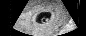
Normally, the fertilized egg is oval in shape and dark gray in color. To fully monitor the formation of the fetus, the following indicators are measured on ultrasound.
The clear visibility of the fetus on the monitor of an ultrasound machine is influenced by many factors, and if the embryo is not visible, do not panic - you should wait two weeks and repeat the study.
At the beginning of pregnancy, the embryo resembles the letter “C”; as it grows, its appearance changes - at 8 weeks you can already see both the head and the distinguished limbs
Why is the fetus not visible on ultrasound when the hCG level is growing?
The membranes of the developing baby produce a special substance - human chorionic gonadotropin, which indicates that conception has taken place. In the first trimester, the amount of this hormone protein in a woman’s circulating blood grows very quickly - in the first weeks its concentration doubles every second day.
Monitoring the dynamics of hCG level growth allows obstetricians-gynecologists to make an accurate conclusion about the development of pregnancy.
If, when assessing the amount of a given biologically active substance, an increase in its amount is observed, the doctor confirms with exact certainty the onset and successful development of pregnancy. Every woman wants to find out about pregnancy early, but the accuracy of ultrasound results in the second week of a missed period is very low - it is better to wait until the fifth week.
If, with positive hCG tests (in the case where the quantitative final data of the tests correspond to the expected gestational age), pregnancy is not detected by ultrasound, then you need to come for an additional examination. An hCG level of more than 1800 mU/ml corresponds to the third week of pregnancy and, if the ultrasound scanner does not observe a fertilized egg in the uterine cavity, the doctor assumes the development of an ectopic pregnancy.
The absence of an increase in hCG levels (negative test) may indicate the fact that the embryo does not develop - either it died, or fertilization of the egg did not occur in this cycle. Not all women are aware of such a phenomenon as biochemical pregnancy or preclinical spontaneous miscarriage. In this case, conception occurs, the fertilized egg attaches to the uterine wall, but when the next period arrives, the pregnancy is terminated.
Emphasis should also be placed on those situations where pregnancy is not visible on an ultrasound, but the test is positive - monitoring the hCG level is of particular importance; it is necessary to take a blood test several times, with an interval of several days. The final data of laboratory tests make it possible to determine whether the hormone concentration corresponds to the norm and its increase.
Practitioners advise future parents to try not to force events; exceptions are possible only when it is necessary to confirm or deny the pregnancy as urgently as possible.
What to do if pregnancy is not detected during an ultrasound scan?
If a situation arises when the ultrasound doctor cannot see the outline of the embryo, and sometimes even the fertilized egg itself, you must try to remain calm and not succumb to false beliefs! This is possible in the absence of gestation or its period is too short to be noticed on the monitor. Without absolute evidence that the pregnancy has been interrupted, curettage of the uterine cavity cannot be performed!
You should go to another clinic and undergo the examination again - it is better to do this using expert-class equipment with high resolution. It is also necessary that the ultrasound be accompanied by blood tests for hCG levels. The examination may need to be completed several times. Future parents should make every effort to ensure that diagnostic errors do not cost the child’s life!