How to calculate fetal weight based on ultrasound results
How to calculate the estimated weight of the fetus online calculator
During 9 months of pregnancy, a woman undergoes at least 3 mandatory ultrasound examinations, or ultrasound screening.
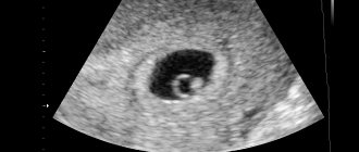
Ultrasound image at 5 weeks of pregnancy
Ultrasound at 5 weeks of pregnancy: what it looks like, why it is done, normal indicators Sign up for a diagnosis or consultation today! You can make an appointment by phone: +7 (812) 901-03-03 Or leave a request Full name Phone number By clicking the “Make an appointment” button, I agree to the terms of the Policy on the processing of personal data Ultrasound in the 5th obstetric week of pregnancy To perform ultrasound screening during During pregnancy, standard periods are established. For the first trimester it is 10-13 weeks, for the second – 20-24, for the third – 32-34 weeks. But in some cases, ultrasound diagnostics can be performed not only during these periods, but also earlier. Quite often, women are prescribed an ultrasound at 5 weeks of pregnancy. This is the period when the embryo can already be visualized and its heart begins to beat. Why is an ultrasound performed at 5 weeks of pregnancy? Ultrasound at the 5th obstetric week of pregnancy is performed with the purpose of: Confirming pregnancy. At 5 weeks, the pregnancy test should already show two lines, although rare errors do occur. To 100% confirm pregnancy, ultrasound screening can be performed. Confirm correct attachment of the fertilized egg. It must be fixed in the uterus, otherwise (for example, when the fertilized egg is fixed in the fallopian tube), an ectopic pregnancy is diagnosed. Assess the vital activity of the embryo in the fertilized egg. Detect multiple pregnancies. To refute the possibility that the increased level of hCG to which the pregnancy test reacted is not a consequence of a pathology of the female reproductive system. Identify threats of miscarriage. As a rule, at such an early stage, ultrasound is prescribed for those women who: Have previously had a cesarean section. In this case, you need to look at how the chorion (which will later become the placenta) is attached. Close attachment to a uterine scar or attachment directly to a scar can become a serious threat of miscarriage. Previously suffered miscarriages. Previously had a frozen or ectopic pregnancy. We became pregnant through the IVF procedure. What happens to the fetus in the fifth week? During the first four weeks of pregnancy, the fetus developed at an incredible rate. In the fifth week, his heart begins to beat, his cardiovascular and nervous systems develop. The formation of the spine and spinal cord also occurs, and the fetal brain now consists of two lobes. By the fifth week, the fruit becomes pear-shaped and reaches approximately 4 mm in length. Such dimensions can already be visualized by ultrasound scanning. The rounded part of the embryo will in the future transform into the baby’s head, and the sharp part into the spine. Also in the fifth week, the formation of the internal organs of the fetus occurs: liver, pancreas, larynx, trachea. How to prepare for an ultrasound? A procedure such as an ultrasound at 5 weeks of pregnancy requires little preparation. 2-3 days before the examination, you need to exclude from your diet foods that lead to excessive gas formation in the intestines. You should also empty your bladder before the ultrasound. How is an ultrasound done in the fifth week of pregnancy? At such an early stage as 5 weeks, an ultrasound is performed transvaginally by inserting a special sensor into the pregnant woman’s vagina. A special medical condom is first placed on the sensor. Transvaginal ultrasound is a painless procedure, but it can cause some discomfort to a pregnant woman. It is not recommended to carry out such a diagnostic procedure if a woman experiences bleeding or complains of abdominal pain. What will an ultrasound show at 5 weeks of pregnancy? An ultrasound in the fifth week of pregnancy will show a fertilized egg with an embryo inside (with a normal, standard course of pregnancy). Also, based on the scan results, it will be possible to judge the attachment of the chorion and the location of the fertilized egg in the uterine cavity. If the fertilized egg has not reached the uterus and is fixed, for example, in the fallopian tube, an ultrasound will show this, and the woman will be diagnosed with an ectopic pregnancy. An ultrasound at the 5th week will show the baby’s heart and will allow you to evaluate the frequency of its beating. Already at such a short period of time, it will be possible to suspect possible pathologies of the fetal heart, although it is difficult to make an unambiguous conclusion about the presence/absence of a heart defect. An ultrasound will show how actively the fetal neural tube, which will later become the baby’s bone marrow, is developing. Movements of the embryo To assess the viability of the fetus and its well-being in the womb, its heart rate is assessed. Norms and interpretation There are several main parameters of fetal development that are taken into account when interpreting ultrasound results: embryo size - standard value is about 4 mm; yolk sac size – standard value 4-5 mm; the size of the fertilized egg - according to the norm, it should be approximately 1 cm; Heart rate is normal from 70 to 100 beats/min. Ultrasound can also be used to estimate the weight of the embryo - it should be approximately 4 g. Ultrasound results are not the only information that should be taken into account when making a diagnosis. It is also important to evaluate blood tests, urine tests, and the results of other additional studies. If the embryo is not visible on an ultrasound If an ultrasound at the 5th obstetric week of pregnancy shows that there is a fertilized egg, but the embryo is not visible in it, this is not yet a reason for serious concern. Probably, the capabilities of the device are not great enough to see the embryo at such an early stage. To confirm pregnancy, you can undergo an ultrasound a little later - at 6-7 weeks. A sadder prognosis is also possible. The absence of visualization of the embryo may indicate a frozen pregnancy or anembryony (that is, the embryo did not form or it died immediately after birth). In such cases, the attending physician must understand the reasons for the death of the embryo and prescribe certain examinations for the woman.
How to prepare for research? Ultrasound at 5 weeks of pregnancy is prescribed in cases where
Photo 301
Read only for those who need to get rid of constipation at 38 weeks of pregnancy!
Signs of impending labor at 38 weeks If an increase in temperature really turns out to be a sign of imminent labor,
Pregnancy pain like menstruation. Aching pain, as during menstruation. Why does your stomach hurt in early pregnancy?
Sometimes it happens, while waiting for the onset of menstruation, you feel a certain discomfort, and critical days are so
pregnancy
What kind of discharge is there at 11 weeks of pregnancy and what to do with it
Fetus (development, size) The laying and formation of organs is still ongoing, but now the baby will be
Does amniotic fluid change during pregnancy?
The amniotic sac in which your unborn baby grows and develops is called the amnion. From the
Intestinal infections in infants: causes and symptoms
Intestinal infection in children is a group of diseases of various etiologies that primarily affect the digestive tract.
Can you feel sick in the evening during early pregnancy?
It is difficult to imagine greater happiness in a woman’s life than the happiness of being a mother. Having learned about interesting things
Drugs that relax the muscles of the uterus (tocolytics)
Women who are approaching their expected delivery date ask a completely natural question: how to relax their cervix
Diarrhea in a 4 year old child: how to treat it
Diarrhea in a child (how to help at home?)
Signs of loose stools in children Doctors do not consider diarrhea a separate disease, including it in
For any suggestions regarding the site: [email protected]
Для любых предложений по сайту: [email protected]