Modern medicine has made enormous strides in the examination of pregnant women, and if previously it was considered quite sufficient to undergo an ultrasound examination of the fetus and undergo some tests, today this category of patients is under the vigilant supervision of specialists and undergoes periodic procedures and examinations. They are mainly aimed at determining the condition of the baby developing in the womb of the mother and timely diagnosing various anomalies. This includes ultrasound detection of such characteristics as the normality of the nasal bone, carried out after ten weeks of pregnancy.
The need to measure the fetal nasal bone
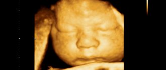
The paired, quadrangular-shaped nasal bone is determined by visualization during an ultrasound procedure at 12 weeks of gestation. Its absence at this time or deviation from generally accepted standards later indicates abnormal intrauterine development of the child, moreover, it may signal the presence of Down syndrome in the baby. In such situations, mandatory additional examination is recommended.
Some specialists, knowing the norms of the first screening, use comparative tabular values, comparing their values with the results of ultrasound. And again, we must not forget that the very fact of the presence of the nasal bone of the fetus is of primary importance during the period of 10-12 weeks of pregnancy, and measurements are not so indicative, because chromosomal ossification abnormalities occur later.
Biochemical analysis
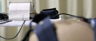
At 10-14 weeks, a woman undergoes a double test during pregnancy. Her blood serum is analyzed for the amount of hCG hormone and Papp a protein.
If the amount of these substances at week 12 deviates from the norm, and in addition, the length of the nasal bone in the table is far from the norm, a pronounced pathology of the fetus is evident.
HCG is the very first marker of chromosomal abnormalities.
This hormone is produced only in women during pregnancy and regulates many processes that begin to occur in the female body after conception and implantation of the fertilized egg into the uterine cavity. First of all, hCG triggers changes in the body of the expectant mother and reduces the functioning of the immune system, which is in a hurry to reject the foreign body.
If the hormone level is low at week 12, this indicates a missed abortion or an ectopic pregnancy.
Also, low hCG may indicate intrauterine growth retardation or a threat of miscarriage.
Often a small amount of indicator indicates placental insufficiency. However, low levels may be associated with the low weight of the expectant mother. But if hCG is high, this indicates a hydatidiform mole or the development of Down syndrome. But it can also indicate a multiple pregnancy or be caused by the woman being too overweight.
Another marker of pathologies is the Papp a protein.
It is produced in the body of absolutely every person, but there is more of it in a woman’s blood during pregnancy, since in this case the protein begins to be produced by the outer layer of the placenta.
In a woman’s body, it doubles in the first weeks, then its growth slows down. In the second trimester, this indicator is no longer informative, while its low content in the blood of the expectant mother may indicate chromosomal abnormalities. An elevated level at 12 weeks is not so critical, but may indicate a threat of miscarriage.
Parameters of the nasal bone at different stages of pregnancy
Certain periods of fetal development and growth are characterized by certain patterns. One of the main parameters is the length of the nasal bone. The normal nasal bone at 12 weeks is about three millimeters in length. Later, by the 21st week, this value reaches 5-5.7 millimeters, and by the 35th week – 9 millimeters.
The accuracy of embryo ultrasound measurements depends not only on the perfection of the equipment, but also on the experience and professionalism of the specialist.
All weeks of pregnancy:
Questions for the article
CTE 57.3 corresponds to 12.2 weeks. pregnancy. Fetal heartbeat...
Weeks and 6 days: KTR-49TVP-1.3 nasal bones-1.9HR-160internal organs in...
05/11/15 - ultrasound examination revealed a square egg of 0.56 cm, a fat sac of 0.2 cm for a period of 4 weeks; 21.05 - flat egg...
Conception. Is this acceptable and normal?...
The runny nose went away, but then reappeared. Temperature 37.2-37.3….
Experts can judge how healthy a child will be born long before he is born. Using ultrasound, doctors carefully study the proportions of the little man to understand whether the size of the embryo corresponds to the norm for obstetric weeks. Are his limbs and organs developing correctly? Are there any obvious markers of chromosomal abnormalities?
The first ultrasound of the expectant mother is prescribed at 10–14 weeks of pregnancy. Already at this time, doctors draw the first conclusions from their observations. Doctors are especially interested in the size of the child’s nasal bones. This parameter allows you to draw some conclusions about the general health of the baby.
Table of basic parameters of the nasal bone
The norm of the nasal bone at 12 weeks of fetal development, as well as in the subsequent months of a woman’s pregnancy, is systematized and serves as a starting point when studying the results of an ultrasound study. Based on the information received, doctors monitor the entire course of the baby’s intrauterine stay and identify the presence or absence of anomalies.
If the nasal bone is not visualized and the collar part is thickened, then the likelihood of having a baby with Down syndrome or congenital facial defects is extremely high.
Knowing this, many parents choose to terminate the pregnancy. Since the birth of a healthy child is a task not only for mothers, but also for doctors, such a significant role is given to such an indicator as the norm of the nasal bone at 12 weeks. The table below provides a clear description of its increase during pregnancy. Normal nasal bone
| Term, weeks | Minimum size, mm | Maximum size, mm |
| 12-13 | 2,0 | 4,2 |
| 14-15 | 2,9 | 4,7 |
| 20-21 | 5,7 | 8,3 |
| 22-23 | 6,0 | 9,2 |
| 32-33 | 8,9 | 13,9 |
| 34-35 | 9,0 | 15,6 |
Fetal pathologies
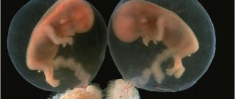
During pregnancy, the child can develop both mild and severe pathologies. Anomalies that were previously considered rare have become increasingly common. The reasons for this are unknown. It is curious that in 1987 there was an outbreak of the birth of children with Down syndrome.
In addition to this chromosomal anomaly, Edwards syndrome is often found - trisomy 18. In addition to blood counts and ultrasound, the doctor can guess about this pathology by the child’s weak activity, polyhydramnios, and small placenta. At birth, such children weigh no more than two kilograms and often die before reaching five months.
Treacher-Collins syndrome is a gene mutation that leads to facial deformities, problems with swallowing, and breathing problems. It is interesting that with this pathology, the child’s development does not lag behind his peers.
In addition to pathologies associated with chromosomal abnormalities, the fetus may experience various defects: gastrointestinal tract, heart, genitourinary system, central nervous system.
The doctor closely monitors the development of the fetus throughout the woman’s pregnancy. In the second trimester, the expectant mother undergoes a second screening, which consists of an ultrasound and a triple test with a blood test for the amount of hCG, free estriol and AFP. At 30-34 weeks, the pregnant woman expects a third screening. In this case, the doctor no longer looks at the anomalies, but checks the maturity of the placenta and the presentation of the baby.
How does one determine the length of the fetal nasal bone?
Already after the tenth week of pregnancy, measurements of the most important characteristics of the fetus can be made. The norm of the nasal bone at 12 weeks must correspond to the required parameters, otherwise you will have to think about possible severe chromosomal problems of the unborn child. Hypoplasia is considered a symptom of such complex diseases as Down syndrome, Edwards syndrome, Patau syndrome, Turner syndrome, etc.
To obtain a more accurate result, an expert ultrasound examination of the fetus is prescribed, and if it is confirmed again that the nasal bone normal does not meet the required values, a genetic analysis of the amniotic fluid is performed. This will allow you to objectively assess the intrauterine state of the baby, since it is in this environment that oxygen, carbon dioxide, hormones, fetal metabolic products, and enzymes accumulate.
Accuracy of modern diagnostics
Convinced from the results of ultrasound diagnostics that the indicators of the nasal bone of the unborn child are less than normal, parents succumb to endless despair. You should not react this way, because this parameter alone is not enough to diagnose Down syndrome or another serious pathological condition. And although the norms of the first screening are indicative to a certain extent, you need to calmly wait for the repeated results of a more in-depth examination.
You also need to understand that each child during intrauterine development is individual, therefore the sizes of individual parts of the body will differ, including the nasal bone at 12 weeks. But if examination of the fetus demonstrates obvious defects of internal organs and shortening of limbs, then in these cases we can confidently talk about congenital defects.
Fetal fetometry by week and a detailed description of fetal development by week.
There is a calendar B on the baby, where everything is written. I also watched a video on YouTube
Tell me, is your article describing obstetric or fetal weeks?
thanks, just what I was looking for!
and also for reassurance... only hypoplasia of the nasal bone can indicate syndromes... and hypoplasia is underdevelopment, i.e. SIZES SMALLER are the norm and not higher, as in the signs you look at on the Internet... anything higher means GROWING WELL)))))
the size of the nose is not important... here it must be visualized rigidly... so if it is visible everything is already fine)))) well, the nose will be big))) and what)
Often, future parents, hearing from a doctor that according to ultrasound, the size of their child’s nasal bone does not correspond to the norm, fall into panic. In fact, the size of the nasal bone alone cannot confirm or refute the presence of Down syndrome in a fetus. For this purpose, additional examination or repeat ultrasound may be prescribed. In addition, do not forget that each person is individual, and therefore the sizes of body parts may differ. Moreover, we can speak with confidence about the presence of developmental defects when the fetus also has shortening of the legs or arms, or there are defects in the internal organs.
- 3 mm – at 12-13 weeks of fetal development;
- 3.4 - 3.6 mm - at 14-15 weeks of fetal development;
- 5 – 5.2 mm – at 18-19 weeks of fetal development;
- 5.5 – 5.7 mm – at 20-21 weeks of fetal development;
- 5.8 – 6.1 mm – at 22-23 weeks of fetal development;
- 6.5 – 6.9 mm – at 24-25 weeks of fetal development;
- 7.2 – 7.6 mm – at 26-27 weeks of fetal development;
- 8.2 – 8.5 mm – at 28-29 weeks of fetal development;
- 8.6 – 8.7 mm – at 30-31 weeks of fetal development;
- 8.9 mm – at 32-33 weeks of fetal development;
- 9 mm – at 34-35 weeks of fetal development.
They didn’t find it for me at all at 12 weeks. They redid the ultrasound a week later, then they found it. All OK. Only at our first screening they don’t measure the nose, they just put a + on it. Even then, of course, I read a lot and I want to tell you that I read that if you are afraid of diabetes in a child, then it seems that with diabetes the nasal bone is not visible at all until the 16th week. So if there is a bone at your term, this is already the norm)
We had 2mm, and I didn’t even attach any importance to it, I was still lying there and thinking, where did you see that, what a damn nose, maybe you will have a small nose, so it’s too early to worry, the main thing is that the doctor said that everything was normal and on time and there's no point in inventing more
At the 2nd screening, my friend’s nasal bone did not match, the doctors scared me about a down. She gave birth to a normal boy with a normal nose. By the way, she was diagnosed with this in Moscow, but in Penza the doctors said that it didn’t mean anything at all.
Don’t panic, if they say it’s normal, it means it’s normal... they don’t want to scare you and they’re doing the right thing. It’s probably not scary, and that’s why they say that everything is fine... But the fact that they checked the box, that’s what will change? You don’t give up on the child... everything is fine. otherwise they probably would have done something.
In my first B, on the contrary, I had a small nose. We didn’t bother, because according to all other indicators everything was ok. The child was born without any syndromes. Do not be nervous! Everything is fine with the baby!
And for a second, at 20 weeks, for example, the norm is 6.6 mm. I walked with my first one at 20 weeks and my nose bone was 6.1mm. You’re doing fine!
You will just have a little nosey baby))) don’t worry! Since the doctor said that everything is fine, that means it is so. What's the point of them hiding something from you? So don't worry about it in vain
There are no norms for children, all children are individual. There are artificially defined tables for doctors, but they have nothing to do with the norms.
Today we just discussed this with a geneticist. She said that no one measures this bone, because it is not possible to measure clearly, the baby is in motion. And those ultrasound specialists who still managed to measure it give very relative data that geneticists don’t look at, the main thing is that this bone is visualized, which means that everything is fine.
I also find fault with everything, but what else?
On topic, the size is ok. In the residential complex they wrote to me that it was visualized, but on the paid one it was already the size.
At 13 weeks, 2 days in conclusion, 2 mm of bone is visualized. The ultrasound specialist said that everything was fine and there was no reason to worry. The analyzes are also good.
What is perinatal screening
Perinatal screening is a unique examination of the expectant mother to detect the danger of congenital and acquired pathological conditions of fetal development, the results of which are compared with average values.
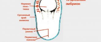
In the first trimester of pregnancy (10-12 weeks), perinatal screening makes it possible to recognize early enough anomalies in the number of chromosomes, a defect in the anterior abdominal wall and pathology of the neural tube, as well as various threats to the normal bearing of a child in general.
Based on the PAPP and hCG indicators characterizing pregnancy, as well as the thickness of the collar space in the embryo, the risk of possible defects is judged. If the norms of the first screening do not coincide with the indicators of the tests performed, the pregnant woman is prescribed a study of the genetics of the unborn child and a chorionic villus biopsy.
What is hypoplasia of the nasal bones in the fetus?
The norm of the nasal bone at 12 weeks during an ultrasound examination allows us to conclude that the fetus is developing well in the early stages of pregnancy. If its length deviates from the accepted standard towards a decrease, then hypoplasia of the nasal bone is present. But there are exceptional cases when a number of examinations conducted do not reveal it at all. Then the question is raised about aplasia of the nasal bone, in other words, the complete absence of the organ.
Fetal fetometry by week and a detailed description of fetal development by week.
There is a calendar B on the baby, where everything is written. I also watched a video on YouTube
Tell me, is your article describing obstetric or fetal weeks?
thanks, just what I was looking for!
and also for reassurance... only hypoplasia of the nasal bone can indicate syndromes... and hypoplasia is underdevelopment, i.e. SIZES SMALLER are the norm and not higher, as in the signs you look at on the Internet... anything higher means GROWING WELL)))))
the size of the nose is not important... here it must be visualized rigidly... so if it is visible everything is already fine)))) well, the nose will be big))) and what)
Often, future parents, hearing from a doctor that according to ultrasound, the size of their child’s nasal bone does not correspond to the norm, fall into panic. In fact, the size of the nasal bone alone cannot confirm or refute the presence of Down syndrome in a fetus. For this purpose, additional examination or repeat ultrasound may be prescribed. In addition, do not forget that each person is individual, and therefore the sizes of body parts may differ. Moreover, we can speak with confidence about the presence of developmental defects when the fetus also has shortening of the legs or arms, or there are defects in the internal organs.
- 3 mm – at 12-13 weeks of fetal development;
- 3.4 - 3.6 mm - at 14-15 weeks of fetal development;
- 5 – 5.2 mm – at 18-19 weeks of fetal development;
- 5.5 – 5.7 mm – at 20-21 weeks of fetal development;
- 5.8 – 6.1 mm – at 22-23 weeks of fetal development;
- 6.5 – 6.9 mm – at 24-25 weeks of fetal development;
- 7.2 – 7.6 mm – at 26-27 weeks of fetal development;
- 8.2 – 8.5 mm – at 28-29 weeks of fetal development;
- 8.6 – 8.7 mm – at 30-31 weeks of fetal development;
- 8.9 mm – at 32-33 weeks of fetal development;
- 9 mm – at 34-35 weeks of fetal development.
They didn’t find it for me at all at 12 weeks. They redid the ultrasound a week later, then they found it. All OK. Only at our first screening they don’t measure the nose, they just put a + on it. Even then, of course, I read a lot and I want to tell you that I read that if you are afraid of diabetes in a child, then it seems that with diabetes the nasal bone is not visible at all until the 16th week. So if there is a bone at your term, this is already the norm)
We had 2mm, and I didn’t even attach any importance to it, I was still lying there and thinking, where did you see that, what a damn nose, maybe you will have a small nose, so it’s too early to worry, the main thing is that the doctor said that everything was normal and on time and there's no point in inventing more
At the 2nd screening, my friend’s nasal bone did not match, the doctors scared me about a down. She gave birth to a normal boy with a normal nose. By the way, she was diagnosed with this in Moscow, but in Penza the doctors said that it didn’t mean anything at all.
Don’t panic, if they say it’s normal, it means it’s normal... they don’t want to scare you and they’re doing the right thing. It’s probably not scary, and that’s why they say that everything is fine... But the fact that they checked the box, that’s what will change? You don’t give up on the child... everything is fine. otherwise they probably would have done something.
In my first B, on the contrary, I had a small nose. We didn’t bother, because according to all other indicators everything was ok. The child was born without any syndromes. Do not be nervous! Everything is fine with the baby!
And for a second, at 20 weeks, for example, the norm is 6.6 mm. I walked with my first one at 20 weeks and my nose bone was 6.1mm. You’re doing fine!
You will just have a little nosey baby))) don’t worry! Since the doctor said that everything is fine, that means it is so. What's the point of them hiding something from you? So don't worry about it in vain
There are no norms for children, all children are individual. There are artificially defined tables for doctors, but they have nothing to do with the norms.
Today we just discussed this with a geneticist. She said that no one measures this bone, because it is not possible to measure clearly, the baby is in motion. And those ultrasound specialists who still managed to measure it give very relative data that geneticists don’t look at, the main thing is that this bone is visualized, which means that everything is fine.
I also find fault with everything, but what else?
On topic, the size is ok. In the residential complex they wrote to me that it was visualized, but on the paid one it was already the size.
At 13 weeks, 2 days in conclusion, 2 mm of bone is visualized. The ultrasound specialist said that everything was fine and there was no reason to worry. The analyzes are also good.
Why do such violations occur?
In fact, there are many reasons for pathological deviations from normal lengths of the paired fetal nasal bones, and they are all of a different nature. This could be, for example, chronic alcoholism of one of the parents or the severe consequences of smoking. Women who have had the flu, colds and other diseases in the first trimester of pregnancy are automatically at risk.
There are other reasons why the nasal bone does not reach the normal level at 12 weeks of fetal development, namely:
- taking antibiotics and other strong medications;
- exposure to gamma radiation;
- exposure to harmful environmental factors on a woman’s body;
- bruises;
- prolonged overheating of a pregnant woman.
Thus, in addition to chromosomal disorders and so-called genetic predispositions, the lifestyle and health of the expectant mother directly affect the intrauterine development of her fetus. And modern medicine, thanks to the latest diagnostic methods, allows us to identify and control chromosomal abnormalities in the baby at a fairly early stage of pregnancy, the consequence of which can be Down syndrome. In any case, it is the attending physician, based on the most accurate tests and studies, who is able to determine the true diagnosis or eliminate possible risks.
Poor results and next steps
As a rule, it is difficult for a pregnant woman to perceive information that the baby is developing a pathology. The expectant mother is lost and often does not know what further steps to take. But it is important to remember that time is very valuable in this case.
Of course, you can get all the tests done at another clinic, but it’s better to make an appointment with a good geneticist.
You should not wait for a scheduled appointment, as in this case you will lose a lot of time.
Most likely, the geneticist will refer you for additional examinations, the results of some of which will take about three weeks to wait. If it later turns out that the fetus really has a serious pathology, and it would be more humane to have an abortion, then it will be too late: at 20 weeks, this is an already formed child who begins to make the first movements. For medical reasons, abortion is best done before 16 weeks.
But it is also important to keep in mind that the results of screening during pregnancy do not guarantee 100% accuracy, although based on the analyzes it is possible to draw conclusions that are quite close to reality.
And yet, both people and equipment make mistakes. In addition, only from mother's blood tests and ultrasound in the first trimester you cannot say for sure whether your baby is sick or not. Therefore, to increase the likelihood, additional studies are prescribed.
In the first trimester, this is a chorionic villus biopsy. This analysis is done up to 13 weeks. The doctor takes chorionic villi, which are genetically identical to the fetus, for examination. That is, by examining the villi, the doctor can tell 99% whether the child has genetic abnormalities or not. However, not all anomalies can be detected by this analysis. For example, the doctor will not tell you whether there is a neural tube defect.
During a chorionic villus biopsy, the specialist carefully punctures the abdomen to obtain the necessary biomaterial.
The villi are collected using a syringe.
On average, the transcript of results is prepared within 10-14 days. It is important to keep in mind that the cost of the analysis is quite high - from 6 to 30 thousand rubles, depending on the clinic. Other procedures to detect abnormalities are carried out later in pregnancy. These include cordocentesis and amniocentesis.
Cordocynthesis is an analysis during which, under ultrasound guidance, a doctor takes blood from the umbilical cord for examination. In this case, the specialist receives the genetic material directly from the baby himself, therefore allowing him to find out the development of the fetus with an accuracy of 99.9%. Cordocentesis is performed not only to detect chromosomal abnormalities. It also allows you to diagnose cystic fibrosis, hemophilia, and fetal infection. The study is carried out starting from 21 weeks. The main difficulty is that the umbilical cord is mobile, and the specialist must be highly qualified to carry it out without harm to the health of the mother and child. Another difficulty lies in obtaining pure baby blood without maternal admixture.
If we compare cordocynthesis with amniocentesis, then the probability of complications during the first analysis is 3%, while during the second it is only 0.5%.
But it also has many advantages. In addition to high accuracy, interpretation of the results is done from several days to 10 days.
Amniocentesis is the removal of amniotic fluid. Such an analysis can be carried out both in the early stages of pregnancy - from 8 to 14 weeks, and after 15 weeks. But if in the first trimester amniocentesis is intended to identify pathologies of fetal development, then in the second trimester the doctor performs amniocentesis to identify intrauterine infections or to understand how mature the lung surfactants are. Deciphering the results takes two weeks.