Blood clotting is one of the body’s defense mechanisms, which helps prevent blood loss in the event of damage to the integrity of the vascular wall. Unfortunately, sometimes the process becomes pathological, which is expressed in the causeless formation of clots inside the vessels, even if they are not damaged. So, what is a blood clot and what does it look like?
A thrombotic blood clot is a specific lump formed intravitally as a result of hyperactivity of the coagulation system from fibrin, formed elements or other plasma components. When answering the question of what a blood clot looks like, it is important to emphasize that blood clots are different, depending on their size, composition, stage of blood clot formation, quality characteristics and location. Currently, there are several types of thrombotic formations, which differ in morphology and localization in the vessels.
Classification by morphology
If we consider blood clots from the point of view of appearance and composition, then the classification will consist of four varieties: white, red, mixed (the most common) and hyaline. Depending on the components of the clot, the effect of medicinal substances may vary, therefore, during the diagnosis, the attending physician determines the type.
White clots
White blood clots are also called gray, agglutination or conglutination. They are formed by platelets - colorless and nuclear-free blood platelets of irregular shape, formed in the red bone marrow. They are directly involved in the coagulation process, and also control blood viscosity, protect vessel walls from damage and promote the dissolution of formed blood clots in healthy people. In some cases, the white clot may be formed by leukocytes (white blood cells) and fibrin (a protein made from fibrinogen synthesized in the liver).
As a rule, this type of clot forms in vessels with good blood flow, mainly in the arteries and cavities of the heart. White blood clots form slowly, perpendicular to the direction of movement of rapidly circulating blood; gradually the growth becomes voluminous and resembles coral. In appearance it is a light gray or white substance, and its surface is embossed. The structure of the clot is crumbling, that is, if it separates from the wall of the vessel, it will disintegrate into small particles.
Red clots
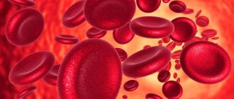
Red blood clots (coagulation) are formed for exactly the opposite reasons - in vessels with slow blood circulation, which at the same time has a high degree of coagulation. The color of clots is determined by the high content of red blood cells, of which in most cases they are composed.
Erythrocytes (red blood cells) are disc-shaped biconcave cells that are saturated with oxygen in the lungs and then carry it to all cells of the body. The red color of these elements is due to the fact that their cytoplasm contains large quantities of hemoglobin, a pigment containing an iron atom. It not only determines the red color of the entire cell, but is also directly involved in the delivery of oxygen, since it is able to bind it.
In appearance, a thrombus of this type is in most cases smooth, but there may also be raised areas. The clot is usually loosely connected to the wall of the vessel, and, unlike white blood clots, it does not form large coral-like formations. Most often, red blood clots affect the veins.
Mixed clots
Mixed blood clots are formed by the fusion of white and red blood clots, so we can say that they are mainly composed of platelets and red blood cells. In this case, such a clot can form in an artery, a vein, or in the cavity of the heart. Mixed thrombi differ in structure from white and red ones; they have three anatomical parts - a conical or flat head, a body (directly mixed thrombus) and a tail. In this case, the head, which is a white blood clot, is attached to the wall of the vessel, and the tail, which is a red blood clot, is freely located in the lumen of the vein or artery and has a loose structure.
Since the head of a mixed thrombus is attached to the vessel wall, the tail poses the greatest danger. It is always localized against the blood circulation, so it can easily come off, clogging the vessel, this process is called thromboembolism.
In rare cases, when the tail reaches a significant size, it can provoke the detachment of the entire blood clot, which can even lead to death if the clot blocks the lumen of vital organs.
It happens that different parts of a mixed thrombus are located in different veins, for example, the head is in the femoral vein, the body is in the external iliac vein, and the tail is in the inferior vena cava.
Hyaline clots
Hyaline blood clots are currently considered the most mysterious by the nature of their occurrence, since researchers do not have a consensus on the mechanism of their formation. They appear predominantly in small vessels of the microcirculatory system and are mostly multiple in nature. Hyaline clots can occur after shocks, burns, disseminated intravascular coagulation (blood clotting disorders due to the release of thromboplastic substances from tissues), electric shocks, extensive skin injuries, etc.
Hyaline blood clots mostly consist of red blood cells, white blood cells, and precipitated plasma proteins glued together or destroyed. Also, the clot may contain a small amount of fibrin protein, but it is not constant, and sometimes the component may disappear completely. The prerequisite for the formation of a hyaline thrombus is often a significant slowdown or complete cessation of blood flow in the vessel.
How blood clots form
The formation of a blood clot is a complex process. Physiological clots form as a result of damage to the vessel. At the same time, substances (thrombin and thromboplastin) are released into the bloodstream, which activate coagulation processes. This occurs as a result of the breakdown of platelets. The following phases of clot formation are distinguished:
Upon activation, the formation of prothrombinase occurs. With its help, the protein thrombin appears. Next comes the coagulation phase. Under the influence of the protein thrombin, fibrinogen is converted into fibrin. The latter is the basis of the resulting blood clot. A mesh is formed in the damaged area, which receives red blood cells, white blood cells and platelets.
This is how a dense fibrin clot is formed. This is the retraction phase. When hemodynamic parameters stabilize, the thrombus resolves. This is a normal blood clotting process in any person. The formation of blood clots, which cause blockage of blood vessels, is no different. Subsequently, such blood clots do not resolve on their own and cause thrombosis.
In the first days, the clots are still poorly fixed. They may break off and enter the main arteries or veins with the development of thromboembolism. This is an even more dangerous condition. The following factors play an important role in the development of thrombosis:
- decrease in circulating blood volume;
- increased blood viscosity;
- valve dysfunction;
- slowing blood flow;
- platelet tendency to aggregation;
- mechanical damage to the walls of blood vessels.
There are people whose blood clotting process is impaired as a result of a lack of special factors. They practically do not form blood clots, which is fraught with large blood loss at the slightest damage. An example would be hemophilia.
Classification by localization in the vessel
In addition to the fact that thrombosis can occur in any part of the human circulatory system, that is, in the area of any internal organ, blood clots also differ in that they can be located differently in the vessel itself. This applies to blood clots of any structure and appearance.
Parietal variety
Parietal clots are quite common; they partially cover the lumen of the vessel. The parietal variety is characteristic of large vessels, as well as elements of the heart - chambers and valves. Often the parietal variety occurs as a result of inflammatory processes, for example thrombophlebitis, which is one of the complications of the last stages of varicose veins.
At first, such clots are not dangerous, since they block the blood flow only partially, but they have the characteristic feature of layering on each other, forming group accumulations. If therapeutic measures are not taken, the formed blood clot will subsequently completely block the blood vessel, which can be fatal.
Obstructive variety
Obstructing, or, in other words, clogging blood clots prevent blood circulation across the entire width of the affected vessel. Moreover, such clots, as opposed to parietal clots, are formed mainly in small blood vessels. The greatest diagnostic difficulty in this case is mixed thrombi, since it becomes difficult to determine where they begin and attach to the vascular wall, and where their tail ends.
Despite the fact that occlusive clots affect small blood vessels, they pose a significant threat to human health and life. If such a phenomenon occurs in the vessel of the spleen, there will be an increase in its size and a partial loss of efficiency; location in the renal branch of the circulatory network will lead to renal infarction, and in the intestine - to gangrene. When an occlusive thrombus is localized in a heart vessel, death is inevitable.
Axial variety
Axial thrombi can be called a kind of middle type between parietal and occlusive. Such clots are attached to the vascular wall in only one anatomical area - the head or part of the body, partially obstructing blood flow. Moreover, if a blood clot comes off, under the influence of blood circulation it can still remain in a suspended state for some time, “collapsing” into a spherical shape.
Treatment methods
In each case, the decision on how to remove blood clots from blood vessels is made individually, depending on the patient’s condition, the form and location of the disease. When the lower extremities are affected, an obligatory component of complex therapy is the use of elastic bandages; this reduces symptoms and prevents complications. All thermal procedures are prohibited.
Conservative treatment begins with diet. The diet includes vegetables and fruits, lean meat, fish, and dairy products. Spicy, salty, fatty dishes, on the contrary. Completely prohibited.
The following groups of drugs are used for drug therapy:
- anticoagulant (Heparin);
- antispasmodics (No-shpa);
- thrombolytic therapy (Streptokinase, Urokinase);
- antiplatelet agents (Aspirin);
- means for improving trophism (Reopoliglyukin);
- sedatives;
- antiarrhythmic drugs for blockage of coronary arteries;
- analgesics.
It is possible to administer drugs that dissolve the blood clot directly into the lesion. This procedure is called thrombolysis. However, it is effective when the clot is not compacted, within 72 hours from the moment of formation.
Surgical therapy is carried out when medication is ineffective or there is a threat to the patient’s life. A thrombectomy operation is performed. The blood clot is removed, and the affected vascular wall is replaced with a prosthesis. In addition, stitching, bypassing and vascular ligation can be used. In patients at high risk of pulmonary embolism, vena cava filters are installed in the inferior vena cava.
Other types
In addition to the main classifications of blood clots, there are several separate types that are specific to certain groups of patients. These varieties include the following types:
- Mirantic. It occurs in weakened elderly people with long-term dehydration. The thrombus is localized mainly in the superficial veins.
- Tumorous. Formed as a result of metastasis, that is, the formation of secondary foci of a malignant tumor. Often such a clot gradually grows towards the right lobes of the heart.
- Septic. Occurs as a result of a local inflammatory process caused by infection. Localized in the veins and on the valve flaps of the heart.
Thus, there are several types of blood clots, each of which will require a specific approach during treatment. The type of blood clot is determined by the doctor during diagnosis.
Clinical manifestations and causes of vascular blockage
You need to know not only the mechanism of blood clot formation, but also the reasons for blockage of blood vessels by blood clots. The veins are most often affected. Most often, this pathology is detected in adults who lead an unhealthy lifestyle and suffer from varicose veins. The following reasons for the development of venous thrombosis are distinguished:
- congenital anomalies;
- varicose veins;
- surgical interventions;
- severe dehydration of the body;
- hormonal disbalance;
- mechanical injuries (bruises, fractures);
- long-term compartment syndrome;
- DIC syndrome;
- septic conditions;
- paralysis;
- physical inactivity;
- bed rest.
Less common is arterial thrombosis. It develops during a stroke, against the background of atherosclerosis, atrial fibrillation, and during organ transplantation. Experienced doctors know not only what a blood clot is, but also the symptoms of vascular blockage. Most often, the process involves the superficial and deep veins of the lower extremities. With thrombosis of the leg veins, the following symptoms are observed:
- swelling;
- heaviness in the legs;
- convulsions;
- bursting pain;
- numbness;
- tingling;
- pale skin;
- fever (when combined with phlebitis).
Sometimes the place where blood clots form is in the veins of the upper extremities and eyes. In the latter case, vision loss may occur. The consequences of the formation of blood clots can be pulmonary embolism, stroke, acute cardiac ischemia, gangrene of the limb, atherosclerosis, and impaired organ function (kidneys, liver, lungs). The most dangerous is blockage of the deep veins.
If a thrombus breaks off and enters the lumen of the pulmonary artery, thromboembolism develops. Its symptoms include pain, decrease or disappearance of the pulse, loss of sensitivity, pallor of the skin in the affected area, cyanosis, and tissue swelling. Against this background, the function of the limb is impaired. In the case of thromboembolism in the mesentery, symptoms of an “acute abdomen” appear.
White blood clots
They are a clot of platelets stuck together and fused to the wall of the vessel. Typically, white blood clots form in vessels with good blood flow, for example, in the heart cavity, aorta, and coronary arteries.
The rate of their formation is slow with fast blood flow. The clumps are located perpendicular to the current, and over time they form entire conglomerates, similar to coral growths.
Externally, a white thrombus is a light gray mass with a corrugated surface, which easily crumbles when trying to separate due to its dry, dense structure. Why is the blood clot red? The reason for the formation of a white blood clot is changes in the endothelium.
Mixed blood clots
They have in their structure both structural elements of white blood clots and fragments of red blood clots. Mixed thrombi have an elongated shape and a variegated red and white color due to the mixing of thrombus elements of different colors.
They start with an elongated head that extends into the body and end with a red tail. A mixed thrombus is located along the bloodstream.
In large vessels it can reach very large sizes, when the head is located, for example, in one branch of the vein, and ends in the opposite side at a distance of several centimeters.
Parietal thrombi
They are located in the lumens of large veins and arteries, for example, in the chamber or on the valves of the heart, and are formed during inflammatory processes (thrombophlebitis, thromboendocarditis).
Wall thrombi are dangerous because over time they layer on top of each other, resulting in complete blockage of the vessel, which can lead to organ failure or even death.
However, the very location of the blood clot along the vessel wall does not block the blood flow, so it can be considered less dangerous. A type of wall thrombus can be considered an extended thrombus, which is attached to the wall along its entire length. It also interferes with blood flow, but does not fill the entire lumen of the vessel.
Prevention of thrombosis
You can avoid blood clots by following these simple recommendations:
- Physical inactivity leads to stagnation of blood in the veins and worsens metabolic processes. Staying in one position for a long time should not be allowed. When working sedentarily, you should take regular breaks and do some exercise;
- It is important to drink more water, at least 1.5 liters. in a day. Water is an excellent blood thinner;
- You should protect yourself from injuries, increase your immunity;
- Avoid alcohol abuse, quit smoking;
- Control your weight;
- Avoid stress if possible;
- Stick to proper nutrition.
Knowing why blood clots form, you can avoid the development of thrombosis and its complications, which without adequate timely treatment can lead to death.
Obstructive thrombi, otherwise called clotting
They completely close the lumen of the vessel and pose a threat to human health and life. If an occluding thrombus is located in the vessels of the brain, this can lead to poor circulation and poor nutrition of the organ.
The presence of a blood clot in the splenic artery leads to splenomegaly, in the renal veins - to renal infarction, and in the intestinal vessels - to intestinal gangrene (find out what intestinal thrombosis is). If a blood clot breaks off in a cardiac artery, this means instant death.
Diagnostics
Diagnosis of thrombosis
Deep vein thrombosis is diagnosed using invasive and non-invasive research techniques, which include:
- ascending venography using a contrast agent (the most accurate method of analysis);
- duplex ultrasound scanning of blood vessels (most often the diagnosis of phlebothrombosis is made after such a study);
- thromboelastography (graphic recording of blood coagulation and fibrinolysis processes);
- radionuclide scanning.
Phlebothrombosis of a mesenteric nature is diagnosed using an abdominal x-ray, a clinical blood test (for the presence of increased leukocytosis), and, in the most severe cases, by laparoscopy, and thrombosis of the cavernous sinus can be seen on a computer thermogram.
