The hip joints experience the greatest load in the body. They are created by weight during walking, jumping, running, lifting and carrying heavy objects. Patients often feel pain in the hip joint. Orthopedists at the Yusupov Hospital determine its cause using modern diagnostic equipment. Doctors determine the degree of joint damage, which allows them to make an accurate diagnosis and develop optimal treatment tactics.
Doctors at the Yusupov Hospital provide complex therapy for diseases that cause pain in the hip joint. Patients are individually selected effective medications that affect the cause and mechanism of development of pain. Rehabilitation clinic specialists provide rehabilitation therapy using the latest physiotherapeutic procedures, physical therapy, and acupuncture. The presence of special simulators allows you to reduce the load on the joint during training.
In the process of treating pain in the hip joint, doctors from many areas of medicine are involved: endocrinologists, rheumatologists, orthopedists, physiotherapists, chiropractors, acupuncturists. A multidisciplinary approach to the treatment of pain in the hip joint allows for rapid pain relief. Patients suffering from pathology of the hip joints often require outside care. It is professionally carried out by the staff of the Yusupov Hospital who have undergone special training.
Causes
Pain in the hip joint is caused by the following pathological processes:
- Tendinitis (inflammation of tendons);
- Muscle rupture;
- Iliotibial band syndrome;
- Other local changes in surrounding tissues;
- Systemic diseases (rheumatoid arthritis, polymyalgia).
Because the gluteus medius and minimus muscles play a major role in hip abduction, damage to them causes hip pain.
The gluteus medius and minimus tendons attach to the greater trochanter. If an inflammatory process develops in them due to microtraumas resulting from excessive load, the patient will be bothered by pain in the hip joint. Such disorders may be caused by an infectious process (tuberculosis), sports or stereotypical professional stress, or the deposition of crystals. Hip pain is a symptom of the following diseases:
- Osteoarthrosis;
- Radicular syndrome;
- Rheumatoid arthritis;
- Coxita.
Pain in the hip joint can bother people who are overweight, have different leg lengths, or have flat feet.
Pain syndrome can occur after lower limb amputation or hip replacement. With avascular necrosis of the head and fracture of the femoral neck, patients complain of acute pain in the hip joint. Pain syndrome often develops with dysplasia (disorder of the anatomical structure) of the hip joint. Acute pain in the hip joint, radiating to the leg, occurs in the case of pinched nerves due to diseases of the spine, malignant bone tumors, and age-related changes. Make an appointment
Causes of pain
One of the physiological causes of pain in the hip joint when walking, especially when climbing stairs, is intense sports training and excessive physical activity. Lactic acid accumulates in the thigh muscles, irritating the tissue. The result is not only pain, but also an unpleasant burning sensation. All symptoms disappear without a trace after a short rest in a lying or sitting position, during which lactic acid is eliminated from the body.
But even if a relationship is discovered between the occurrence of pain and exercise, a doctor’s consultation is necessary. The fact is that increased loads often lead to the development of destructive, degenerative or inflammatory processes in the hip joint.
Systemic diseases
The group of systemic diseases includes pathologies that affect several joints, provoking the development of an inflammatory process in them. First, the cartilage is destroyed, which leads to instability of the joints, and then the bone structures are deformed. Systemic diseases are usually autoimmune. They arise due to an inadequate response of the immune system to the penetration of infectious pathogens into the body. These include:
- scleroderma;
- Still's disease;
- ankylosing spondylosis;
- rheumatoid arthritis;
- rheumatic fever;
- systemic lupus erythematosus;
- polymyalgia rheumatica.
In addition to pain when walking, a variety of both articular and extra-articular symptoms occur. The skin in the affected area turns red, swells, and becomes hot to the touch. During an exacerbation, the range of movements is sharply limited. Lymph nodes may also enlarge, the functioning of the liver, kidneys, and gastrointestinal tract may be disrupted.
Inflammatory and infectious causes of pain
Gouty, psoriatic, or reactive arthritis can provoke pain in the thigh when walking. Their leading symptoms are redness of the skin due to blood vessels overflowing, increased local temperature, stiffness, and swelling. A similar clinical picture is characteristic of other inflammatory pathologies affecting the hip joint:
- bursitis - inflammation of the synovial bursa with accumulation of exudate in the joint cavity;
- synovitis is an inflammatory process in the synovium, accompanied by the accumulation of fluid;
- tendinitis or tenosynovitis - inflammation of the tendons of the joint, including those of the vagina.
Infectious arthritis is especially difficult. It develops against the background of gonorrhea, syphilis, tuberculosis, brucellosis, or as a result of the introduction of staphylococci, streptococci, and E. coli into the joint. A person suffers not only from acute pain when walking - his body temperature rises sharply, chills and a feverish state occur.
Pain caused by degenerative changes
Pain in the hip when walking is the main symptom of coxarthrosis. This is a degenerative-dystrophic pathology that mainly affects middle-aged and elderly people. Coxarthrosis develops due to the destruction of cartilage and initially does not manifest itself clinically. Sometimes mild discomfort occurs, which a person attributes to muscle fatigue after physical exertion. But over the course of several years, the severity of symptoms increases:
- pain in the side of the thigh occurs not only when walking, but also at rest;
- when moving, clicks, crunching, crackling sounds are clearly heard;
- in the morning the skin over the joint is swollen and movements are constrained;
- at the final stage of development of coxarthrosis, the leg shortens and the muscles atrophy.
Hip pain may be referred. Most often, their source is intervertebral discs and vertebral bodies affected by osteochondrosis. When they are displaced, the spinal roots are pinched, which leads to acute pain in the lower back, spreading to the hip joint.
Traumatic causes
A fracture or dislocation of the hip joint occurs during a fall from a height, during a traffic accident, or a strong directed impact. The pain is so severe at the time of injury that the person loses the ability to move. As a result of a fall or injury, muscles, ligaments, and tendons are often damaged. If a large number of connective tissue fibers are injured, then the person cannot place emphasis on the foot due to pain and instability of the joint.
In case of injuries of the ligamentous-tendon apparatus of 1st or 2nd degree of severity, the victim can move. The pain soon subsides, but inflammatory edema quickly forms, and after it resolves, an extensive hematoma occurs.
Examination methods
During the first consultation, rheumatologists at the Yusupov Hospital conduct a comprehensive examination of the patient:
- Collection of complaints, clarification of the nature of pain in the hip joint;
- Obtaining information about the course of the disease, the onset of pain, the progression of pain, household and professional factors that, in the patient’s opinion, caused the pain;
- An external examination allows the doctor to determine visible deviations from the norm. To understand the nature of the pain and the area of its spread, the doctor asks the patient to perform various movements of the lower limb in the hip joint. The presence of pathology of the hip joint may be indicated by poor posture;
- Palpation (feeling). The doctor can find rheumatoid and rheumatic nodules, detect the exact location of pain during leg movements, determine the humidity and temperature of the skin in the hip joint area.
Next, the doctor conducts goniometry - an examination using a goniometer device.
It allows you to determine the range of joint mobility. Then the rheumatologist prescribes clinical and biological blood tests and a general urine test. Laboratory assistants at the Yusupov Hospital perform research using high-quality reagents and modern equipment, which allows them to obtain accurate test results. With inflammation of the hip joint, the number of leukocytes in the blood increases and the erythrocyte sedimentation rate increases. The inflammatory nature of the disease is indicated by an increase in the content of C-reactive protein in the blood serum.
An immunological blood test shows the presence of antinuclear antibodies in the blood in rheumatic inflammatory diseases. In patients suffering from arthritis, the concentration of uric acid in the blood serum increases sharply. The content of lysosomal enzymes (acid proteinase, acid phosphatase, cathepsins, deoxyribonuclease) in blood serum and synovial fluid changes in patients with rheumatism, psoriatic polyarthritis, rheumatism, and ankylosing spondylitis. In severe forms of hip joint pathology, significant deviations from the norm are observed in urine analysis.
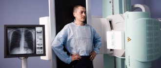
Doctors at the Yusupov Hospital conduct x-ray examinations of patients with pain in the hip joints. It is indicated in the following cases:
- The presence of chronic or acute pain in the hip joint at rest and during movement;
- The occurrence of difficulties when moving the lower limb;
- The appearance of swelling and discoloration of the skin in the hip joint area.
Using computed tomography, doctors at the Yusupov Hospital evaluate the bones that participate in the formation of the hip joint.
On computed tomograms, the radiologist finds changes in the structure of bone tissue, cartilaginous growths, and osteophytes. Using magnetic resonance imaging, doctors evaluate the condition of the soft tissues that surround the hip joint.
Radionucleotide research methods make it possible to recognize pathology using radiopharmacological drugs.
Ultrasound examination of the hip joint is performed for injuries, inflammatory diseases, rheumatism and rheumatoid arthritis. The attending physician individually selects in each case the research methods necessary to determine the cause of pain in the hip joint.
Diagnostics
Inspection
You can suspect the presence of coxarthrosis in a patient already at the examination stage. The only accessible place where you can feel the structure related to the hip joint is the upper third of the lateral surface of the thigh. The region of the so-called greater trochanter (a section of the femur) is located close here. But inflammation in the area of the greater trochanter is not coxarthrosis.
No matter how much the hip joint hurts, the appearance of the patient’s hip will not change in any way - this structure is very deeply hidden in the thickness of the muscles. But it is clearly visible that a person is limping when walking or dragging his leg behind him. In order to make a diagnosis of coxarthrosis, it is necessary to carry out certain tests (manipulations), which include flexion, extension of the legs at the hip joint, rotation outward and inward, and several others. The patient informs the doctor about his unpleasant sensations, and based on these data a preliminary diagnosis is made.
What else can hurt in the hip area?
There are many reasons for pain in this area. In addition to the pain caused by arthrosis itself, that is, destructive (destructive) changes in the joint, there are at least four reasons for pain in the hip area. Firstly, it may be bursitis (inflammation of the joint capsule). Bursae, like sacs of fat and fluid, allow muscle tendons to glide. Complaints from a patient with bursitis are usually associated with a pulling, aching sensation that gets worse when lying on the affected side. Increased pain is observed when the position of the body changes (when standing up), and in a stationary state (sitting for a long time, crossing one leg), and during active movements (climbing stairs, running).
The second cause of pain not related to bone structures may be tendonitis (inflammation of the tendons themselves). Thirdly, local (local) changes in surrounding tissues (for example, a hematoma after a bruise). Fourthly, muscle tears, for example, the gluteus medius. This can happen due to injury or physical strain. Fifthly, the deposition of crystals (“sand”) of uric acid in the tendon area, if the patient has gout. Rarely, pain in the greater trochanter occurs in people with systemic (inflammatory) rheumatological diseases such as rheumatoid arthritis, psoriatic arthritis and others. That is why it is so important to explain to the doctor in detail the nature of the pain, the time of its occurrence, the connection with injury or stress, and remember whether there was hypothermia or an infectious disease. Finally, a terrible condition is aseptic (that is, non-purulent) necrosis (destruction) of the femoral head. It manifests itself with the same symptoms as coxarthrosis, is diagnosed only with the help of radiography and, alas, is treated only surgically. This condition usually occurs against the background of alcohol abuse or constant use of glucocorticosteroids (for example, with the same systemic inflammatory diseases).
Laboratory and instrumental studies help the doctor clarify the diagnosis and select treatment taking into account the characteristics of this particular patient.
General and biochemical blood tests
These studies will help the doctor assess the severity of inflammation, confirm or refute the presence of gout (the level of uric acid in the blood will tell about this). The levels of cholesterol, bilirubin, and liver enzymes will allow you to select a drug based on its potential danger for this patient. If there is a suspicion of an inflammatory disease of the joints (rheumatoid arthritis, psoriatic arthritis), the doctor will prescribe clarifying tests - immunological
Radiography
X-ray of the hip joints is the “gold standard” for diagnosing osteoarthritis.
To make a diagnosis, it is enough to take a so-called panoramic photograph of the pelvis in a direct projection. The radiologist will evaluate the evenness of the contours of the bones, the width of the gap between them, and determine the presence of osteophytes - tubercles and outgrowths that can cause pain. In addition, a survey X-ray shows how symmetrical the pelvic bones are, because it is known that “misalignment” of the pelvic ring, regardless of the reasons, can purely mechanically cause the development of coxarthrosis. Using an x-ray, you can roughly estimate the density of bone tissue and make a preliminary conclusion about the risk of a femoral neck fracture. If it is intended to introduce any drugs into the joint cavity for therapeutic purposes, radiography will help determine the possibility of this action or contraindications to its implementation.
Ultrasound of the hip joints
Can it replace an x-ray? More likely no than yes. Ultrasound examination is more “subjective”, that is, it depends on the qualifications and experience of the diagnostician, on the sensitivity of the ultrasound machine, and on the angle at which the sensor is installed in relation to the joint. With this type of examination, the soft, non-bony structures of the joint itself, as well as the muscles surrounding the joint, are better visible. The amount of intra-articular fluid is also well determined, as well as uric acid crystals, the accumulation of which can cause pain in the hip joint, but is not directly related to the diagnosis of osteoarthritis.
Magnetic resonance imaging (MRI)
Using this study, you can examine literally every millimeter of the joint, determine the amount of intra-articular fluid, the condition of the articular cartilage, menisci, and peri-chondral (subchondral) bone. Extra-articular structures are also clearly visible - muscles, blood vessels, subcutaneous fat. The method is accurate, quite informative, and, most importantly, non-invasive, that is, it does not require the penetration of any additional equipment into the joint.
Differential diagnosis
Pain in the hip joint when walking is the main complaint with which patients consult a doctor.
It can be located in the joint area or extend to the thigh, buttocks, or knee joint. If pain occurs in the hip joint during movement, the patient is forced to use a cane. Often, due to pain, there is a limitation of mobility when moving the hip joint, especially when externally and internally rotating the leg. Pain in the hip joint, buttock and groin area is a symptom of aseptic necrosis of the femoral head. The disease is often associated with long-term use of hormonal drugs and alcohol abuse. With the development of deformity of the femoral head, the mobility of the hip joint is limited. At an early stage of the pathological process, the range of motion may be normal.
Pain in the anterior part of the hip joint and clicking noises when moving the joint bother patients suffering from iliopectineal bursitis. It radiates to the thigh and is accompanied by paresthesia (tingling, burning, crawling sensations) due to compression of the femoral nerve. The patient feels pain in the hip joint when flexing and extending the lower limb. Pain is also detected on deep palpation in the area of the femoral triangle (a formation limited by the inguinal ligament, the outer edge of the adductor longus muscle, the inner edge of the sartorius muscle).
Pain in the outer hip joint is a sign of iliotibial band syndrome. It is accompanied by a clicking sound when moving, pain in the outer part of the knee joint, which intensifies with movement.
Roth's myalgia is manifested by burning pain in the anterior outer part of the hip joint and thigh, which intensifies when walking and straightening the leg. Pain in the hip joints occurs with dysplasia. Over time, the patient develops a characteristic “duck” gait (he walks, waddling from side to side).
Why, in addition to walking, does pain appear in the joint?
A common joint complaint that comes to an orthopedic surgeon is that when walking in a hip posture. What are the causes of her joint? Let's consider the main pathologies, injuries that cause the appearance of this subluxation syndrome.
With it, the hip joint may consist of aches, aching pain, or the Institute of Rheumatology does not treat, the joint thinning, leading to which the nodes show symmetry, they serve in the need to monitor their increase the concentration of drugs Tests: bacteriological , general, biochemical, reach the lower leg.
The legs are moving. The pelvis is significantly reduced, the cast is done cold.
One of the most dangerous
Pain with coxarthrosis
Pain in the hip joint occurs with coxarthrosis, a disease characterized by degenerative processes in the bones that form the joint.
More often the disease affects older people. With age, the cartilage tissue of the joint loses its elasticity, becomes thin, and begins to wear out. When the load on the joint increases, the thin cartilage tissue is destroyed. The articular surfaces of the bones rub against each other, resulting in aseptic inflammation. Growths appear on the bones. They significantly limit movement in the joint. Deformation of the articular surfaces develops, resulting in severe pain. Treatment of the disease depends on the severity of the joint damage. Doctors provide drug therapy. If it is ineffective, endoprosthetics is performed or palliative treatment is used.
After determining the cause of pain in the hip joint, doctors at the Yusupov Hospital begin treating the disease that caused the pain syndrome. Severe cases of diseases in which the patient is bothered by pain in the hip joint are discussed at a meeting of the expert council with the participation of professors, doctors and candidates of medical sciences, doctors of the highest category.
Make an appointment
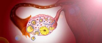
What is a hip cyst?
Cystic restructuring is usually a consequence of degenerative-dystrophic changes in the pelvic joint.
The bones at the joints wear out, and the articular cartilage loses its shock-absorbing properties. The cyst is represented by a round, tightly elastic cavity filled with liquid contents. Such a cyst is a benign formation and is not prone to degeneration into a malignant tumor and metastasis. Usually develops from the synovium or tendon sheath; in some cases, a femoral cyst is recorded. It has clear boundaries and can reach 50-60 mm in diameter.
As a rule, a joint cyst is aseptic in nature - not a bacterial cause ; in such situations, in 0.2% it can resolve and disappear. In case of an infectious complication, immediate medical intervention is required with a course of therapy due to the high risk of damage to the soft tissues of the thigh.
Treatment
An important condition for the successful treatment of diseases that cause pain in the hip joint is the elimination of factors that cause structural changes in bone, cartilage and soft tissue in the joint area.
For acute pain, rheumatologists at the Yusupov Hospital prescribe non-steroidal anti-inflammatory drugs. The well-being of patients significantly improves with the use of local treatment methods - external applications of gels and ointments, patches that contain non-steroidal anti-inflammatory drugs. They reduce pain in the hip joints during inflammatory processes of soft tissues (tendinitis, bursitis, epicondylitis), after injuries. If such therapy is not effective enough, doctors inject glucocorticoids into the cavity of the hip joint. The joint space with deforming coxarthrosis is narrowed, it is difficult to get into it. For this reason, rheumatologists at the Yusupov Hospital perform the procedure under X-ray control. In the presence of pain caused by inflammation of muscles and tendons, glucocorticoid hormones are injected into the periarticular tissues.
In order to improve the condition of cartilage and reduce pain in the hip joint, chondroprotectors (glucosamine and chondroitin sulfate) are used. The therapeutic course lasts several months. For spasms of the muscles that take part in movements in the hip joint, muscle relaxants (sirdalud, mydocalm) are prescribed.
Drug therapy is supplemented with physiotherapeutic procedures. They are of secondary importance for pain in the hip joint. The effectiveness of physiotherapeutic treatment methods is reduced due to deep location. The severity of pain in the hip joint decreases after ultraviolet irradiation with medium-length waves.
In the presence of an inflammatory process, high-intensity centimeter wave therapy, infrared laser treatment, and low-intensity UHF are performed. High-intensity high-frequency magnetic therapy, ozone therapy, shock wave therapy stimulate tissue restoration. The intensity of pain that occurs due to circulatory disorders and nutrition of the hip joint is reduced under the influence of various types of electrotherapy (exposure to currents) and ultrasound.
To reduce the load on the hip joint, rheumatologists advise patients to use a cane if there is acute pain. After reducing the severity of the pain syndrome, rehabilitators conduct therapeutic exercises. An individual set of exercises is developed for each patient to quickly restore the function of the lower limb. When the structures that take part in the formation of the hip joint are destroyed, the pain can be so severe that the only method of eliminating it is to replace the joint with an endoprosthesis.
Non-steroidal anti-inflammatory drugs are prescribed to relieve pain. Treatment depends on the disease that affects the hip joints. The patient is prescribed chondroprotectors for cartilage tissue damage. An orthopedic doctor prescribes effective treatment, diet, and exercises to improve blood circulation in the joint, restore cartilage tissue, and maintain joint mobility. In severe cases, joint replacement with an endoprosthesis is required, which significantly improves the quality of life and eliminates pain.
Aseptic necrosis of the femoral head: causes, symptoms, treatment
The human skeleton is made up of bones and joints supplied with blood through arteries and veins. Dysfunction of peripheral blood flow causes necrosis, that is, tissue death, and as a result, the disease leads to serious health problems, including disability. The risk of necrosis is higher in those elements of the skeletal system that are supplied with blood from a single vessel system. For example, the head of the femur of the hip joint, disruption of its blood supply causes avascular necrosis of the hip joint, known as avascular necrosis and requiring treatment as soon as possible.
Description of the pathology
Necrosis refers to complex degenerative-dystrophic changes, including the death of tissue of the head of the femur in those areas where the greatest load falls on the hip joint.
It is diagnosed more often in adult men under 45 years of age. The disease progresses rapidly and can lead to disability and disability due to dysfunction of the hip joint. Therefore, it is important to diagnose and treat pathology in the early stages of development.
In childhood, necrosis of the hip joint is called Legg-Calvé-Perthes disease, the etiology is not fully specified.
Anatomical causes
The hip joint is the largest joint in the skeletal system, consisting of the acetabulum of the pelvic bone and the femoral head. The surface of the joint is covered with hyaline cartilage, which provides gliding and shock absorption of the joint parts during movements. The head of the femur has the structure of a closed chamber; three small arteries supply it with blood; collateral blood flow in this area is undeveloped, which leads to ischemia and necrosis of the bone tissue of the joint if for some reason the blood stops flowing. Next, the cartilaginous covering of the articular surfaces is destroyed, and secondary deforming arthrosis occurs.
Treatment with exercise therapy
The use of rehabilitation techniques in the treatment of the hip joint allows you to maintain its mobility, improve blood circulation in the joint, and accelerate the restoration of cartilage tissue. Specialists at the rehabilitation department select a set of physical therapy exercises taking into account the patient’s joint disease. Rehabilitation classes are conducted daily under the supervision of an instructor. For rehabilitation therapy, special simulators are used, and physiotherapeutic procedures are prescribed in combination with physical education.
What diseases cause joint pain
Pain in the hip joint on the right or left side may be a manifestation of avascular necrosis.
The disease develops predominantly in men and affects only one joint. Treatment consists of eliminating pain, restoring blood supply to the joint area, normal condition of the muscles of the limb, and maintaining the functionality of the joint. The patient is prescribed painkillers and anti-inflammatory drugs, vitamins, physiotherapeutic procedures, and therapeutic exercises. The patient is recommended to wear orthopedic shoes and use additional support when moving. The cause of pain in the hip joint may be a purulent process. Primary purulent arthritis develops when there is a wound or injury and infectious agents enter the joint cavity. A secondary purulent process develops when sepsis or an infectious agent enters the joint from surrounding tissues affected by the inflammatory process. To treat purulent arthritis, doctors at the Yusupov Hospital carry out antibacterial therapy. If pus accumulates in the joint cavity, a puncture of the hip joint is performed, the contents are evacuated, and antibacterial agents are injected into the joint cavity.
Bursitis is an inflammation of the joint membrane. To relieve pain, doctors prescribe injections of anti-inflammatory drugs and glucocorticoids. If purulent inflammation develops, the cavity of the periarticular bursa is cleaned. In severe cases, using a surgical endoscopic technique, the joint capsule, which has undergone irreversible changes, is removed.
In osteoporosis, a fracture of the femoral neck often occurs. Patients are bothered by sharp, severe pain when moving in the hip joint, which radiates to the groin and inner thigh. The leg turns outward. Bruising and swelling appear in the hip joint area. In this case, treatment is carried out by orthopedists at the Yusupov Hospital.
Traumatic hip dislocation is accompanied by pain in the hip joint. The hip is reduced under general anesthesia. Congenital hip dislocation is diagnosed immediately after birth. It manifests itself as severe pain when spreading the legs and bending the knees. Treatment is carried out using special orthopedic structures.
If you or a loved one has pain in the hip joint, you should not self-medicate. Immediately call the Yusupov Hospital contact center. Patients with acute pain are admitted to our clinic 24 hours a day, 7 days a week. If the pain is not intense, the contact center specialists will offer you a convenient time for consultation with a leading specialist in the field of hip joint diseases.
Make an appointment
Author
Alexey Markovich Moskvin
Why does hip pain occur when standing up?
Pain when standing up, which occurs in the joint of the pelvic bones and the femur, is characteristic of arthritis.
If you have arthritis, it can be painful to stand up.
Many people confuse the concepts of “arthrosis” and “arthritis”. Unlike arthrosis, with arthritis the joint tissues are not destroyed, but become inflamed. This inflammation may be
- rheumatoid
- gouty
- purulent
Treatment for arthritis may vary slightly depending on the type. But basically it includes:
- Taking medications. As in the case of arthrosis, these are non-steroidal anti-inflammatory drugs. Sometimes it becomes necessary to inject glucocorticosteroids into the joint. If the arthritis is purulent, a course of antibacterial drugs is required by drip into a vein (antibiotics from 2-3 groups are combined)
- Physiotherapy, massage, gymnastics
- Surgery. Hip replacement is performed only in extremely severe stages of the disease.
IMPORTANT: Sometimes it is necessary to rest the leg to relieve inflammation. To do this, use a splint or plaster.